|
Longitudinal Striae
In human neuroanatomy, the longitudinal striae (also striae lancisi or nerves of Lancisi) are two bundles of fibres embedded in the indusium griseum running along the corpus callosum of the brain. They were originally described by Italian physician, epidemiologist and anatomist Giovanni Maria Lancisi. The striae are categorized as medial longitudinal stria and lateral longitudinal stria; the area between the striae is a useful neurosurgical mark of the middle of the corpus callosum. After the indisium griseum curves along the rostrum of the corpus callosum the combined striae continue toward the amygdala as part of the diagonal band of Broca The diagonal band of Broca is one of the basal forebrain structures that are derived from the ventral telencephalon during development. This structure forms the medial margin of the anterior perforated substance. This brain region was described b .... References External links UMichAtlas [...More Info...] [...Related Items...] OR: [Wikipedia] [Google] [Baidu] |
Neuroanatomy
Neuroanatomy is the study of the structure and organization of the nervous system. In contrast to animals with radial symmetry, whose nervous system consists of a distributed network of cells, animals with bilateral symmetry have segregated, defined nervous systems. Their neuroanatomy is therefore better understood. In vertebrates, the nervous system is segregated into the internal structure of the brain and spinal cord (together called the central nervous system, or CNS) and the routes of the nerves that connect to the rest of the body (known as the peripheral nervous system, or PNS). The delineation of distinct structures and regions of the nervous system has been critical in investigating how it works. For example, much of what neuroscientists have learned comes from observing how damage or "lesions" to specific brain areas affects behavior or other neural functions. For information about the composition of non-human animal nervous systems, see nervous system. For information ab ... [...More Info...] [...Related Items...] OR: [Wikipedia] [Google] [Baidu] |
Indusium Griseum
The indusium griseum, (supracallosal gyrus, gyrus epicallosus) consists of a thin membranous layer of grey matter in contact with the upper surface of the corpus callosum and continuous laterally with the grey matter of the cingulate cortex and inferiorly with the hippocampus. It is vestigial in humans and is a remnant of the former position of the hippocampus in lower animals. On either side of the midline of the indusium griseum are two ridges formed by bands of longitudinally directed fibers known as the medial and lateral longitudinal striae. The indusium griseum is prolonged around the splenium of the corpus callosum as a delicate layer, the fasciolar gyrus, which is continuous below with the surface of the dentate gyrus. Toward the genu of the corpus callosum it curves down along the rostrum to form the subcallosal gyrus The subcallosal gyrus (paraterminal gyrus, peduncle of the corpus callosum) is a narrow lamina on the medial surface of the hemisphere in front of the lam ... [...More Info...] [...Related Items...] OR: [Wikipedia] [Google] [Baidu] |
Corpus Callosum
The corpus callosum (Latin for "tough body"), also callosal commissure, is a wide, thick nerve tract, consisting of a flat bundle of commissural fibers, beneath the cerebral cortex in the brain. The corpus callosum is only found in placental mammals. It spans part of the longitudinal fissure, connecting the left and right cerebral hemispheres, enabling communication between them. It is the largest white matter structure in the human brain, about in length and consisting of 200–300 million axonal projections. A number of separate nerve tracts, classed as subregions of the corpus callosum, connect different parts of the hemispheres. The main ones are known as the genu, the rostrum, the trunk or body, and the splenium. Structure The corpus callosum forms the floor of the longitudinal fissure that separates the two cerebral hemispheres. Part of the corpus callosum forms the roof of the lateral ventricles. The corpus callosum has four main parts – individual nerve ... [...More Info...] [...Related Items...] OR: [Wikipedia] [Google] [Baidu] |
Brain
The brain is an organ that serves as the center of the nervous system in all vertebrate and most invertebrate animals. It consists of nervous tissue and is typically located in the head ( cephalization), usually near organs for special senses such as vision, hearing and olfaction. Being the most specialized organ, it is responsible for receiving information from the sensory nervous system, processing those information (thought, cognition, and intelligence) and the coordination of motor control (muscle activity and endocrine system). While invertebrate brains arise from paired segmental ganglia (each of which is only responsible for the respective body segment) of the ventral nerve cord, vertebrate brains develop axially from the midline dorsal nerve cord as a vesicular enlargement at the rostral end of the neural tube, with centralized control over all body segments. All vertebrate brains can be embryonically divided into three parts: the forebrain (prosencep ... [...More Info...] [...Related Items...] OR: [Wikipedia] [Google] [Baidu] |
Giovanni Maria Lancisi
Giovanni Maria Lancisi (26 October 1654 – 20 January 1720) was an Italian physician, epidemiologist and anatomist who made a correlation between the presence of mosquitoes and the prevalence of malaria. He was also known for his studies about cardiovascular diseases, an examination of the corpus callosum of the brain, and is remembered in the eponymous Lancisi's sign. He also studied rinderpest during an outbreak of the disease in Europe. Biography Giovanni Maria Lancisi (Latin name: Johannes Maria Lancisius) was born in Rome. His mother died shortly after his birth and he was raised by his aunt in Orvieto. He was educated at the Collegio Romano and the University of Rome, where he qualified in medicine aged 18. He worked at Santo Spirito and trained at the Picentine College, Lauro. In 1684 he went to Sapienza University and held the chair of anatomy for thirteen years. He served as physician to Popes Innocent XI, Clement XI and Innocent XII. He was given the lost anatomical ... [...More Info...] [...Related Items...] OR: [Wikipedia] [Google] [Baidu] |
Amygdala
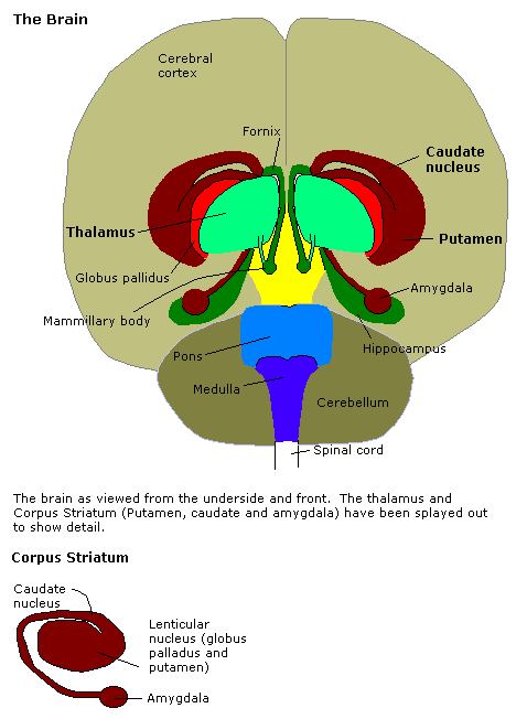
The amygdala (; plural: amygdalae or amygdalas; also '; Latin from Greek, , ', 'almond', 'tonsil') is one of two almond-shaped clusters of nuclei located deep and medially within the temporal lobes of the brain's cerebrum in complex vertebrates, including humans. Shown to perform a primary role in the processing of memory, decision making, and emotional responses (including fear, anxiety, and aggression), the amygdalae are considered part of the limbic system. The term "amygdala" was first introduced by Karl Friedrich Burdach in 1822. Structure The regions described as amygdala nuclei encompass several structures of the cerebrum with distinct connectional and functional characteristics in humans and other animals. Among these nuclei are the basolateral complex, the cortical nucleus, the medial nucleus, the central nucleus, and the intercalated cell clusters. The basolateral complex can be further subdivided into the lateral, the basal, and the accessory ba ... [...More Info...] [...Related Items...] OR: [Wikipedia] [Google] [Baidu] |
Diagonal Band Of Broca
The diagonal band of Broca is one of the basal forebrain structures that are derived from the ventral telencephalon during development. This structure forms the medial margin of the anterior perforated substance. This brain region was described by the French neuroanatomist Paul Broca. Structure It consists of fibers that are said to arise in the parolfactory area, the gyrus subcallosus and the anterior perforated substance, and course backward in the longitudinal striae to the dentate gyrus and the hippocampal region. This is a cholinergic bundle of nerve fibers posterior to the anterior perforated substance. It interconnects the subcallosal gyrus in the septal area with the hippocampus and lateral olfactory area. Nuclei Two structures are often described in this brain regions, namely the nuclei of the vertical and horizontal limbs of the diagonal band of Broca (nvlDBB and nhlDBB, respectively). nvlDBB projects to the hippocampal formation through the fornix and it is ... [...More Info...] [...Related Items...] OR: [Wikipedia] [Google] [Baidu] |


