|
Lockwood's Suspensory Ligament
The suspensory ligament of eyeball (or Lockwood's ligament) forms a hammock stretching below the eyeball between the medial and lateral check ligaments and enclosing the inferior rectus and inferior oblique muscles of the eye. It is a thickening of Tenon's capsule, the dense connective tissue capsule surrounding the globe and separating it from orbital fat. This ligament is responsible for maintaining and supporting the position of the eyeball in its normal upward and forward position within the orbit, and prevents downward displacement of the eyeball. It can be considered a part of the bulbar sheath. It is named for Charles Barrett Lockwood Charles Barrett Lockwood (23 September 1856 – 8 November 1914) was a British surgeon and anatomist who practiced surgery at St. Bartholomew's Hospital in London. Lockwood was a member of the Royal College of Surgeons. Lockwood is remembere .... References Human eye anatomy {{Eye-stub ... [...More Info...] [...Related Items...] OR: [Wikipedia] [Google] [Baidu] |
Inferior Rectus
{{disambiguation ...
Inferior may refer to: * Inferiority complex * An anatomical term of location * Inferior angle of the scapula, in the human skeleton * ''Inferior'' (book), by Angela Saini * ''The Inferior'', a 2007 novel by Peadar Ó Guilín See also *Junior (other) Junior or Juniors may refer to: Arts and entertainment Music * ''Junior'' (Junior Mance album), 1959 * ''Junior'' (Röyksopp album), 2009 * ''Junior'' (Kaki King album), 2010 * ''Junior'' (LaFontaines album), 2019 Films * ''Junior'' (1994 ... [...More Info...] [...Related Items...] OR: [Wikipedia] [Google] [Baidu] |
Inferior Oblique
The inferior oblique muscle or obliquus oculi inferior is a thin, narrow muscle placed near the anterior margin of the floor of the orbit. The inferior oblique is one of the extraocular muscles, and is attached to the maxillary bone (origin) and the posterior, inferior, lateral surface of the eye (insertion). The inferior oblique is innervated by the inferior branch of the oculomotor nerve. Structure The inferior oblique arises from the orbital surface of the maxilla, lateral to the lacrimal groove. Unlike the other extraocular muscles (recti and superior oblique), the inferior oblique muscle does ''not'' originate from the common tendinous ring ( annulus of Zinn). Passing lateralward, backward, and upward, between the inferior rectus and the floor of the orbit, and just underneath the lateral rectus muscle, the inferior oblique inserts onto the scleral surface between the inferior rectus and lateral rectus. In humans, the muscle is about 35 mm long. Innervation Th ... [...More Info...] [...Related Items...] OR: [Wikipedia] [Google] [Baidu] |
Tenon Capsule
Tenon's capsule (), also known as the Tenon capsule, fascial sheath of the eyeball () or the fascia bulbi, is a thin membrane which envelops the eyeball from the optic nerve to the corneal limbus, separating it from the orbital fat and forming a socket in which it moves. The inner surface of Tenon's capsule is smooth and is separated from the outer surface of the sclera by the periscleral lymph space. This lymph space is continuous with the subdural and subarachnoid cavities and is traversed by delicate bands of connective tissue which extend between the capsule and the sclera. The capsule is perforated behind by the ciliary vessels and nerves and fuses with the sheath of the optic nerve and with the sclera around the entrance of the optic nerve. In front it adheres to the conjunctiva, and both structures are attached to the ciliary region of the eyeball. The structure was named after Jacques-René Tenon (1724–1816), a French surgeon and pathologist. Structure Relations Te ... [...More Info...] [...Related Items...] OR: [Wikipedia] [Google] [Baidu] |
Orbit (anatomy)
In anatomy, the orbit is the cavity or socket of the skull in which the eye and its appendages are situated. "Orbit" can refer to the bony socket, or it can also be used to imply the contents. In the adult human, the volume of the orbit is , of which the eye occupies . The orbital contents comprise the eye, the orbital and retrobulbar fascia, extraocular muscles, cranial nerves II, III, IV, V, and VI, blood vessels, fat, the lacrimal gland with its sac and duct, the eyelids, medial and lateral palpebral ligaments, cheek ligaments, the suspensory ligament, septum, ciliary ganglion and short ciliary nerves. Structure The orbits are conical or four-sided pyramidal cavities, which open into the midline of the face and point back into the head. Each consists of a base, an apex and four walls."eye, human."Encyclopædia Britannica from Encyclopædia Britannica 2006 Ultimate Reference Suite DVD 2009 Openings There are two important foramina, or windows, two important fissu ... [...More Info...] [...Related Items...] OR: [Wikipedia] [Google] [Baidu] |
Ligament
A ligament is the fibrous connective tissue that connects bones to other bones. It is also known as ''articular ligament'', ''articular larua'', ''fibrous ligament'', or ''true ligament''. Other ligaments in the body include the: * Peritoneal ligament: a fold of peritoneum or other membranes. * Fetal remnant ligament: the remnants of a fetal tubular structure. * Periodontal ligament: a group of fibers that attach the cementum of teeth to the surrounding alveolar bone. Ligaments are similar to tendons and fasciae as they are all made of connective tissue. The differences among them are in the connections that they make: ligaments connect one bone to another bone, tendons connect muscle to bone, and fasciae connect muscles to other muscles. These are all found in the skeletal system of the human body. Ligaments cannot usually be regenerated naturally; however, there are periodontal ligament stem cells located near the periodontal ligament which are involved in the adult regener ... [...More Info...] [...Related Items...] OR: [Wikipedia] [Google] [Baidu] |
Bulbar Sheath
Tenon's capsule (), also known as the Tenon capsule, fascial sheath of the eyeball () or the fascia bulbi, is a thin membrane which envelops the eyeball from the optic nerve to the corneal limbus, separating it from the orbital fat and forming a socket in which it moves. The inner surface of Tenon's capsule is smooth and is separated from the outer surface of the sclera by the periscleral lymph space. This lymph space is continuous with the subdural and subarachnoid cavities and is traversed by delicate bands of connective tissue which extend between the capsule and the sclera. The capsule is perforated behind by the ciliary vessels and nerves and fuses with the sheath of the optic nerve and with the sclera around the entrance of the optic nerve. In front it adheres to the conjunctiva, and both structures are attached to the ciliary region of the eyeball. The structure was named after Jacques-René Tenon (1724–1816), a French surgeon and pathologist. Structure Relations Te ... [...More Info...] [...Related Items...] OR: [Wikipedia] [Google] [Baidu] |
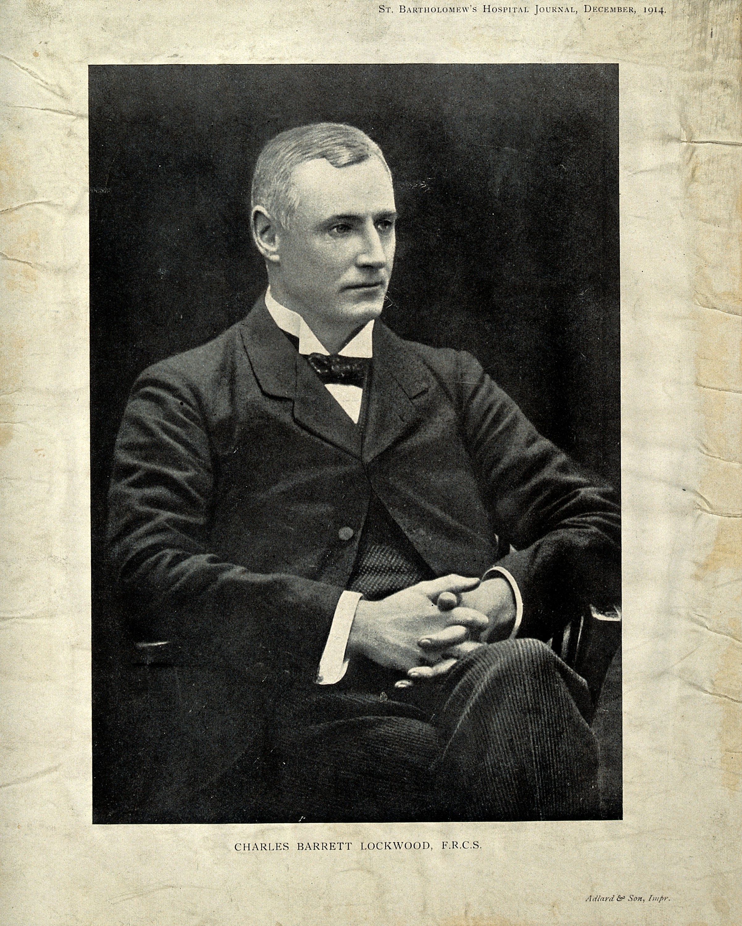
Charles Barrett Lockwood
Charles Barrett Lockwood (23 September 1856 – 8 November 1914) was a British surgeon and anatomist who practiced surgery at St. Bartholomew's Hospital in London. Lockwood was a member of the Royal College of Surgeons. Lockwood is remembered for his surgical work with femoral and inguinal hernias. He developed an infra- inguinal approach for femoral hernia operations that is known today as the "low approach" or "Lockwood's operation". In 1893, he published an important book titled "Radical Cure of Femoral and Inguinal Hernia". He first conceived the idea of an Anatomical Society in 1887, acted as their first treasurer and was elected president of the society for 1901 to 1903. The " Lockwood's suspensory ligament" of the eye is named after him. This structure is the thickened area of contact between Tenon's capsule and the sheaths of the inferior rectus and inferior oblique muscles. This ligament is responsible for maintaining the position of the eyeball in its norma ... [...More Info...] [...Related Items...] OR: [Wikipedia] [Google] [Baidu] |


