|
List Of MeSH Codes (A10)
The following is a partial list of the "A" codes for Medical Subject Headings (MeSH), as defined by the United States National Library of Medicine The United States National Library of Medicine (NLM), operated by the United States federal government, is the world's largest medical library. Located in Bethesda, Maryland, the NLM is an institute within the National Institutes of Health. Its ... (NLM). This list continues the information at List of MeSH codes (A09). Codes following these are found at List of MeSH codes (A11). For other MeSH codes, see List of MeSH codes. The source for this content is the set o2006 MeSH Treesfrom the NLM. – Biological tissue, tissues – connective tissue * – adipose tissue * – abdominal fat * – intra-abdominal fat * – subcutaneous fat, abdominal * – brown fat * – subcutaneous fat * – subcutaneous fat, abdominal * – bone and bones * – bone matrix * – bony callus * – haversian system * – periosteum * – ... [...More Info...] [...Related Items...] OR: [Wikipedia] [Google] [Baidu] |
Medical Subject Headings
Medical Subject Headings (MeSH) is a comprehensive controlled vocabulary for the purpose of indexing journal articles and books in the life sciences. It serves as a thesaurus that facilitates searching. Created and updated by the United States National Library of Medicine (NLM), it is used by the MEDLINE/PubMed article database and by NLM's catalog of book holdings. MeSH is also used by ClinicalTrials.gov registry to classify which diseases are studied by trials registered in ClinicalTrials. MeSH was introduced in the 1960s, with the NLM's own index catalogue and the subject headings of the Quarterly Cumulative Index Medicus (1940 edition) as precursors. The yearly printed version of MeSH was discontinued in 2007; MeSH is now available only online. It can be browsed and downloaded free of charge through PubMed. Originally in English, MeSH has been translated into numerous other languages and allows retrieval of documents from different origins. Structure MeSH vocabulary is divi ... [...More Info...] [...Related Items...] OR: [Wikipedia] [Google] [Baidu] |
Periosteum
The periosteum is a membrane that covers the outer surface of all bones, except at the articular surfaces (i.e. the parts within a joint space) of long bones. Endosteum lines the inner surface of the medullary cavity of all long bones. Structure The periosteum consists of an outer fibrous layer, and an inner cambium layer (or osteogenic layer). The fibrous layer is of dense irregular connective tissue, containing fibroblasts, while the cambium layer is highly cellular containing progenitor cells that develop into osteoblasts. These osteoblasts are responsible for increasing the width of a long bone and the overall size of the other bone types. After a bone fracture, the progenitor cells develop into osteoblasts and chondroblasts, which are essential to the healing process. The outer fibrous layer and the inner cambium layer is differentiated under electron micrography. As opposed to osseous tissue, the periosteum has nociceptors, sensory neurons that make it very sensitive to ... [...More Info...] [...Related Items...] OR: [Wikipedia] [Google] [Baidu] |
Endothelium
The endothelium is a single layer of squamous endothelial cells that line the interior surface of blood vessels and lymphatic vessels. The endothelium forms an interface between circulating blood or lymph in the lumen and the rest of the vessel wall. Endothelial cells form the barrier between vessels and tissue and control the flow of substances and fluid into and out of a tissue. Endothelial cells in direct contact with blood are called vascular endothelial cells whereas those in direct contact with lymph are known as lymphatic endothelial cells. Vascular endothelial cells line the entire circulatory system, from the heart to the smallest capillaries. These cells have unique functions that include fluid filtration, such as in the glomerulus of the kidney, blood vessel tone, hemostasis, neutrophil recruitment, and hormone trafficking. Endothelium of the interior surfaces of the heart chambers is called endocardium. An impaired function can lead to serious health issues throug ... [...More Info...] [...Related Items...] OR: [Wikipedia] [Google] [Baidu] |
Bruch Membrane
Bruch's membrane is the innermost layer of the choroid of the eye. It is also called the ''vitreous lamina'' or ''Membrane vitriae'', because of its glassy microscopic appearance. It is 2–4 μm thick. Layers Bruch's membrane consists of five layers (from inside to outside): #the basement membrane of the retinal pigment epithelium #the inner collagenous zone #a central band of elastic fibers #the outer collagenous zone #the basement membrane of the choriocapillaris The retinal pigment epithelium transports metabolic waste from the photoreceptors across Bruch's membrane to the choroid. Embryology Bruch's membrane is present by midterm in fetal development as an elastic sheet. Pathology Bruch's membrane thickens with age, slowing the transport of metabolites. This may lead to the formation of drusen in age-related macular degeneration. There is also a buildup of deposits (Basal Linear Deposits or BLinD and Basal Lamellar Deposits BLamD) on and within the membrane, primarily ... [...More Info...] [...Related Items...] OR: [Wikipedia] [Google] [Baidu] |
Basement Membrane
The basement membrane is a thin, pliable sheet-like type of extracellular matrix that provides cell and tissue support and acts as a platform for complex signalling. The basement membrane sits between Epithelium, epithelial tissues including mesothelium and endothelium, and the underlying connective tissue. Structure As seen with the electron microscope, the basement membrane is composed of two layers, the basal lamina and the reticular lamina. The underlying connective tissue attaches to the basal lamina with collagen VII anchoring fibrils and fibrillin microfibrils. The basal lamina layer can further be subdivided into two layers based on their visual appearance in electron microscopy. The lighter-colored layer closer to the epithelium is called the lamina lucida, while the denser-colored layer closer to the connective tissue is called the lamina densa. The Electron microscope, electron-dense lamina densa layer is about 30–70 nanometers thick and consists of an underlying ... [...More Info...] [...Related Items...] OR: [Wikipedia] [Google] [Baidu] |
Epithelium
Epithelium or epithelial tissue is one of the four basic types of animal tissue, along with connective tissue, muscle tissue and nervous tissue. It is a thin, continuous, protective layer of compactly packed cells with a little intercellular matrix. Epithelial tissues line the outer surfaces of organs and blood vessels throughout the body, as well as the inner surfaces of cavities in many internal organs. An example is the epidermis, the outermost layer of the skin. There are three principal shapes of epithelial cell: squamous (scaly), columnar, and cuboidal. These can be arranged in a singular layer of cells as simple epithelium, either squamous, columnar, or cuboidal, or in layers of two or more cells deep as stratified (layered), or ''compound'', either squamous, columnar or cuboidal. In some tissues, a layer of columnar cells may appear to be stratified due to the placement of the nuclei. This sort of tissue is called pseudostratified. All glands are made up of epithe ... [...More Info...] [...Related Items...] OR: [Wikipedia] [Google] [Baidu] |
Subcutaneous Tissue
The subcutaneous tissue (), also called the hypodermis, hypoderm (), subcutis, superficial fascia, is the lowermost layer of the integumentary system in vertebrates. The types of cells found in the layer are fibroblasts, adipose cells, and macrophages. The subcutaneous tissue is derived from the mesoderm, but unlike the dermis, it is not derived from the mesoderm's dermatome region. It consists primarily of loose connective tissue, and contains larger blood vessels and nerves than those found in the dermis. It is a major site of fat storage in the body. In arthropods, a hypodermis can refer to an epidermal layer of cells that secretes the chitinous cuticle. The term also refers to a layer of cells lying immediately below the epidermis of plants. Structure * Fibrous bands anchoring the skin to the deep fascia * Collagen and elastin fibers attaching it to the dermis * Fat is absent from the eyelids, clitoris, penis, much of pinna, and scrotum * Blood vessels on route to the der ... [...More Info...] [...Related Items...] OR: [Wikipedia] [Google] [Baidu] |
Cicatrix
''Cicatrix'' is a genus of wasp found in Australia. It was erected in 2011 following a revision of ''Mikeius''. Species within ''Cicatrix'': * '' Cicatrix pilosiscutum'' (Girault, 1929) * '' Cicatrix neumanoides'' Paretas-Martínez & Restrepo-Ortiz, 2011 * ''Cicatrix schauffi ''Cicatrix schauffi'' is a species of wasp found in Australia. It was initially described as ''Mikeius schauffi'' in 2008 by Matthew Buffington. In a 2011 revision, the species was moved from '' Mikeius'' to the newly erected genus ''Cicatrix ...'' (Girault, 1929) References Hymenoptera genera Insects described in 2011 Cynipoidea {{Wasp-stub ... [...More Info...] [...Related Items...] OR: [Wikipedia] [Google] [Baidu] |
Granulation Tissue
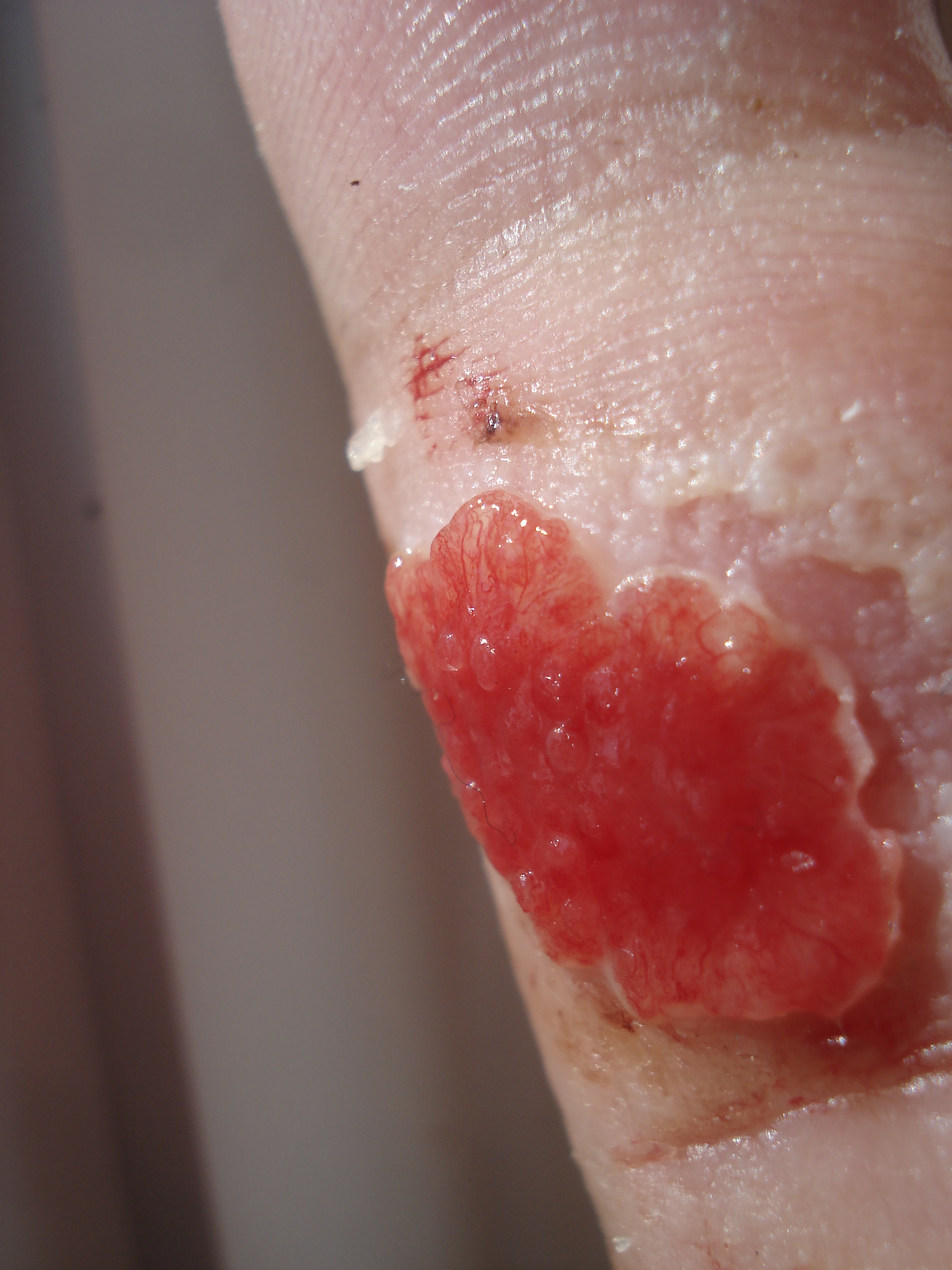
Granulation tissue is new connective tissue and microscopic blood vessels that form on the surfaces of a wound during the healing process. Granulation tissue typically grows from the base of a wound and is able to fill wounds of almost any size. Examples of granulation tissue can be seen in pyogenic granulomas and pulp polyps. Its histological appearance is characterized by proliferation of fibroblasts and new thin-walled, delicate capillaries (angiogenesis), infiltrated inflammatory cells in a loose extracellular matrix. Appearance During the migratory phase of wound healing, granulation tissue is: * light red or dark pink, being perfused with new capillary loops or "buds"; * soft to the touch; * moist; * bumpy (granular) in appearance, due to punctate hemorrhages; * pulsatile on palpation; * painless when healthy; Structure Granulation tissue is composed of tissue matrix supporting a variety of cell types, most of which can be associated with one of the following functions: ... [...More Info...] [...Related Items...] OR: [Wikipedia] [Google] [Baidu] |
Elastic Tissue
Elastic fibers (or yellow fibers) are an essential component of the extracellular matrix composed of bundles of proteins ( elastin) which are produced by a number of different cell types including fibroblasts, endothelial, smooth muscle, and airway epithelial cells. These fibers are able to stretch many times their length, and snap back to their original length when relaxed without loss of energy. Elastic fibers include elastin, elaunin and oxytalan. Elastic tissue is classified as "connective tissue proper". Elastic fibers are formed via elastogenesis, a highly complex process involving several key proteins including fibulin-4, fibulin-5, latent transforming growth factor β binding protein 4, and microfibril associated protein 4. In this process tropoelastin, the soluble monomeric precursor to elastic fibers is produced by elastogenic cells and chaperoned to the cell surface. Following excretion from the cell, tropoelastin self associates into ~200 nm particles by coac ... [...More Info...] [...Related Items...] OR: [Wikipedia] [Google] [Baidu] |
Hyaline Cartilage
Hyaline cartilage is the glass-like (hyaline) and translucent cartilage found on many joint surfaces. It is also most commonly found in the ribs, nose, larynx, and trachea. Hyaline cartilage is pearl-gray in color, with a firm consistency and has a considerable amount of collagen. It contains no nerves or blood vessels, and its structure is relatively simple. Structure Hyaline cartilage is covered externally by a fibrous membrane known as the perichondrium or, when it's along articulating surfaces, the synovial membrane. This membrane contains vessels that provide the cartilage with nutrition through diffusion. Hyaline cartilage matrix is primarily made of type II collagen and chondroitin sulphate, both of which are also found in elastic cartilage. Hyaline cartilage exists on the sternal ends of the ribs, in the larynx, trachea, and bronchi, and on the articulating surfaces of bones. It gives the structures a definite but pliable form. The presence of collagen fibres makes suc ... [...More Info...] [...Related Items...] OR: [Wikipedia] [Google] [Baidu] |
Triangular Fibrocartilage
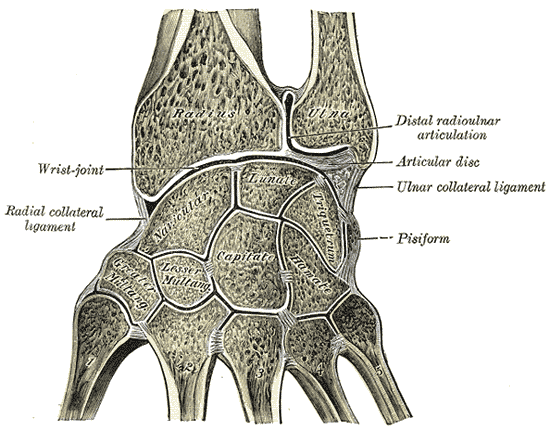
The triangular fibrocartilage complex (TFCC) is formed by the triangular fibrocartilage discus (TFC), the radioulnar ligaments (RULs) and the ulnocarpal ligaments (UCLs). Structure Triangular fibrocartilage disc The triangular fibrocartilage disc (TFC) is an articular discus that lies on the pole of the distal ulna. It has a triangular shape and a biconcave body; the periphery is thicker than its center. The central portion of the TFC is thin and consists of chondroid fibrocartilage; this type of tissue is often seen in structures that can bear compressive loads. This central area is often so thin that it is translucent and in some cases it is even absent. The peripheral portion of the TFC is well vascularized, while the central portion has no blood supply. This discus is attached by thick tissue to the base of the ulnar styloid and by thinner tissue to the edge of the radius just proximal to the radiocarpal articular surface. Radioulnar ligaments The radioulnar ligaments ... [...More Info...] [...Related Items...] OR: [Wikipedia] [Google] [Baidu] |


