|
Lipoma Ultrasound 110322120428 1206550
A lipoma is a benign tumor made of fat tissue. They are generally soft to the touch, movable, and painless. They usually occur just under the skin, but occasionally may be deeper. Most are less than in size. Common locations include upper back, shoulders, and abdomen. It is possible to have a number of lipomas. The cause is generally unclear. Risk factors include family history, obesity, and lack of exercise. Diagnosis is typically based on a physical exam. Occasionally medical imaging or tissue biopsy is used to confirm the diagnosis. Treatment is typically by observation or surgical removal. Rarely, the condition may recur following removal, but this can generally be managed with repeat surgery. They are not generally associated with a future risk of cancer. Lipomas have a prevalence of roughly 2 out of every 100 people. Lipomas typically occur in adults between 40 and 60 years of age. Males are more often affected than females. They are the most common noncancerous soft-t ... [...More Info...] [...Related Items...] OR: [Wikipedia] [Google] [Baidu] |
Forearm
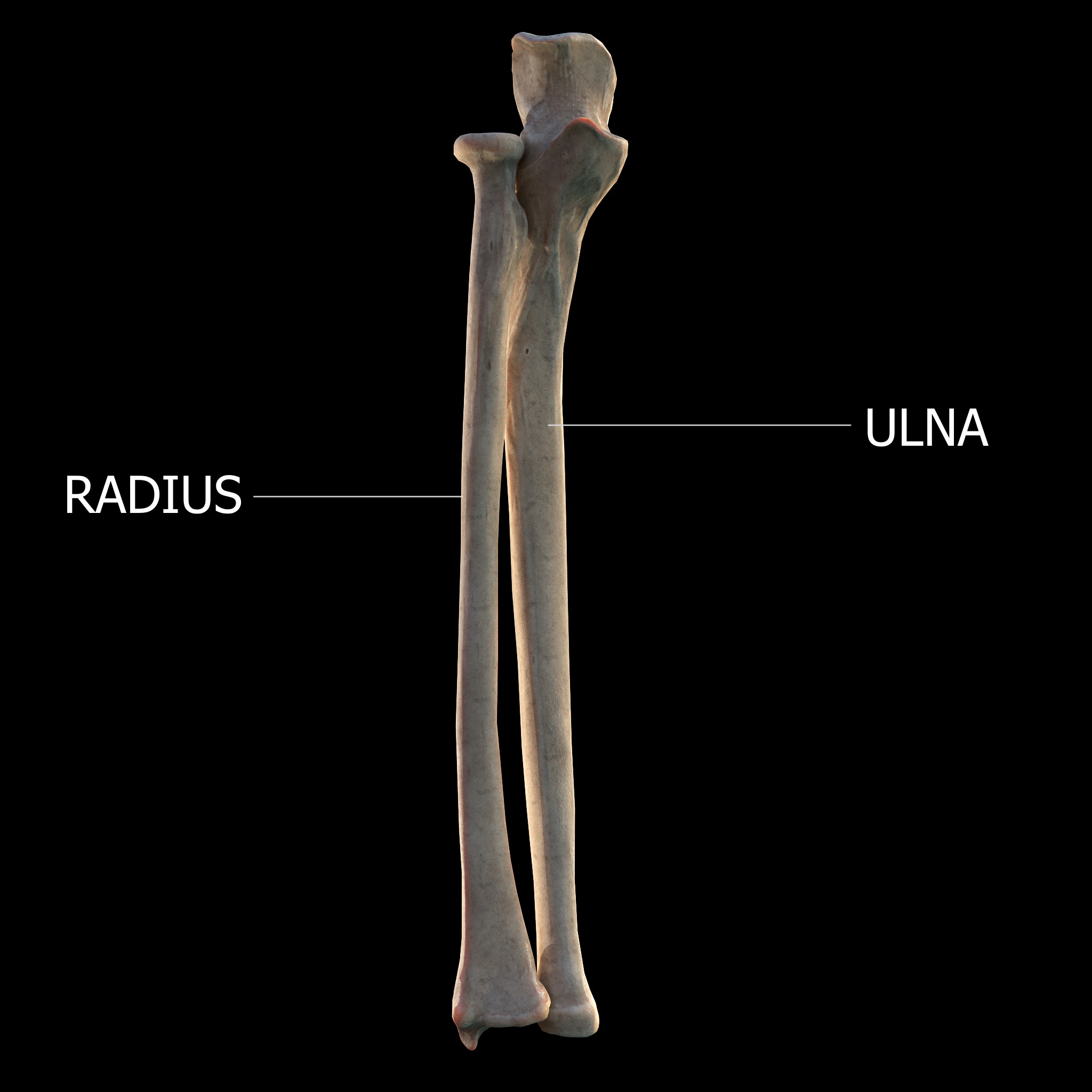
The forearm is the region of the upper limb between the elbow and the wrist. The term forearm is used in anatomy to distinguish it from the arm, a word which is most often used to describe the entire appendage of the upper limb, but which in anatomy, technically, means only the region of the upper arm, whereas the lower "arm" is called the forearm. It is homologous to the region of the leg that lies between the knee and the ankle joints, the crus. The forearm contains two long bones, the radius and the ulna, forming the two radioulnar joints. The interosseous membrane connects these bones. Ultimately, the forearm is covered by skin, the anterior surface usually being less hairy than the posterior surface. The forearm contains many muscles, including the flexors and extensors of the wrist, flexors and extensors of the digits, a flexor of the elbow (brachioradialis), and pronators and supinators that turn the hand to face down or upwards, respectively. In cross-section, the for ... [...More Info...] [...Related Items...] OR: [Wikipedia] [Google] [Baidu] |
Histopathology Of A Fibrolipoma
Histopathology (compound of three Greek words: ''histos'' "tissue", πάθος ''pathos'' "suffering", and -λογία ''-logia'' "study of") refers to the microscopic examination of tissue in order to study the manifestations of disease. Specifically, in clinical medicine, histopathology refers to the examination of a biopsy or surgical specimen by a pathologist, after the specimen has been processed and histological sections have been placed onto glass slides. In contrast, cytopathology examines free cells or tissue micro-fragments (as "cell blocks"). Collection of tissues Histopathological examination of tissues starts with surgery, biopsy, or autopsy. The tissue is removed from the body or plant, and then, often following expert dissection in the fresh state, placed in a fixative which stabilizes the tissues to prevent decay. The most common fixative is 10% neutral buffered formalin (corresponding to 3.7% w/v formaldehyde in neutral buffered water, such as phosphate buff ... [...More Info...] [...Related Items...] OR: [Wikipedia] [Google] [Baidu] |
Intradermal Spindle Cell Lipoma
Intradermal spindle cell lipoma is distinct in that it most commonly affects women, and has a wide distribution, occurring with relatively equal frequency on the head and neck, trunk, and upper and lower extremities.James, William; Berger, Timothy; Elston, Dirk (2005). ''Andrews' Diseases of the Skin: Clinical Dermatology''. (10th ed.). Saunders. . See also * Spindle cell lipoma * List of cutaneous conditions Many skin conditions affect the human integumentary system—the organ system covering the entire surface of the body and composed of skin, hair, nails, and related muscle and glands. The major function of this system is as a barrier against t ... References Dermal and subcutaneous growths {{Dermal-growth-stub ... [...More Info...] [...Related Items...] OR: [Wikipedia] [Google] [Baidu] |
Brown Fat
Brown adipose tissue (BAT) or brown fat makes up the adipose organ together with white adipose tissue (or white fat). Brown adipose tissue is found in almost all mammals. Classification of brown fat refers to two distinct cell populations with similar functions. The first shares a common embryological origin with muscle cells, found in larger "classic" deposits. The second develops from white adipocytes that are stimulated by the sympathetic nervous system. These adipocytes are found interspersed in white adipose tissue and are also named 'beige' or 'brite' (for "brown in white"). Brown adipose tissue is especially abundant in newborns and in hibernating mammals. It is also present and metabolically active in adult humans, but its prevalence decreases as humans age. Its primary function is thermoregulation. In addition to heat produced by shivering muscle, brown adipose tissue produces heat by non-shivering thermogenesis. The therapeutic targeting of brown fat for the treatment ... [...More Info...] [...Related Items...] OR: [Wikipedia] [Google] [Baidu] |
Hibernoma
A hibernoma is a benign neoplasm of vestigial brown fat. The term was originally used by the French anatomist Louis Gery in 1914. Signs and symptoms Patients present with a slow-growing, painless, solitary mass, usually of the subcutaneous tissues. It is much less frequently noted in the intramuscular tissue. It is not uncommon for symptoms to be present for years. Benign neoplasm with brown fat is noted. Diagnosis Imaging findings In general, imaging studies show a well-defined, heterogeneous mass, usually showing a mass which is hypointense to subcutaneous fat on magnetic resonance T1-weight images. Serpentine, thin, low signal bands (septations or vessels) are often seen throughout the tumor. Pathology findings From a macroscopic perspective, there is a well-defined, encapsulated or circumscribed mass, showing a soft, yellow tan to deep brown mass. The size ranges from 1 to 27 cm, although the mean is about 10 cm. The tumors histologically resemble brown fat. ... [...More Info...] [...Related Items...] OR: [Wikipedia] [Google] [Baidu] |
Commissure
A commissure () is the location at which two objects abut or are joined. The term is used especially in the fields of anatomy and biology. * The most common usage of the term refers to the brain's commissures, of which there are five. Such a commissure is a bundle of commissural fibers as a tract that crosses the midline at its level of origin or entry (as opposed to a decussation of fibers that cross obliquely). The five are the anterior commissure, posterior commissure, corpus callosum, commissure of fornix (hippocampal commissure), and habenular commissure. They consist of fibre tracts that connect the two cerebral hemispheres and span the longitudinal fissure. In the spinal cord there are the anterior white commissure, and the gray commissure. ''Commissural neurons'' refer to neuronal cells that grow their axons across the midline of the nervous system within the brain and the spinal cord. * ''Commissure'' also often refers to cardiac anatomy of heart valves. In the heart, a co ... [...More Info...] [...Related Items...] OR: [Wikipedia] [Google] [Baidu] |
Corpus Callosum
The corpus callosum (Latin for "tough body"), also callosal commissure, is a wide, thick nerve tract, consisting of a flat bundle of commissural fibers, beneath the cerebral cortex in the brain. The corpus callosum is only found in placental mammals. It spans part of the longitudinal fissure, connecting the left and right cerebral hemispheres, enabling communication between them. It is the largest white matter structure in the human brain, about in length and consisting of 200–300 million axonal projections. A number of separate nerve tracts, classed as subregions of the corpus callosum, connect different parts of the hemispheres. The main ones are known as the genu, the rostrum, the trunk or body, and the splenium. Structure The corpus callosum forms the floor of the longitudinal fissure that separates the two cerebral hemispheres. Part of the corpus callosum forms the roof of the lateral ventricles. The corpus callosum has four main parts – individual nerve tracts ... [...More Info...] [...Related Items...] OR: [Wikipedia] [Google] [Baidu] |
Chondroid Lipoma
Chondroid lipomas are deep-seated, firm, yellow tumors that characteristically occur on the legs of women. They exhibit a characteristic genetic translocation t(11;16) with a resulting C11orf95-MKL2 fusion oncogene.James, William; Berger, Timothy; Elston, Dirk (2005). ''Andrews' Diseases of the Skin: Clinical Dermatology''. (10th ed.). Saunders. .Genes Chromosomes Cancer. 2010 Sep;49(9):810-8. doi: 10.1002/gcc.20788. C11orf95-MKL2 is the resulting fusion oncogene of t(11;16)(q13;p13) in chondroid lipoma. Huang D1, Sumegi J, Dal Cin P, Reith JD, Yasuda T, Nelson M, Muirhead D, Bridge JA. See also *Lipoma *Skin lesion *List of cutaneous conditions Many skin conditions affect the human integumentary system—the organ system covering the entire surface of the body and composed of skin, hair, nails, and related muscle and glands. The major function of this system is as a barrier agai ... References External links Dermal and subcutaneous growths {{Dermal-gro ... [...More Info...] [...Related Items...] OR: [Wikipedia] [Google] [Baidu] |
Internal Auditory Canal
The internal auditory meatus (also meatus acusticus internus, internal acoustic meatus, internal auditory canal, or internal acoustic canal) is a canal within the petrous part of the temporal bone of the skull between the posterior cranial fossa and the inner ear. Structure The opening to the meatus is called the porus acusticus internus or internal acoustic opening. It is located inside the posterior cranial fossa of the skull, near the center of the posterior surface of the petrous part of the temporal bone. The size varies considerably. Its outer margins are smooth and rounded. The canal which comprises the internal auditory meatus is short (about 1 cm) and runs laterally into the bone. The lateral (outer) aspect of the canal is known as the fundus. The fundus is subdivided by two thin crests of bone to form three separate canals, through which course the facial and vestibulocochlear nerve branches. The falciform crest first divides the meatus into superior and inferior ... [...More Info...] [...Related Items...] OR: [Wikipedia] [Google] [Baidu] |
Cerebellar Pontine Angle
The cerebellum (Latin for "little brain") is a major feature of the hindbrain of all vertebrates. Although usually smaller than the cerebrum, in some animals such as the mormyrid fishes it may be as large as or even larger. In humans, the cerebellum plays an important role in motor control. It may also be involved in some cognitive functions such as attention and language as well as emotional control such as regulating fear and pleasure responses, but its movement-related functions are the most solidly established. The human cerebellum does not initiate movement, but contributes to coordination, precision, and accurate timing: it receives input from sensory systems of the spinal cord and from other parts of the brain, and integrates these inputs to fine-tune motor activity. Cerebellar damage produces disorders in fine movement, equilibrium, posture, and motor learning in humans. Anatomically, the human cerebellum has the appearance of a separate structure attached to the bottom ... [...More Info...] [...Related Items...] OR: [Wikipedia] [Google] [Baidu] |
Angiolipoma
Angiolipoma is a subcutaneous nodule with vascular structure, having all other features of a typical lipoma. They are commonly painful.James, William; Berger, Timothy; Elston, Dirk (2005). ''Andrews' Diseases of the Skin: Clinical Dermatology''. (10th ed.). Saunders. . Pathology Histopathology File:Angiolipoma (4670135502).jpg, Small vessels in adipose tissue File:Angiolipoma (4669511063).jpg, Small vessels in adipose tissue File:Skin Tumors-PA030897.jpg, Small vessels in adipose tissue File:Skin Tumors-PA030898.jpg, Small vessels in adipose tissue File:Histopathology of angiolipoma.jpg, The vessels typically contain hyaline or fibrin (pictured) thrombi Topic Completed: 1 August 2012. Minor changes: 20 March 2019 Diagnosis Differential diagnosis * Kaposi sarcoma * Angiosarcoma See also * Lipoma * Skin lesion A skin condition, also known as cutaneous condition, is any medical condition that affects the integumentary system—the organ system that encloses the bod ... [...More Info...] [...Related Items...] OR: [Wikipedia] [Google] [Baidu] |


