|
Lipofibromatosis
Lipofibromatosis (LPF) is an extremely rare soft tissue tumor which was first clearly described in 2000 by Fetsch et al as a strictly pediatric, locally invasive, and often recurrent (at the site of its surgical removal) tumor. It is nonetheless a non- metastasizing, i.e. benign, tumor. While even the more recent literature has sometimes regarded LPF as a strictly childhood disorder, rare cases of LPF has been diagnosed in adults. The diagnosis of lipofibromatosis should not be automatically discarded because of an individual's age. Based primarily on histopathologic (i.e. microscopic appearance of specially prepared tissue) analyses, lipofibromatosis was initially regarded as either a type of, or very similar to, aponeurotic fibroma (also termed calcifying aponeurotic fibroma), fibrous hamartoma of infancy, ''EWSRI-SMAD3''-rearranged fibroblastic tumor (also termed ''EWSR1-SMAD3''-positive fibroblastic tumor), or infantile digital fibromatosis. However, further analyses of these ... [...More Info...] [...Related Items...] OR: [Wikipedia] [Google] [Baidu] |
Lipofibromatosis-like Neural Tumor
Lipofibromatosis-like neural tumor (LPF-NT) is an extremely rare soft tissue tumor first described by Agaram ''et al'' in 2016. As of mid-2021, at least 39 cases of LPF-NT have been reported in the literature. LPF-NT tumors have several features that resemble lipofibromatosis (LPF) tumors, malignant peripheral nerve sheath tumors, spindle cell sarcomas, low-grade Nervous tissue#tumours, neural tumors, peripheral nerve sheath tumors, and other less clearly defined tumors; Prior to the Agaram ''at al'' report, LPF-NTs were likely diagnosed as variants or atypical forms of these tumors. The analyses of Agaram ''at al'' and subsequent studies uncovered critical differences between LPF-NT and the other tumor forms which suggest that it is a distinct tumor entity differing not only from lipofibromatosis but also the other tumor forms. LPF-NTs are locally invasive, are commonly treated by surgical excision, and have a relatively high rate of local recurrence if their surgical excisions ar ... [...More Info...] [...Related Items...] OR: [Wikipedia] [Google] [Baidu] |
Fibroblastic And Myofibroblastic Tumors
Fibroblastic and myofibroblastic tumors (FMTs) develop from the mesenchymal stem cells which differentiate into fibroblasts (the most common cell type in connective tissue) and/or the myocytes/myoblasts that differentiate into muscle cells. FMTs are a heterogeneous group of soft tissue neoplasms (i.e. abnormal and excessive tissue growths). The World Health Organization (2020) defined tumors as being FMTs based on their morphology and, more importantly, newly discovered abnormalities in the expression levels of key gene products made by these tumors' neoplastic cells. Histopathologically, FMTs consist of neoplastic connective tissue cells which have differented into cells that have microscopic appearances resembling fibroblasts and/or myofibroblasts. The fibroblastic cells are characterized as spindle-shaped cells with inconspicuous nucleoli that express vimentin, an intracellular protein typically found in mesenchymal cells, and CD34, a cell surface membrane glycoprotein. Myo ... [...More Info...] [...Related Items...] OR: [Wikipedia] [Google] [Baidu] |
Pediatrics
Pediatrics ( also spelled ''paediatrics'' or ''pædiatrics'') is the branch of medicine that involves the medical care of infants, children, adolescents, and young adults. In the United Kingdom, paediatrics covers many of their youth until the age of 18. The American Academy of Pediatrics recommends people seek pediatric care through the age of 21, but some pediatric subspecialists continue to care for adults up to 25. Worldwide age limits of pediatrics have been trending upward year after year. A medical doctor who specializes in this area is known as a pediatrician, or paediatrician. The word ''pediatrics'' and its cognates mean "healer of children," derived from the two Greek words: (''pais'' "child") and (''iatros'' "doctor, healer"). Pediatricians work in clinics, research centers, universities, general hospitals and children's hospitals, including those who practice pediatric subspecialties (e.g. neonatology requires resources available in a NICU). History The ear ... [...More Info...] [...Related Items...] OR: [Wikipedia] [Google] [Baidu] |
Lipoblast
A lipoblast is a precursor cell for an adipocyte. Alternate terms include adipoblast and preadipocyte. Early stages are almost indistinguishable from fibroblasts. File:Lipoblasts and lipocytes.jpg, Lipoblasts (white arrow) and lipocytes (black arrow), in a case of lipoblastoma File:Dedifferentiated liposarcoma - cropped - very high mag.jpg, Micrograph showing a lipoblast (left-bottom of image) in a liposarcoma. H&E stain. Liposarcoma Lipoblasts are seen in liposarcoma and characteristically have abundant multivacuolated clear cytoplasm and a dark staining (hyperchromatic), indented nucleus. See also * Adipogenesis * Adipose differentiation-related protein * Lipoblastoma *List of human cell types derived from the germ layers This is a list of cells in humans derived from the three embryonic germ layers – ectoderm, mesoderm, and endoderm. Cells derived from ectoderm Surface ectoderm Skin * Trichocyte * Keratinocyte Anterior pituitary * Gonadotrope * Corticotro ... ... [...More Info...] [...Related Items...] OR: [Wikipedia] [Google] [Baidu] |
Myofibril
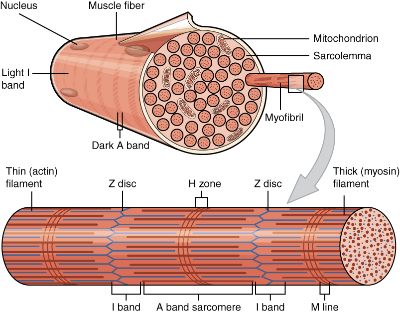
A myofibril (also known as a muscle fibril or sarcostyle) is a basic rod-like organelle of a muscle cell. Skeletal muscles are composed of long, tubular cells known as muscle fibers, and these cells contain many chains of myofibrils. Each myofibril has a diameter of 1–2 micrometres. They are created during embryonic development in a process known as myogenesis. Myofibrils are composed of long proteins including actin, myosin, and titin, and other proteins that hold them together. These proteins are organized into thick, thin, and elastic myofilaments, which repeat along the length of the myofibril in sections or units of contraction called sarcomeres. Muscles contract by sliding the thick myosin, and thin actin myofilaments along each other. Structure Each myofibril has a diameter of between 1 and 2 micrometres (μm). The filaments of myofibrils, myofilaments, consist of three types, thick, thin, and elastic filaments. *Thin filaments consist primarily of the protein acti ... [...More Info...] [...Related Items...] OR: [Wikipedia] [Google] [Baidu] |
Adipose Tissue
Adipose tissue, body fat, or simply fat is a loose connective tissue composed mostly of adipocytes. In addition to adipocytes, adipose tissue contains the stromal vascular fraction (SVF) of cells including preadipocytes, fibroblasts, vascular endothelial cells and a variety of immune cells such as adipose tissue macrophages. Adipose tissue is derived from preadipocytes. Its main role is to store energy in the form of lipids, although it also cushions and insulates the body. Far from being hormonally inert, adipose tissue has, in recent years, been recognized as a major endocrine organ, as it produces hormones such as leptin, estrogen, resistin, and cytokines (especially TNFα). In obesity, adipose tissue is also implicated in the chronic release of pro-inflammatory markers known as adipokines, which are responsible for the development of metabolic syndrome, a constellation of diseases including, but not limited to, type 2 diabetes, cardiovascular disease and atherosclerosis. T ... [...More Info...] [...Related Items...] OR: [Wikipedia] [Google] [Baidu] |
Adipocyte
Adipocytes, also known as lipocytes and fat cells, are the cells that primarily compose adipose tissue, specialized in storing energy as fat. Adipocytes are derived from mesenchymal stem cells which give rise to adipocytes through adipogenesis. In cell culture, adipocyte progenitors can also form osteoblasts, myocytes and other cell types. There are two types of adipose tissue, white adipose tissue (WAT) and brown adipose tissue (BAT), which are also known as white and brown fat, respectively, and comprise two types of fat cells. Structure White fat cells White fat cells contain a single large lipid droplet surrounded by a layer of cytoplasm, and are known as unilocular. The nucleus is flattened and pushed to the periphery. A typical fat cell is 0.1 mm in diameter with some being twice that size, and others half that size. However, these numerical estimates of fat cell size depend largely on the measurement method and the location of the adipose tissue. The fat stored i ... [...More Info...] [...Related Items...] OR: [Wikipedia] [Google] [Baidu] |
Immunohistochemistry
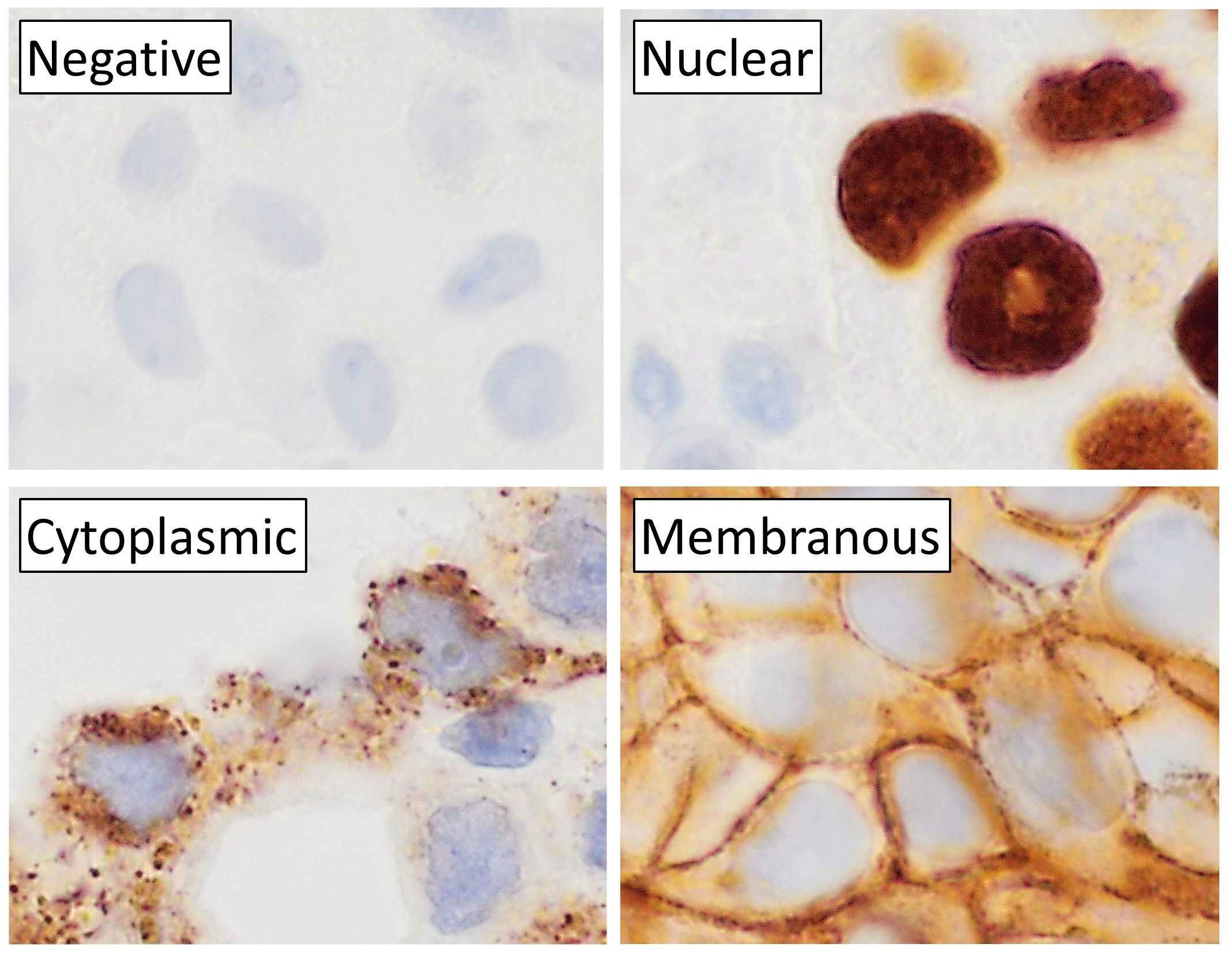
Immunohistochemistry (IHC) is the most common application of immunostaining. It involves the process of selectively identifying antigens (proteins) in cells of a tissue section by exploiting the principle of antibodies binding specifically to antigens in biological tissues. IHC takes its name from the roots "immuno", in reference to antibodies used in the procedure, and "histo", meaning tissue (compare to immunocytochemistry). Albert Coons conceptualized and first implemented the procedure in 1941. Visualising an antibody-antigen interaction can be accomplished in a number of ways, mainly either of the following: * ''Chromogenic immunohistochemistry'' (CIH), wherein an antibody is conjugated to an enzyme, such as peroxidase (the combination being termed immunoperoxidase), that can catalyse a colour-producing reaction. * '' Immunofluorescence'', where the antibody is tagged to a fluorophore, such as fluorescein or rhodamine. Immunohistochemical staining is widely used in the dia ... [...More Info...] [...Related Items...] OR: [Wikipedia] [Google] [Baidu] |
Fibroblast
A fibroblast is a type of cell (biology), biological cell that synthesizes the extracellular matrix and collagen, produces the structural framework (Stroma (tissue), stroma) for animal Tissue (biology), tissues, and plays a critical role in wound healing. Fibroblasts are the most common cells of connective tissue in animals. Structure Fibroblasts have a branched cytoplasm surrounding an elliptical, speckled cell nucleus, nucleus having two or more nucleoli. Active fibroblasts can be recognized by their abundant Endoplasmic reticulum#Rough endoplasmic reticulum, rough endoplasmic reticulum. Inactive fibroblasts (called fibrocytes) are smaller, spindle-shaped, and have a reduced amount of rough endoplasmic reticulum. Although disjointed and scattered when they have to cover a large space, fibroblasts, when crowded, often locally align in parallel clusters. Unlike the epithelial cells lining the body structures, fibroblasts do not form flat monolayers and are not restricted by a ... [...More Info...] [...Related Items...] OR: [Wikipedia] [Google] [Baidu] |
Tumor Marker
A tumor marker is a biomarker found in blood, urine, or body tissues that can be elevated by the presence of one or more types of cancer. There are many different tumor markers, each indicative of a particular disease process, and they are used in oncology to help detect the presence of cancer. An elevated level of a tumor marker can indicate cancer; however, there can also be other causes of the elevation (false positive values). Tumor markers can be produced directly by the tumor or by non-tumor cells as a response to the presence of a tumor. Although mammography, ultrasonography, computed tomography, magnetic resonance imaging scans, and tumor marker assays help in the staging and treatment of the cancer, they are usually not definitive diagnostic tests. The diagnosis is mostly confirmed by biopsy. Classification On the basis of their chemical nature, tumor markers can be proteins, conjugated proteins, peptides and carbohydrates. Proteins or conjugated proteins may be enzy ... [...More Info...] [...Related Items...] OR: [Wikipedia] [Google] [Baidu] |
CD99
CD99 antigen (Cluster of differentiation 99), also known as MIC2 or single-chain type-1 glycoprotein, is a heavily O-glycosylated transmembrane protein that is encoded by the ''CD99'' gene in humans. The protein has a mass of 32 kD. Unusually for a gene present on the X chromosome, the CD99 gene does not undergo X inactivation, and it was the first such pseudoautosomal gene to be discovered in humans. Expression It is expressed on all leukocytes but highest on thymocytes and is believed to augment T-cell adhesion and apoptosis of double positive T cells. It has been found in endothelial cells and in the periodontium, including gingival fibroblasts and gingival epithelial cells. It also participates in migration and activation. There is also experimental evidence that it binds to cyclophilin A. It is found on the cell surface of Ewing's sarcoma tumors and is positive in granulosa cell tumors. It is more expressed in malignant gliomas than in the brain, and such overexpression re ... [...More Info...] [...Related Items...] OR: [Wikipedia] [Google] [Baidu] |


.jpg)