|
Lepiota Maculans
''Lepiota maculans'' is a rare species of agaric fungus in the family Agaricaceae. It was originally collected in Missouri, and then 105 years later in eastern Tennessee. It is the only member of ''Lepiota'' known to have a pink spore print instead of the usual white or cream color. The fruit bodies have caps up to in diameter, with brownish, sparsely scaled centers. The gills are closely spaced, not attached to the stipe, and discolor reddish at the edges. Taxonomy The species was first described in 1905 by American mycologist Charles Horton Peck. The type collection was made by physician and amateur botanist Noah Miller Glatfelter from St. Louis, Missouri. Peck characterized it as a "small but pretty species, easily known by the flesh of both pileus and stem changing to a reddish color where wounded and by the lamellae assuming a reddish or pink color with age or in drying." The mushroom was found again 105 years later in a survey of macrofungi in the Great Smoky Moun ... [...More Info...] [...Related Items...] OR: [Wikipedia] [Google] [Baidu] |
Charles Horton Peck
Charles Horton Peck (March 30, 1833 – July 11, 1917) was an American mycologist of the 19th and early 20th centuries. He was the New York State Botanist from 1867 to 1915, a period in which he described over 2,700 species of North American fungi. Biography Charles Horton Peck was born on March 30, 1833, in the northeastern part of the town Sand Lake, New York, now called Averill Park. After suffering a light stroke early in November 1912 and then a severe stroke in 1913, he died at his house in Menands, New York, on July 11, 1917. In 1794, Eleazer Peck (his great grandfather) moved from Farmington, Conn. to Sand Lake, NY attracted by oak timber that was manufactured for the Albany market. Later on, Pamelia Horton Peck married Joel B., both from English descent, and became Charles Peck parents (Burnham 1919; Atkinson 1918). Even though his family was rich and locally prominent, his education was provincial (Haines 1986). During his childhood, he used to enjoy fishing and h ... [...More Info...] [...Related Items...] OR: [Wikipedia] [Google] [Baidu] |
Staining
Staining is a technique used to enhance contrast in samples, generally at the microscopic level. Stains and dyes are frequently used in histology (microscopic study of biological tissues), in cytology (microscopic study of cells), and in the medical fields of histopathology, hematology, and cytopathology that focus on the study and diagnoses of diseases at the microscopic level. Stains may be used to define biological tissues (highlighting, for example, muscle fibers or connective tissue), cell populations (classifying different blood cells), or organelles within individual cells. In biochemistry, it involves adding a class-specific ( DNA, proteins, lipids, carbohydrates) dye to a substrate to qualify or quantify the presence of a specific compound. Staining and fluorescent tagging can serve similar purposes. Biological staining is also used to mark cells in flow cytometry, and to flag proteins or nucleic acids in gel electrophoresis. Light microscopes are used for viewin ... [...More Info...] [...Related Items...] OR: [Wikipedia] [Google] [Baidu] |
Clamp Connection
A clamp connection is a hook-like structure formed by growing hyphal cells of certain fungi. It is a characteristic feature of Basidiomycetes fungi. It is created to ensure that each cell, or segment of hypha separated by septa (cross walls), receives a set of differing nuclei, which are obtained through mating of hyphae of differing sexual types. It is used to maintain genetic variation within the hypha much like the mechanisms found in crozier (hook) during sexual reproduction. Formation Clamp connections are formed by the terminal hypha during elongation. Before the clamp connection is formed this terminal segment contains two nuclei. Once the terminal segment is long enough it begins to form the clamp connection. At the same time, each nucleus undergoes mitotic division to produce two daughter nuclei. As the clamp continues to develop it uptakes one of the daughter (green circle) nuclei and separates it from its sister nucleus. While this is occurring the remaining nuclei ... [...More Info...] [...Related Items...] OR: [Wikipedia] [Google] [Baidu] |
Cap Cuticle
The pileipellis is the uppermost layer of hyphae in the pileus of a fungal fruit body. It covers the trama, the fleshy tissue of the fruit body. The pileipellis is more or less synonymous with the cuticle, but the cuticle generally describes this layer as a macroscopic feature, while pileipellis refers to this structure as a microscopic layer. Pileipellis type is an important character in the identification of fungi. Pileipellis types include the cutis, trichoderm, epithelium, and hymeniderm types. Types Cutis A cutis is a type of pileipellis characterized by hyphae that are repent, that is, that run parallel to the pileus surface. In an ixocutis, the hyphae are gelatinous. Trichoderm In a trichoderm, the outermost hyphae emerge roughly parallel, like hairs, perpendicular to the cap surface. The prefix "tricho-" comes from a Greek word for "hair". In an ixotrichodermium, the outermost hyphae are gelatinous. Epithelium An epithelium is a pileipellis consisting of rounded ce ... [...More Info...] [...Related Items...] OR: [Wikipedia] [Google] [Baidu] |
Cystidia
A cystidium (plural cystidia) is a relatively large cell found on the sporocarp of a basidiomycete (for example, on the surface of a mushroom gill), often between clusters of basidia. Since cystidia have highly varied and distinct shapes that are often unique to a particular species or genus, they are a useful micromorphological characteristic in the identification of basidiomycetes. In general, the adaptive significance of cystidia is not well understood. Classification of cystidia By position Cystidia may occur on the edge of a lamella (or analogous hymenophoral structure) (cheilocystidia), on the face of a lamella (pleurocystidia), on the surface of the cap (dermatocystidia or pileocystidia), on the margin of the cap (circumcystidia) or on the stipe (caulocystidia). Especially the pleurocystidia and cheilocystidia are important for identification within many genera. Sometimes the cheilocystidia give the gill edge a distinct colour which is visible to the naked eye or wit ... [...More Info...] [...Related Items...] OR: [Wikipedia] [Google] [Baidu] |
Basidia
A basidium () is a microscopic sporangium (a spore-producing structure) found on the hymenophore of fruiting bodies of basidiomycete fungi which are also called tertiary mycelium, developed from secondary mycelium. Tertiary mycelium is highly-coiled secondary myceliuma dikaryon. The presence of basidia is one of the main characteristic features of the Basidiomycota. A basidium usually bears four sexual spores called basidiospores; occasionally the number may be two or even eight. In a typical basidium, each basidiospore is borne at the tip of a narrow prong or horn called a sterigma (), and is forcibly discharged upon maturity. The word ''basidium'' literally means "little pedestal", from the way in which the basidium supports the spores. However, some biologists suggest that the structure more closely resembles a club. An immature basidium is known as a basidiole. Structure Most basidiomycota have single celled basidia (holobasidia), but in some groups basidia can be multice ... [...More Info...] [...Related Items...] OR: [Wikipedia] [Google] [Baidu] |
Micrometre
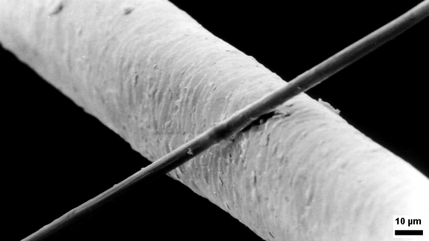
The micrometre ( international spelling as used by the International Bureau of Weights and Measures; SI symbol: μm) or micrometer (American spelling), also commonly known as a micron, is a unit of length in the International System of Units (SI) equalling (SI standard prefix "micro-" = ); that is, one millionth of a metre (or one thousandth of a millimetre, , or about ). The nearest smaller common SI unit is the nanometre, equivalent to one thousandth of a micrometre, one millionth of a millimetre or one billionth of a metre (). The micrometre is a common unit of measurement for wavelengths of infrared radiation as well as sizes of biological cells and bacteria, and for grading wool by the diameter of the fibres. The width of a single human hair ranges from approximately 20 to . The longest human chromosome, chromosome 1, is approximately in length. Examples Between 1 μm and 10 μm: * 1–10 μm – length of a typical bacterium * 3–8 μm – width of ... [...More Info...] [...Related Items...] OR: [Wikipedia] [Google] [Baidu] |
Spore
In biology, a spore is a unit of sexual or asexual reproduction that may be adapted for dispersal and for survival, often for extended periods of time, in unfavourable conditions. Spores form part of the life cycles of many plants, algae, fungi and protozoa. Bacterial spores are not part of a sexual cycle, but are resistant structures used for survival under unfavourable conditions. Myxozoan spores release amoeboid infectious germs ("amoebulae") into their hosts for parasitic infection, but also reproduce within the hosts through the pairing of two nuclei within the plasmodium, which develops from the amoebula. In plants, spores are usually haploid and unicellular and are produced by meiosis in the sporangium of a diploid sporophyte. Under favourable conditions the spore can develop into a new organism using mitotic division, producing a multicellular gametophyte, which eventually goes on to produce gametes. Two gametes fuse to form a zygote which develops into a new s ... [...More Info...] [...Related Items...] OR: [Wikipedia] [Google] [Baidu] |
Annulus (mycology)
An annulus is the ring-like or collar-like structure sometimes found on the stipe of some species of mushrooms. The annulus represents the remnants of the partial veil, after it has ruptured to expose the gills or other spore-producing surface. It can also be called a ring which is what the Latin word annulus directly translates as. The modern usage of the Latin word originates from the early days of botany and mycology when species descriptions were only written in Latin. Outside of the formal setting of scientific publications which still have a Latin requirement, it will often just be referred to as a ring or stem ring in field guide A field guide is a book designed to help the reader identify wildlife (flora or fauna) or other objects of natural occurrence (e.g. rocks and minerals). It is generally designed to be brought into the "field" or local area where such objects exi ...s and on identification websites. Ring descriptions The way in which the structure and appea ... [...More Info...] [...Related Items...] OR: [Wikipedia] [Google] [Baidu] |
Partial Veil
In mycology, a partial veil (also called an inner veil, to differentiate it from the "outer", or universal veil) is a temporary structure of tissue found on the fruiting bodies of some basidiomycete fungi, typically agarics. Its role is to isolate and protect the developing spore-producing surface, represented by gills or tubes, found on the lower surface of the cap. A partial veil, in contrast to a universal veil, extends from the stem surface to the cap edge. The partial veil later disintegrates, once the fruiting body has matured and the spores are ready for dispersal. It might then give rise to a stem ring, or fragments attached to the stem or cap edge. In some mushrooms, both a partial veil and a universal veil may be present. Structure In the immature fruit bodies of some basidiomycete fungi, the partial veil extends from the stem surface to the cap margin and shields the gills during development, and later breaks to expose the mature gills. The presence, absence, or struct ... [...More Info...] [...Related Items...] OR: [Wikipedia] [Google] [Baidu] |
Trama (mycology)
In mycology, the term trama is used in two ways. In the broad sense, it is the inner, fleshy portion of a mushroom's basidiocarp, or fruit body. It is distinct from the outer layer of tissue, known as the pileipellis or cuticle, and from the spore-bearing tissue layer known as the hymenium. In essence, the trama is the tissue that is commonly referred to as the "flesh" of mushrooms and similar fungi.Largent D, Johnson D, Watling R. 1977. ''How to Identify Mushrooms to Genus III: Microscopic Features''. Arcata, CA: Mad River Press. . pp. 60–70. The second use is more specific, and refers to the "hymenophoral trama" that supports the hymenium. It is similarly interior, connective tissue, but it is more specifically the central layer of hyphae running from the underside of the mushroom cap to the lamella or gill, upon which the hymenium rests. Various types have been classified by their structure, including trametoid, cantharelloid, boletoid, and agaricoid, with agaricoid the ... [...More Info...] [...Related Items...] OR: [Wikipedia] [Google] [Baidu] |
Umbo (mycology)
'' Cantharellula umbonata'' has an umbo. The cap of '' Psilocybe makarorae'' is acutely papillate.">papillate.html" ;"title="Psilocybe makarorae'' is acutely papillate">Psilocybe makarorae'' is acutely papillate. An umbo is a raised area in the center of a mushroom cap. pileus (mycology), Caps that possess this feature are called ''umbonate''. Umbos that are sharply pointed are called ''acute'', while those that are more rounded are ''broadly umbonate''. If the umbo is elongated, it is ''cuspidate'', and if the umbo is sharply delineated but not elongated (somewhat resembling the shape of a human areola The human areola (''areola mammae'', or ) is the pigmented area on the breast around the nipple. Areola, more generally, is a small circular area on the body with a different histology from the surrounding tissue, or other small circular ar ...), it is called '' mammilate'' or ''papillate''. References {{reflist Fungal morphology and anatomy Mycology ... [...More Info...] [...Related Items...] OR: [Wikipedia] [Google] [Baidu] |


