|
Lenticulostriate Artery
The lenticulostriate arteries, anterolateral central arteries, or antero-lateral ganglionic branches are a group of small arteries arising from the initial part M1 of the middle cerebral artery that supply the basal ganglia. Structure The lenticulostriate arteries are also known as the lateral striate arteries that arise from the middle cerebral artery. The other striate artery is the medial striate artery known as the recurrent artery of Heubner that arises from the anterior cerebral artery. The lenticulostriate arteries originate from the initial segment ( M1) of the middle cerebral artery (MCA). They are small perforating arteries, which enter the underside of the brain at the anterior perforated substance to supply blood to part of the basal ganglia and posterior limb of the internal capsule. The lenticulostriate perforators are end arteries. The name of these arteries is derived from some of the structures they supply, namely the lentiform nucleus and the striatum. Clinic ... [...More Info...] [...Related Items...] OR: [Wikipedia] [Google] [Baidu] |
Middle Cerebral Artery
The middle cerebral artery (MCA) is one of the three major paired cerebral artery, cerebral arteries that supply blood to the cerebrum. The MCA arises from the internal carotid artery and continues into the lateral sulcus where it then branches and projects to many parts of the lateral cerebral cortex. It also supplies blood to the anterior temporal lobes and the insular cortex, insular cortices. The left and right MCAs rise from trifurcations of the internal carotid artery, internal carotid arteries and thus are connected to the anterior cerebral artery, anterior cerebral arteries and the posterior communicating artery, posterior communicating arteries, which connect to the posterior cerebral artery, posterior cerebral arteries. The MCAs are not considered a part of the Circle of Willis. Structure The middle cerebral artery divides into four segments, named by the region they supply as opposed to order of branching as the latter can be somewhat variable: *M1: The ''sphenoidal' ... [...More Info...] [...Related Items...] OR: [Wikipedia] [Google] [Baidu] |
Basal Ganglia
The basal ganglia (BG), or basal nuclei, are a group of subcortical nuclei, of varied origin, in the brains of vertebrates. In humans, and some primates, there are some differences, mainly in the division of the globus pallidus into an external and internal region, and in the division of the striatum. The basal ganglia are situated at the base of the forebrain and top of the midbrain. Basal ganglia are strongly interconnected with the cerebral cortex, thalamus, and brainstem, as well as several other brain areas. The basal ganglia are associated with a variety of functions, including control of voluntary motor movements, procedural learning, habit learning, conditional learning, eye movements, cognition, and emotion. The main components of the basal ganglia – as defined functionally – are the striatum, consisting of both the dorsal striatum (caudate nucleus and putamen) and the ventral striatum (nucleus accumbens and olfactory tubercle), the globus pallidus, ... [...More Info...] [...Related Items...] OR: [Wikipedia] [Google] [Baidu] |
Recurrent Artery Of Heubner
The recurrent artery of Heubner, Heubner's artery or distal medial striate artery is an artery in the head. It is named after the German paediatrician Otto Heubner. It is a branch of the anterior cerebral artery. Its vascular territory is the anteromedial section of the caudate nucleus and the anterioinferior section of the internal capsule, as well as parts of the putamen and septal nuclei. Structure The recurrent artery of Heubner is a branch of the anterior cerebral artery. It has a mean diameter of 0.8 mm, and a mean length of 2.4 mm. It is also known together with the lenticulostriate arteries as a striate artery. The lenticulostriate arteries arise from the middle cerebral artery. Variation The recurrent artery of Heubner usually arises from the A1-A2 junction (between 44% and 62% of the time), but may arise from the proximal A2 segment (between 23% and 43%), or more rarely from the A1 segment (maybe up to 14% of the time). The recurrent artery of Heubner has a very var ... [...More Info...] [...Related Items...] OR: [Wikipedia] [Google] [Baidu] |
Anterior Cerebral Artery
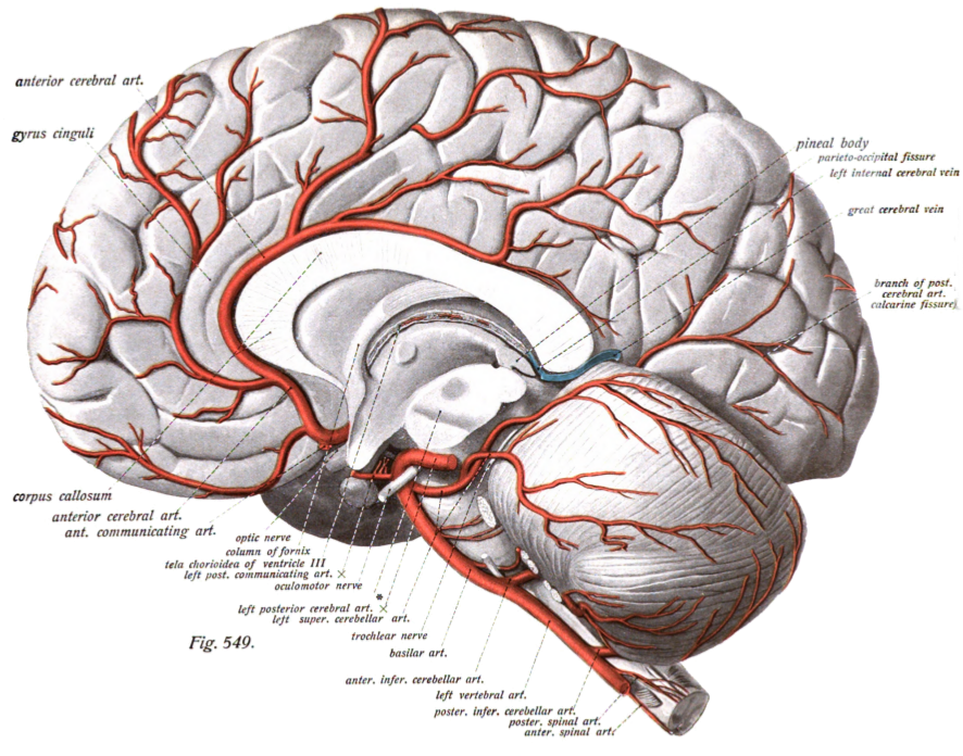
The anterior cerebral artery (ACA) is one of a pair of cerebral arteries that supplies oxygenated blood to most midline portions of the frontal lobes and superior medial parietal lobes of the brain. The two anterior cerebral arteries arise from the internal carotid artery and are part of the circle of Willis. The left and right anterior cerebral arteries are connected by the anterior communicating artery. Anterior cerebral artery syndrome refers to symptoms that follow a stroke occurring in the area normally supplied by one of the arteries. It is characterized by weakness and sensory loss in the lower leg and foot opposite to the lesion and behavioral changes. Structure The anterior cerebral artery is divided into 5 segments. Its smaller branches: the callosal (supracallosal) arteries are considered to be the A4 and A5 segments. *A1 originates from the internal carotid artery and extends to the ''anterior communicating artery'' (AComm). The ''anteromedial central'' (medial lent ... [...More Info...] [...Related Items...] OR: [Wikipedia] [Google] [Baidu] |
Anterior Perforated Substance
The anterior perforated substance is a part of the brain. It is bilateral. It is irregular and quadrilateral. It lies in front of the optic tract and behind the olfactory trigone. Structure The anterior perforated substance is bilateral. It lies in front of the optic tract. It lies behind the olfactory trigone, separated by the fissure prima. Medially and in front, it is continuous with the subcallosal gyrus. Laterally, it is bounded by the lateral stria of the olfactory tract, and is continued into the uncus. Its gray substance is confluent above with that of the corpus striatum, and is perforated anteriorly by numerous small blood vessels that supply such areas as the internal capsule. The anterior cerebral artery arises just below the anterior perforated substance. The middle cerebral artery passes through its lateral two thirds. Blood supply The anterior perforated substance is supplied by lenticulostriate arteries, which branch from the middle cerebral artery. It i ... [...More Info...] [...Related Items...] OR: [Wikipedia] [Google] [Baidu] |
Internal Capsule
The internal capsule is a white matter structure situated in the inferomedial part of each cerebral hemisphere of the brain. It carries information past the basal ganglia, separating the caudate nucleus and the thalamus from the putamen and the globus pallidus. The internal capsule contains both ascending and descending axons, going to and coming from the cerebral cortex. It also separates the caudate nucleus and the putamen in the dorsal striatum, a brain region involved in motor and reward pathways. The corticospinal tract constitutes a large part of the internal capsule, carrying motor information from the primary motor cortex to the lower motor neurons in the spinal cord. Above the basal ganglia the corticospinal tract is a part of the corona radiata. Below the basal ganglia the tract is called cerebral crus (a part of the cerebral peduncle) and below the pons it is referred to as the corticospinal tract. Structure The internal capsule consists of three parts and is V-shap ... [...More Info...] [...Related Items...] OR: [Wikipedia] [Google] [Baidu] |
End Artery
An end artery, or terminal artery is an artery that is the only supply of oxygenated blood to a portion of tissue Arteries which do not anastomose with their neighbors are called end arteries. There is no collateral circulation present besides the end arteries. Examples of an end artery include the splenic artery that supplies the spleen and the renal artery that supplies the kidneys. End arteries are of particular interest to medicine where they supply the heart or brain because if the arteries are occluded, the tissue is completely cut off, leading to a myocardial infarction or an ischaemic stroke. Other end arteries supply all or parts of the liver, intestines, fingers, toes, ears, nose, retina, penis, and other organs. Because vital tissues such as the brain or heart muscle are vulnerable to ischaemia, arteries often form anastomoses to provide alternative supplies of fresh blood. End arteries can exist when no anastomosis exists or when an anastomosis exists but is incap ... [...More Info...] [...Related Items...] OR: [Wikipedia] [Google] [Baidu] |
Lentiform Nucleus
The lentiform nucleus, or lenticular nucleus, comprises the putamen and the globus pallidus within the basal ganglia. With the caudate nucleus, it forms the dorsal striatum. It is a large, lens-shaped mass of gray matter just lateral to the internal capsule. Structure When divided horizontally, it exhibits, to some extent, the appearance of a biconvex lens, while a coronal section of its central part presents a somewhat triangular outline. It is shorter than the caudate nucleus and does not extend as far forward. Boundaries It is lateral to the caudate nucleus and thalamus, and is seen only in sections of the hemisphere. It is bounded laterally by a lamina of white substance called the external capsule, and lateral to this is a thin layer of gray substance termed the claustrum. Its anterior end is continuous with the lower part of the head of the caudate nucleus and with the anterior perforated substance. Components In a coronal section through the middle of the lentiform nu ... [...More Info...] [...Related Items...] OR: [Wikipedia] [Google] [Baidu] |
Striatum
The striatum, or corpus striatum (also called the striate nucleus), is a nucleus (a cluster of neurons) in the subcortical basal ganglia of the forebrain. The striatum is a critical component of the motor and reward systems; receives glutamatergic and dopaminergic inputs from different sources; and serves as the primary input to the rest of the basal ganglia. Functionally, the striatum coordinates multiple aspects of cognition, including both motor and action planning, decision-making, motivation, reinforcement, and reward perception. The striatum is made up of the caudate nucleus and the lentiform nucleus. The lentiform nucleus is made up of the larger putamen, and the smaller globus pallidus. Strictly speaking the globus pallidus is part of the striatum. It is common practice, however, to implicitly exclude the globus pallidus when referring to striatal structures. In primates, the striatum is divided into a ventral striatum, and a dorsal striatum, subdivisions that are ... [...More Info...] [...Related Items...] OR: [Wikipedia] [Google] [Baidu] |
Lacunar Infarcts
Lacunar stroke or lacunar cerebral infarct (LACI) is the most common type of ischemic stroke, resulting from the occlusion of small penetrating arteries that provide blood to the brain's deep structures. Patients who present with symptoms of a lacunar stroke, but who have not yet had diagnostic imaging performed, may be described as having lacunar stroke syndrome (LACS). Much of the current knowledge of lacunar strokes comes from C. Miller Fisher's cadaver dissections of post-mortem stroke patients. He observed "lacunae" (empty spaces) in the deep brain structures after occlusion of 200–800 μm penetrating arteries and connected them with five classic syndromes. These syndromes are still noted today, though lacunar infarcts are diagnosed based on clinical judgment and radiologic imaging. Signs and symptoms Each of the five classical lacunar syndromes has a relatively distinct symptom complex. Symptoms may occur suddenly, progressively, or in a fluctuating (e.g., the cap ... [...More Info...] [...Related Items...] OR: [Wikipedia] [Google] [Baidu] |
Arteriolosclerosis
Arteriolosclerosis is a form of cardiovascular disease involving hardening and loss of elasticity of arterioles or small arteries and is most often associated with hypertension and diabetes mellitus. Types include hyaline arteriolosclerosis and hyperplastic arteriolosclerosis, both involved with vessel wall thickening and luminal narrowing that may cause downstream ischemic injury. The following two terms whilst similar, are distinct in both spelling and meaning and may easily be confused with arteriolosclerosis. * Arteriosclerosis is any hardening (and loss of elasticity) of medium or large arteries (from the Greek '' arteria'', meaning ''artery'', and '' sclerosis'', meaning ''hardening'') * Atherosclerosis is a hardening of an artery specifically due to an atheromatous plaque. The term ''atherogenic'' is used for substances or processes that cause atherosclerosis. Hyaline arteriolosclerosis Also arterial hyalinosis and arteriolar hyalinosis refers to thickening of the walls ... [...More Info...] [...Related Items...] OR: [Wikipedia] [Google] [Baidu] |


