|
Lens Capsule
The lens capsule is a component of the globe of the eye. It is a clear, membrane-like structure composed of collagen IV and laminin that is quite elastic, a quality that keeps it under constant tension. As a result, the lens naturally tends towards a rounder or more globular configuration, a shape it must assume for the eye to focus at a near distance. Lens capsule is the thickest basement membrane in the body. Normally, the lens capsule serves as a diffusion barrier. It is permeable to low molecular weight compounds but restricts the movement of large colloidal particles. Anatomy The lens capsule is a transparent membrane that surrounds the entire lens. The capsule is thinnest at the posterior pole with approximate thickness of 3.5μm. Average thickness at the equator is 7μm. Anterior pole thickness increases with age from 11-15μm. The thickest portion of is annular region surrounding the anterior pole. This will also increases with age (from 13.5-16μm). Even though the ... [...More Info...] [...Related Items...] OR: [Wikipedia] [Google] [Baidu] |
|
 |
Human Eye
The human eye is a sensory organ, part of the sensory nervous system, that reacts to visible light and allows humans to use visual information for various purposes including seeing things, keeping balance, and maintaining circadian rhythm. The eye can be considered as a living optical device. It is approximately spherical in shape, with its outer layers, such as the outermost, white part of the eye (the sclera) and one of its inner layers (the pigmented choroid) keeping the eye essentially light tight except on the eye's optic axis. In order, along the optic axis, the optical components consist of a first lens (the cornea—the clear part of the eye) that accomplishes most of the focussing of light from the outside world; then an aperture (the pupil) in a diaphragm (the iris—the coloured part of the eye) that controls the amount of light entering the interior of the eye; then another lens (the crystalline lens) that accomplishes the remaining focussing of light in ... [...More Info...] [...Related Items...] OR: [Wikipedia] [Google] [Baidu] |
 |
Ciliary Muscle
The ciliary muscle is an intrinsic muscle of the eye formed as a ring of smooth muscleSchachar, Ronald A. (2012). "Anatomy and Physiology." (Chapter 4) . in the eye's middle layer, uvea (vascular layer). It controls accommodation for viewing objects at varying distances and regulates the flow of aqueous humor into Schlemm's canal. It also changes the shape of the lens within the eye but not the size of the pupil which is carried out by the sphincter pupillae muscle and dilator pupillae. Structure Development The ciliary muscle develops from mesenchyme within the choroid and is considered a cranial neural crest derivative.Dudek RW, Fix JD (2004). "Eye" (chapter 9). ''Embryology - Board Review Series'' (3rd edition, illustrated). Lippincott Williams & Wilkins. p. 92. , . Books.Google.com. Retrieved on 2010-01-17 from https://books.google.com/books?id=MmoJQWsJteoC. Nerve supply The ciliary muscle receives parasympathetic fibers from the short ciliary nerves that arise fro ... [...More Info...] [...Related Items...] OR: [Wikipedia] [Google] [Baidu] |
 |
Posterior Capsular Opacification
Cataract surgery, also called lens replacement surgery, is the removal of the natural lens of the eye (also called "crystalline lens") that has developed an opacification, which is referred to as a cataract, and its replacement with an intraocular lens. Metabolic changes of the crystalline lens fibers over time lead to the development of the cataract, causing impairment or loss of vision. Some infants are born with congenital cataracts, and certain environmental factors may also lead to cataract formation. Early symptoms may include strong glare from lights and small light sources at night, and reduced acuity at low light levels. During cataract surgery, a patient's cloudy natural cataract lens is removed, either by emulsification in place or by cutting it out. An artificial intraocular lens (IOL) is implanted in its place. Cataract surgery is generally performed by an ophthalmologist in an ambulatory setting at a surgical center or hospital rather than an inpatient settin ... [...More Info...] [...Related Items...] OR: [Wikipedia] [Google] [Baidu] |
 |
Intraocular Lens
Intraocular lens (IOL) is a lens (optics), lens implanted in the human eye, eye as part of a treatment for cataracts or myopia. If the natural lens is left in the eye, the IOL is known as Phakic intraocular lens, phakic, otherwise it is a pseudophakic, or false lens. Such a lens is typically implanted during cataract surgery, after the eye's cloudy lens (anatomy), natural lens (cataract) has been removed. The pseudophakic IOL provides the same light-focusing function as the natural crystalline lens. The phakic type of IOL is placed over the existing natural lens and is used in refractive surgery to change the eye's optical power as a treatment for myopia (nearsightedness). This is an alternative to LASIK. IOLs usually consist of a small plastic lens with plastic side struts, called haptics, to hold the lens in place in the capsular bag inside the eye. IOLs were conventionally made of an inflexible material (Polymethyl methacrylate, PMMA), although this has largely been superseded ... [...More Info...] [...Related Items...] OR: [Wikipedia] [Google] [Baidu] |
|
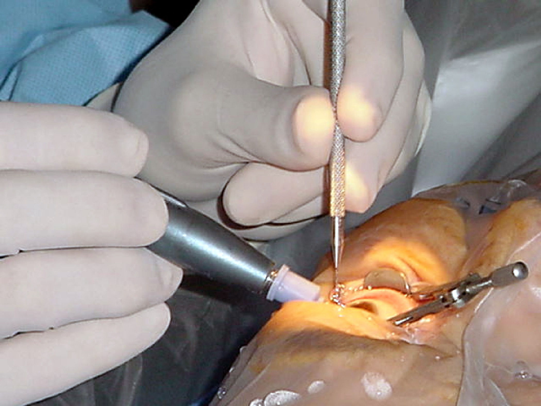
Phacoemulsification
Phacoemulsification is a modern cataract surgery method in which the eye's internal lens is emulsified with an ultrasonic handpiece and aspirated from the eye. Aspirated fluids are replaced with irrigation of balanced salt solution to maintain the anterior chamber. Etymology The term originated from phaco- (Greek ''phako-'', comb. form of ''phakós'', lentil; see lens) + emulsification. Preparation and precautions Proper anesthesia is essential for ocular surgery. Topical anesthesia is most commonly employed, typically by the instillation of a local anesthetic such as tetracaine or lidocaine. Alternatively, lidocaine and/or longer-acting bupivacaine anesthetic may be injected into the area surrounding (peribulbar block) or behind ( retrobulbar block) the eye muscle cone to more fully immobilize the extraocular muscles and minimize pain sensation. A facial nerve block using lidocaine and bupivacaine may occasionally be performed to reduce lid squeezing. General anest ... [...More Info...] [...Related Items...] OR: [Wikipedia] [Google] [Baidu] |
|
 |
Cataract Surgery
Cataract surgery, also called lens replacement surgery, is the removal of the natural lens of the eye (also called "crystalline lens") that has developed an opacification, which is referred to as a cataract, and its replacement with an intraocular lens. Metabolic changes of the crystalline lens fibers over time lead to the development of the cataract, causing impairment or loss of vision. Some infants are born with congenital cataracts, and certain environmental factors may also lead to cataract formation. Early symptoms may include strong glare from lights and small light sources at night, and reduced acuity at low light levels. During cataract surgery, a patient's cloudy natural cataract lens is removed, either by emulsification in place or by cutting it out. An artificial intraocular lens (IOL) is implanted in its place. Cataract surgery is generally performed by an ophthalmologist in an ambulatory setting at a surgical center or hospital rather than an inpatient setting. ... [...More Info...] [...Related Items...] OR: [Wikipedia] [Google] [Baidu] |
|
Accommodation (eye)
Accommodation is the process by which the vertebrate eye changes optical power to maintain a clear image or focus on an object as its distance varies. In this, distances vary for individuals from the far point—the maximum distance from the eye for which a clear image of an object can be seen, to the near point—the minimum distance for a clear image. Accommodation usually acts like a reflex, including part of the accommodation-vergence reflex, but it can also be consciously controlled. Mammals, birds and reptiles vary their eyes' optical power by changing the form of the elastic lens using the ciliary body (in humans up to 15 dioptres in the mean). Fish and amphibians vary the power by changing the distance between a rigid lens and the retina with muscles. The young human eye can change focus from distance (infinity) to as near as 6.5 cm from the eye. This dramatic change in focal power of the eye of approximately 15 dioptres (the reciprocal of focal length in metr ... [...More Info...] [...Related Items...] OR: [Wikipedia] [Google] [Baidu] |
|
|
Zonule Of Zinn
The zonule of Zinn () (Zinn's membrane, ciliary zonule) (after Johann Gottfried Zinn) is a ring of fibrous strands forming a zonule (little band) that connects the ciliary body with the crystalline lens of the eye. These fibers are sometimes collectively referred to as the suspensory ligaments of the lens, as they act like suspensory ligaments. Development The ciliary epithelial cells of the eye probably synthesize portions of the zonules. Anatomy The zonule of Zinn is split into two layers: a thin layer, which lines the hyaloid fossa, and a thicker layer, which is a collection of zonular fibers. Together, the fibers are known as the suspensory ligament of the lens. The zonules are about 1–2 μm in diameter. The zonules attach to the lens capsule 2 mm anterior and 1 mm posterior to the equator, and arise of the ciliary epithelium from the pars plana region as well as from the valleys between the ciliary processes in the pars plicata. When colour granules are displaced fro ... [...More Info...] [...Related Items...] OR: [Wikipedia] [Google] [Baidu] |
|
 |
Gestational Age (obstetrics)
In obstetrics, gestational age is a measure of the age of a pregnancy which is taken from the beginning of the woman's last menstrual period (LMP), or the corresponding age of the gestation as estimated by a more accurate method if available. Such methods include adding 14 days to a known duration since fertilization (as is possible in in vitro fertilization), or by obstetric ultrasonography. The popularity of using this definition of gestational age is that menstrual periods are essentially always noticed, while there is usually a lack of a convenient way to discern when fertilization occurred. Gestational age is contrasted with fertilization age which takes the date of fertilization as the start date of gestation. The initiation of pregnancy for the calculation of gestational age can differ from definitions of initiation of pregnancy in context of the abortion debate or beginning of human personhood. Methods According to American College of Obstetricians and Gynecologists, th ... [...More Info...] [...Related Items...] OR: [Wikipedia] [Google] [Baidu] |
|
Globe (human Eye)
The globe of the eye, or bulbus oculi, is the eyeball apart from its appendages. A hollow structure, the bulbus oculi is composed of a wall enclosing a cavity filled with fluid with three coats: the sclera, choroid, and the retina. Normally, the bulbus oculi is bulb-like structure. However, the bulbus oculi is not completely spherical. Its anterior surface, transparent and more curved, is known as the cornea of the bulbus oculi. See also * Sclera The sclera, also known as the white of the eye or, in older literature, as the tunica albuginea oculi, is the opaque, fibrous, protective, outer layer of the human eye containing mainly collagen and some crucial elastic fiber. In humans, and som ... * Choroid * Retina References {{DEFAULTSORT:Globe (Human Eye) Human eye anatomy ... [...More Info...] [...Related Items...] OR: [Wikipedia] [Google] [Baidu] |
|
|
Hyaloid Artery
The hyaloid artery is a branch of the ophthalmic artery, which is itself a branch of the internal carotid artery. It is contained within the optic stalk of the eye and extends from the optic disc through the vitreous humor to the lens. Usually fully regressed before birth, its purpose is to supply nutrients to the developing lens in the growing fetus. During the tenth week of development in humans (time varies depending on species), the lens grows independent of a blood supply and the hyaloid artery usually regresses. Its proximal portion remains as the central artery of the retina. Regression of the hyaloid artery leaves a clear central zone through the vitreous humor, called the hyaloid canal or Cloquet's canal. Cloquet's canal is named after the French physician Jules Germain Cloquet (1790–1883) who first described it. Occasionally the artery may not fully regress, resulting in the condition ''persistent hyaloid artery''. More commonly, small remnants of the artery may ... [...More Info...] [...Related Items...] OR: [Wikipedia] [Google] [Baidu] |
|
|
Mesenchyme
Mesenchyme () is a type of loosely organized animal embryonic connective tissue of undifferentiated cells that give rise to most tissues, such as skin, blood or bone. The interactions between mesenchyme and epithelium help to form nearly every organ in the developing embryo. Vertebrates Structure Mesenchyme is characterized morphologically by a prominent ground substance matrix containing a loose aggregate of reticular fibers and unspecialized mesenchymal stem cells. Mesenchymal cells can migrate easily (in contrast to epithelial cells, which lack mobility), are organized into closely adherent sheets, and are polarized in an apical-basal orientation. Development The mesenchyme originates from the mesoderm. From the mesoderm, the mesenchyme appears as an embryologically primitive "soup". This "soup" exists as a combination of the mesenchymal cells plus serous fluid plus the many different tissue proteins. Serous fluid is typically stocked with the many serous elements, su ... [...More Info...] [...Related Items...] OR: [Wikipedia] [Google] [Baidu] |