|
Left Coronary
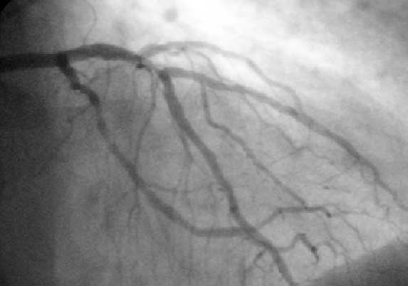
The left coronary artery (LCA) is a coronary artery that arises from the aorta above the left cusp of the aortic valve, and feeds blood to the left side of the heart muscle. It is also known as the left main coronary artery (LMCA) and the left main stem coronary artery (LMS). Branching The left coronary artery typically runs for 10 to 25 mm, and then bifurcates into the left anterior descending artery (also called the widow maker) and the left circumflex artery. Sometimes, an additional artery arises at the bifurcation of the left main artery, forming a trifurcation; this extra artery is called the ''ramus'' or ''intermediate artery''. The part that is between the aorta and the bifurcation only is known as the left main artery (LM), while the term "LCA" might refer to just the left main, or to the left main and all its eventual branches. A "first septal branch" is sometimes described. Additional images File:Coronary arteries 1.jpg, Left coronary artery File:Cardiac vess ... [...More Info...] [...Related Items...] OR: [Wikipedia] [Google] [Baidu] |
Ascending Aorta
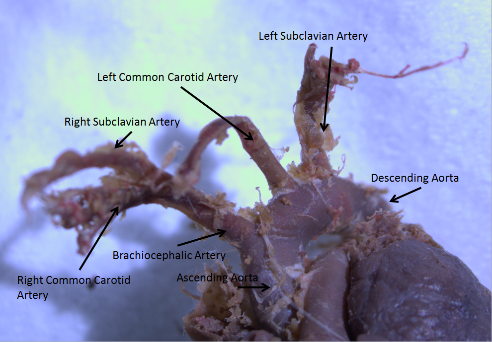
The ascending aorta (AAo) is a portion of the aorta commencing at the upper part of the base of the left ventricle, on a level with the lower border of the third costal cartilage behind the left half of the sternum. Structure It passes obliquely upward, forward, and to the right, in the direction of the heart's axis, as high as the upper border of the second right costal cartilage, describing a slight curve in its course, and being situated, about behind the posterior surface of the sternum. The total length is about . Components The aortic root is the portion of the aorta beginning at the aortic annulus and extending to the sinotubular junction. It is sometimes regarded as a part of the ascending aorta, and sometimes regarded as a separate entity from the rest of the ascending aorta. Between each commissure of the aortic valve and opposite the cusps of the aortic valve, three small dilatations called the aortic sinuses. The sinotubular junction is the point in the ascendi ... [...More Info...] [...Related Items...] OR: [Wikipedia] [Google] [Baidu] |
Left Circumflex Artery
The circumflex branch of left coronary artery, or left circumflex artery or circumflex artery, is a branch of the left coronary artery. Description The left circumflex artery follows the left part of the coronary sulcus, running first to the left and then to the right, reaching nearly as far as the posterior longitudinal sulcus. There have been multiple anomalies described, for example the left circumflex having an aberrant course from the right coronary artery. Branches The circumflex artery curves to the left around the heart within the coronary sulcus, giving rise to one or more left marginal arteries (also called obtuse marginal branches) as it curves toward the posterior surface of the heart. It helps form the posterior left ''ventricular branch'' or posterolateral artery. The circumflex artery ends at the point where it joins to form to the posterior interventricular artery in 15% of all cases, which lies in the posterior interventricular sulcus. In the other 85% of all ... [...More Info...] [...Related Items...] OR: [Wikipedia] [Google] [Baidu] |
Coronary Circulation
Coronary circulation is the circulation of blood in the blood vessels that supply the heart muscle (myocardium). Coronary arteries supply oxygenated blood to the heart muscle. Cardiac veins then drain away the blood after it has been deoxygenated. Because the rest of the body, and most especially the brain, needs a steady supply of oxygenated blood that is free of all but the slightest interruptions, the heart is required to function continuously. Therefore its circulation is of major importance not only to its own tissues but to the entire body and even the level of consciousness of the brain from moment to moment. Interruptions of coronary circulation quickly cause heart attacks (myocardial infarctions), in which the heart muscle is damaged by oxygen starvation. Such interruptions are usually caused by coronary ischemia linked to coronary artery disease, and sometimes to embolism from other causes like obstruction in blood flow through vessels. Structure Coronary arteri ... [...More Info...] [...Related Items...] OR: [Wikipedia] [Google] [Baidu] |
Gray's Anatomy
''Gray's Anatomy'' is a reference book of human anatomy written by Henry Gray, illustrated by Henry Vandyke Carter, and first published in London in 1858. It has gone through multiple revised editions and the current edition, the 42nd (October 2020), remains a standard reference, often considered "the doctors' bible". Earlier editions were called ''Anatomy: Descriptive and Surgical'', ''Anatomy of the Human Body'' and ''Gray's Anatomy: Descriptive and Applied'', but the book's name is commonly shortened to, and later editions are titled, ''Gray's Anatomy''. The book is widely regarded as an extremely influential work on the subject. Publication history Origins The English anatomist Henry Gray was born in 1827. He studied the development of the endocrine glands and spleen and in 1853 was appointed Lecturer on Anatomy at St George's Hospital Medical School in London. In 1855, he approached his colleague Henry Vandyke Carter with his idea to produce an inexpensive and ac ... [...More Info...] [...Related Items...] OR: [Wikipedia] [Google] [Baidu] |
Ostium Primum
In the developing heart, the atria are initially open to each other, with the opening known as the primary interatrial foramen or ostium primum (or interatrial foramen primum). The foramen lies beneath the edge of septum primum and the endocardial cushions. It progressively decreases in size as the septum grows downwards, and disappears with the formation of the atrial septum. Structure The foramen lies beneath the edge of septum primum and the endocardial cushions. It progressively decreases in size as the septum grows downwards, and disappears with the formation of the atrial septum. Closure The septum primum, a which grows down to separate the primitive atrium into the left atrium and right atrium, grows in size over the course of heart development. The primary interatrial foramen is the gap between the septum primum and the septum intermedium, which gets progressively smaller until it closes. Clinical significance Failure of the septum primum to fuse with the endocardial c ... [...More Info...] [...Related Items...] OR: [Wikipedia] [Google] [Baidu] |
Autopsy
An autopsy (post-mortem examination, obduction, necropsy, or autopsia cadaverum) is a surgical procedure that consists of a thorough examination of a corpse by dissection to determine the cause, mode, and manner of death or to evaluate any disease or injury that may be present for research or educational purposes. (The term "necropsy" is generally reserved for non-human animals). Autopsies are usually performed by a specialized medical doctor called a pathologist. In most cases, a medical examiner or coroner can determine the cause of death. However, only a small portion of deaths require an autopsy to be performed, under certain circumstances. Purposes of performance Autopsies are performed for either legal or medical purposes. Autopsies can be performed when any of the following information is desired: * Determine if death was natural or unnatural * Injury source and extent on the corpse * Manner of death must be determined * Post mortem interval * Determining the deceas ... [...More Info...] [...Related Items...] OR: [Wikipedia] [Google] [Baidu] |
Coronary Angiogram
A coronary catheterization is a minimally invasive procedure to access the coronary circulation and blood filled chambers of the heart using a catheter. It is performed for both diagnostic and interventional (treatment) purposes. Coronary catheterization is one of the several cardiology diagnostic tests and procedures. Specifically, through the injection of a liquid radiocontrast agent and illumination with X-rays, angiocardiography allows the recognition of occlusion, stenosis, restenosis, thrombosis or aneurysmal enlargement of the coronary artery lumens; heart chamber size; heart muscle contraction performance; and some aspects of heart valve function. Important internal heart and lung blood pressures, not measurable from outside the body, can be accurately measured during the test. The relevant problems that the test deals with most commonly occur as a result of advanced atherosclerosis – atheroma activity within the wall of the coronary arteries. Less frequently, va ... [...More Info...] [...Related Items...] OR: [Wikipedia] [Google] [Baidu] |
Aortic Arch
The aortic arch, arch of the aorta, or transverse aortic arch () is the part of the aorta between the ascending and descending aorta. The arch travels backward, so that it ultimately runs to the left of the trachea. Structure The aorta begins at the level of the upper border of the second/third sternocostal articulation of the right side, behind the ventricular outflow tract and pulmonary trunk. The right atrial appendage overlaps it. The first few centimeters of the ascending aorta and pulmonary trunk lies in the same pericardial sheath. and runs at first upward, arches over the pulmonary trunk, right pulmonary artery, and right main bronchus to lie behind the right second coastal cartilage. The right lung and sternum lies anterior to the aorta at this point. The aorta then passes posteriorly and to the left, anterior to the trachea, and arches over left main bronchus and left pulmonary artery, and reaches to the left side of the T4 vertebral body. Apart from T4 vertebral body ... [...More Info...] [...Related Items...] OR: [Wikipedia] [Google] [Baidu] |
Widow Maker (medicine)
The left anterior descending artery (also LAD, anterior interventricular branch of left coronary artery, or anterior descending branch) is a branch of the left coronary artery. Blockage of this artery is often called the ''widow-maker infarction'' due to a high death risk. Structure It passes at first behind the pulmonary artery and then comes forward between that vessel and the left atrium to reach the anterior interventricular sulcus, along which it descends to the notch of cardiac apex. Although rare, multiple anomalous courses of the LAD have been described. These include the origin of the artery from the right aortic sinus. In 78% of cases, it reaches the apex of the heart. Branches The LAD gives off two types of branches: ''septals'' and ''diagonals''. * Septals originate from the LAD at 90 degrees to the surface of the heart, perforating and supplying the anterior 2/3 of the interventricular septum. * Diagonals run along the surface of the heart and supply the lateral ... [...More Info...] [...Related Items...] OR: [Wikipedia] [Google] [Baidu] |
Anterior Interventricular Branch Of Left Coronary Artery
The left anterior descending artery (also LAD, anterior interventricular branch of left coronary artery, or anterior descending branch) is a branch of the left coronary artery. Blockage of this artery is often called the ''widow-maker infarction'' due to a high death risk. Structure It passes at first behind the pulmonary artery and then comes forward between that vessel and the left atrium to reach the anterior interventricular sulcus, along which it descends to the notch of cardiac apex. Although rare, multiple anomalous courses of the LAD have been described. These include the origin of the artery from the right aortic sinus. In 78% of cases, it reaches the apex of the heart. Branches The LAD gives off two types of branches: ''septals'' and ''diagonals''. * Septals originate from the LAD at 90 degrees to the surface of the heart, perforating and supplying the anterior 2/3 of the interventricular septum. * Diagonals run along the surface of the heart and supply the lateral ... [...More Info...] [...Related Items...] OR: [Wikipedia] [Google] [Baidu] |
Heart Muscle
Cardiac muscle (also called heart muscle, myocardium, cardiomyocytes and cardiac myocytes) is one of three types of vertebrate muscle tissues, with the other two being skeletal muscle and smooth muscle. It is an involuntary, striated muscle that constitutes the main tissue of the wall of the heart. The cardiac muscle (myocardium) forms a thick middle layer between the outer layer of the heart wall (the pericardium) and the inner layer (the endocardium), with blood supplied via the coronary circulation. It is composed of individual cardiac muscle cells joined by intercalated discs, and encased by collagen fibers and other substances that form the extracellular matrix. Cardiac muscle contracts in a similar manner to skeletal muscle, although with some important differences. Electrical stimulation in the form of a cardiac action potential triggers the release of calcium from the cell's internal calcium store, the sarcoplasmic reticulum. The rise in calcium causes the cell's ... [...More Info...] [...Related Items...] OR: [Wikipedia] [Google] [Baidu] |
Aortic Valve
The aortic valve is a valve in the heart of humans and most other animals, located between the left ventricle and the aorta. It is one of the four valves of the heart and one of the two semilunar valves, the other being the pulmonary valve. The aortic valve normally has three cusps or leaflets, although in 1–2% of the population it is found to congenitally have two leaflets. The aortic valve is the last structure in the heart the blood travels through before stopping the flow through the systemic circulation. Structure The aortic valve normally has three cusps however there is some discrepancy in their naming. They may be called the left coronary, right coronary and non-coronary cusp. Some sources also advocate they be named as a left, right and posterior cusp. Anatomists have traditionally named them the left posterior (origin of left coronary), anterior (origin of the right coronary) and right posterior. The three cusps, when the valve is closed, contain a sinus called an a ... [...More Info...] [...Related Items...] OR: [Wikipedia] [Google] [Baidu] |


