|
Lateral Sural Cutaneous Nerve
The lateral sural cutaneous nerve of the lumbosacral plexus supplies the skin on the posterior and lateral surfaces of the leg. The lateral sural cutaneous nerve originates from the common fibular nerve''(L4-S2)'' and is the terminal branch of the common fibular nerve. Sural communicating branch One branch, the '' sural communicating nerve'' or colloquially known as the ''peroneal anastomotic'' (n. communicans fibularis), arises from sciatic origins near the head of the fibula, crosses the lateral head of the gastrocnemius to the middle of the leg, and joins with the medial sural cutaneous nerve to form the sural nerve Variation Another branch observed, that is mentioned in passing in previous literature is the medial branch of the lateral sural cutaneous nerve. In a 2021 study bSteele et al.''(Annals of Anatomy)'', a medial branch of the lateral sural cutaneous nerve was observed in approximately 36% of lower extremities dissected (''n''=208) with an average diameter of 1. ... [...More Info...] [...Related Items...] OR: [Wikipedia] [Google] [Baidu] |
Common Peroneal Nerve
The common fibular nerve (also known as the common peroneal nerve, external popliteal nerve, or lateral popliteal nerve) is a nerve in the lower leg that provides sensation over the posterolateral part of the leg and the knee joint. It divides at the knee into two terminal branches: the superficial fibular nerve and deep fibular nerve, which innervate the muscles of the lateral and anterior compartments of the leg respectively. When the common fibular nerve is damaged or compressed, foot drop can ensue. Structure The common fibular nerve is the smaller terminal branch of the sciatic nerve. The common fibular nerve has root values of L4, L5, S1, and S2. It arises from the superior angle of the popliteal fossa and extends to the lateral angle of the popliteal fossa, along the medial border of the biceps femoris. It then winds around the neck of the fibula to pierce the fibularis longus and divides into terminal branches of the superficial fibular nerve and the deep fibular nerve. Bef ... [...More Info...] [...Related Items...] OR: [Wikipedia] [Google] [Baidu] |
Lumbosacral Plexus
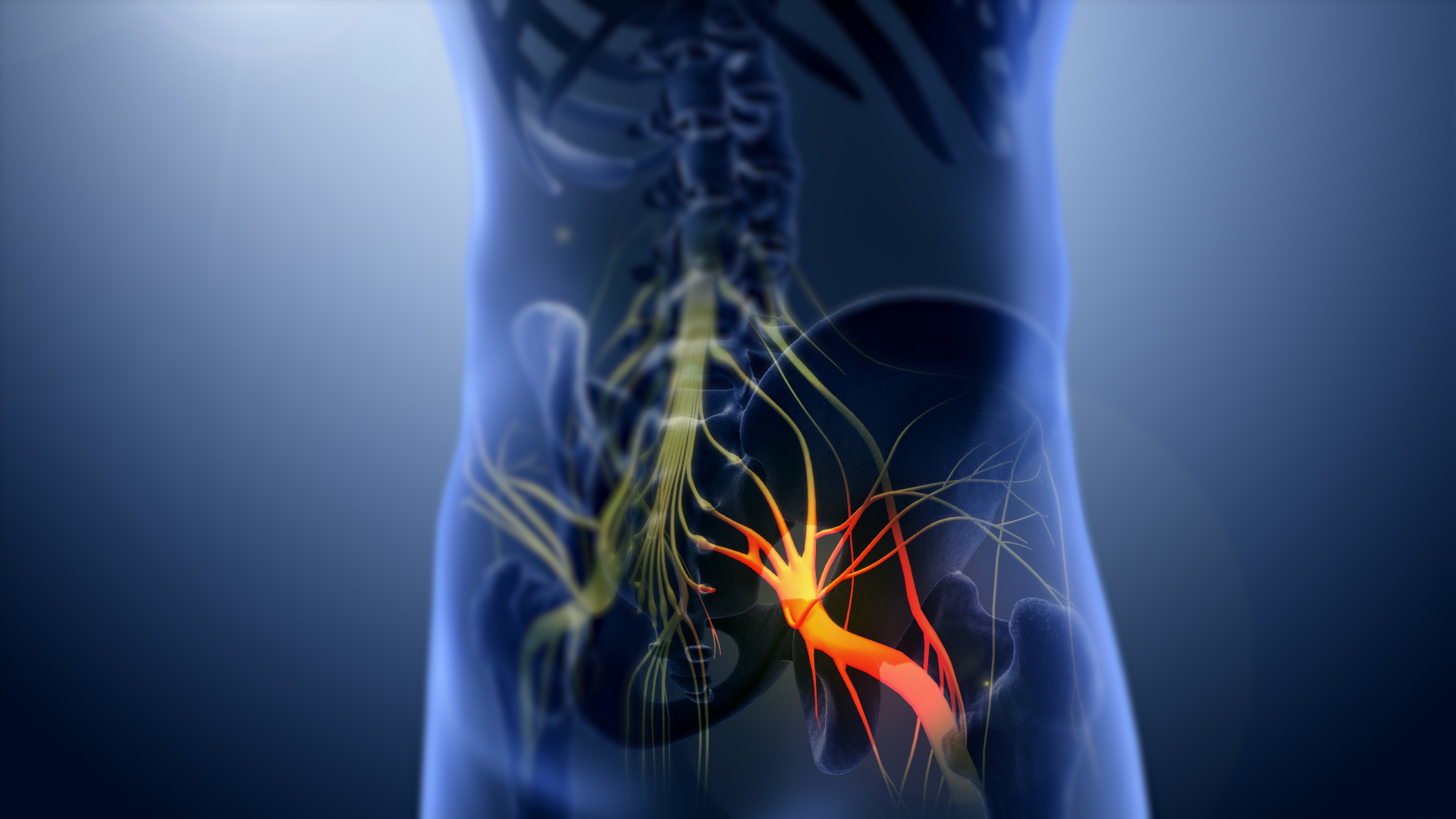
The anterior divisions of the lumbar nerves, sacral nerves, and coccygeal nerve form the lumbosacral plexus, the first lumbar nerve being frequently joined by a branch from the twelfth thoracic. For descriptive purposes this plexus is usually divided into three parts: * lumbar plexus * sacral plexus * pudendal plexus Injuries to the lumbosacral plexus are predominantly witnessed as bone injuries. Lumbosacral trunk and sacral plexus In human anatomy, the sacral plexus is a nerve plexus which provides motor and sensory nerves for the posterior thigh, most of the lower leg and foot, and part of the pelvis. It is part of the lumbosacral plexus and emerges from the lumbar verte ... palsies are common injury patterns. References External links * - "Lumbosacral Plexus" Additional Images File:Slide2Anat.JPG, Lumbosacral plexus Deep dissection. File:Slide4Anat.JPG, Lumbosacral plexus Deep dissection. Nerve plexus Nerves of the lower limb and lower torso {{Neuroana ... [...More Info...] [...Related Items...] OR: [Wikipedia] [Google] [Baidu] |
Sural Communicating Branch Of Common Peroneal Nerve
The sural communicating nerve ''(SCN)'' (''peroneal communicating branch'' of the common fibular nerve) is a separate and independent nerve from both the medial and lateral sural cutaneous nerves, often arising from a common trunk of the common fibular nerve The primary purpose of the sural communicating branch is to provide the structural path for transferring tibial nerve fascicular components to the sural nerve. Anatomy The ''sural communicating nerve'' (colloquially the peroneal communicating nerve) is one of the components of thsural nerve complex( MSCN, LSCN,SCN). It travels in an inferomedial direction from its origins either as a terminal component of the LSCN or is considered a nerve that originates along a common trunk of the lateral sural cutaneous nerve. This particular nerve is best described by Dr. Huelke in 1958. ''Dr. Huelke'' took the original modern cadaveric studies (from the turn of the 20th century) and wrote a treatise titled "The origin of the peroneal co ... [...More Info...] [...Related Items...] OR: [Wikipedia] [Google] [Baidu] |
Sciatic
The sciatic nerve, also called the ischiadic nerve, is a large nerve in humans and other vertebrate animals which is the largest branch of the sacral plexus and runs alongside the hip joint and down the lower limb. It is the longest and widest single nerve in the human body, going from the top of the leg to the foot on the posterior aspect. The sciatic nerve has no cutaneous branches for the thigh. This nerve provides the connection to the nervous system for the skin of the lateral leg and the whole foot, the muscles of the back of the thigh, and those of the leg and foot. It is derived from spinal nerves L4 to S3. It contains fibers from both the anterior and posterior divisions of the lumbosacral plexus. Structure In humans, the sciatic nerve is formed from the L4 to S3 segments of the sacral plexus, a collection of nerve fibres that emerge from the sacral part of the spinal cord. The lumbosacral trunk from the L4 and L5 roots descends between the sacral promontory and ala and ... [...More Info...] [...Related Items...] OR: [Wikipedia] [Google] [Baidu] |
Fibula
The fibula or calf bone is a leg bone on the lateral side of the tibia, to which it is connected above and below. It is the smaller of the two bones and, in proportion to its length, the most slender of all the long bones. Its upper extremity is small, placed toward the back of the head of the tibia, below the knee joint and excluded from the formation of this joint. Its lower extremity inclines a little forward, so as to be on a plane anterior to that of the upper end; it projects below the tibia and forms the lateral part of the ankle joint. Structure The bone has the following components: * Lateral malleolus * Interosseous membrane connecting the fibula to the tibia, forming a syndesmosis joint * The superior tibiofibular articulation is an arthrodial joint between the lateral condyle of the tibia and the head of the fibula. * The inferior tibiofibular articulation (tibiofibular syndesmosis) is formed by the rough, convex surface of the medial side of the lower end of the f ... [...More Info...] [...Related Items...] OR: [Wikipedia] [Google] [Baidu] |
Gastrocnemius
The gastrocnemius muscle (plural ''gastrocnemii'') is a superficial two-headed muscle that is in the back part of the lower leg of humans. It runs from its two heads just above the knee to the heel, a three joint muscle (knee, ankle and subtalar joints). The muscle is named via Latin, from Greek γαστήρ (''gaster'') 'belly' or 'stomach' and κνήμη (''knḗmē'') 'leg', meaning 'stomach of the leg' (referring to the bulging shape of the calf). Structure The gastrocnemius is located with the soleus in the posterior (back) compartment of the leg. The lateral head originates from the lateral condyle of the femur, while the medial head originates from the medial condyle of the femur. Its other end forms a common tendon with the soleus muscle; this tendon is known as the calcaneal tendon or Achilles tendon and inserts onto the posterior surface of the calcaneus, or heel bone. It is considered a superficial muscle as it is located directly under skin, and its shape may often b ... [...More Info...] [...Related Items...] OR: [Wikipedia] [Google] [Baidu] |
Medial Sural Cutaneous Nerve
The medial sural cutaneous nerve ''(L4-S3)'' is a sensory nerve of the leg. It supplies cutaneous innervation the posteromedial leg. Structure The medial sural cutaneous nerve originates from the posterior aspect of the tibial nerve of the sciatic nerve. It descends between the two heads of the gastrocnemius muscle. Around the middle of the back of the leg, it pierces the deep fascia to become superficial. It unites with the lateral sural cutaneous nerve to form the sural nerve The sural nerve ''(L4-S1)'' is generally considered a pure cutaneous nerve of the posterolateral leg to the lateral ankle. The sural nerve originates from a combination of either the sural communicating branch and medial sural cutaneous nerve, or .... Morphometric properties According to a large cadaveric study in which 208 sural nerves were dissected in their native position (bSteele et al. the medial sural cutaneous nerve was consistently present in most lower extremities. This information aligns ... [...More Info...] [...Related Items...] OR: [Wikipedia] [Google] [Baidu] |
Sural Nerve
The sural nerve ''(L4-S1)'' is generally considered a pure cutaneous nerve of the posterolateral leg to the lateral ankle. The sural nerve originates from a combination of either the sural communicating branch and medial sural cutaneous nerve, or the lateral sural cutaneous nerve. This group of nerves is termed the sural nerve complex. There are eight documented variations of the sural nerve complex. Once formed the sural nerve takes its course midline posterior to posterolateral around the lateral malleolus. The sural nerve terminates as the lateral dorsal cutaneous nerve. Anatomy The sural nerve ''(L4-S1)'' is a cutaneous sensory nerve of the posterolateral calf with cutaneous innervation to the distal one-third of the lower leg. Formation of the ''sural nerve'' is the result of either anastomosis of the medial sural cutaneous nerve and the sural communicating nerve, or it may be found as a continuation of the lateral sural cutaneous nerve traveling parallel to the medial ... [...More Info...] [...Related Items...] OR: [Wikipedia] [Google] [Baidu] |
Annals Of Anatomy
''Annals of Anatomy'' is a peer-reviewed scientific journal covering the field of anatomy, published by Elsevier under its "Urban and Fischer" imprint. It was established in 1886 by Karl von Bardeleben and until 1991 was published under the title ''Anatomischer Anzeiger'' () by Gustav Fischer Verlag. According to the ''Journal Citation Reports'', the journal has a 2020 impact factor The impact factor (IF) or journal impact factor (JIF) of an academic journal is a scientometric index calculated by Clarivate that reflects the yearly mean number of citations of articles published in the last two years in a given journal, as i ... of 2.698, ranking it fifth out of 21 journals in the category "Anatomy & Morphology". References External links * {{DEFAULTSORT:Annals Of Anatomy Publications established in 1886 Elsevier academic journals Anatomy journals English-language journals Bimonthly journals 1886 establishments in Germany ... [...More Info...] [...Related Items...] OR: [Wikipedia] [Google] [Baidu] |
Subcutaneous Tissue
The subcutaneous tissue (), also called the hypodermis, hypoderm (), subcutis, superficial fascia, is the lowermost layer of the integumentary system in vertebrates. The types of cells found in the layer are fibroblasts, adipose cells, and macrophages. The subcutaneous tissue is derived from the mesoderm, but unlike the dermis, it is not derived from the mesoderm's dermatome region. It consists primarily of loose connective tissue, and contains larger blood vessels and nerves than those found in the dermis. It is a major site of fat storage in the body. In arthropods, a hypodermis can refer to an epidermal layer of cells that secretes the chitinous cuticle. The term also refers to a layer of cells lying immediately below the epidermis of plants. Structure * Fibrous bands anchoring the skin to the deep fascia * Collagen and elastin fibers attaching it to the dermis * Fat is absent from the eyelids, clitoris, penis, much of pinna, and scrotum * Blood vessels on route to the der ... [...More Info...] [...Related Items...] OR: [Wikipedia] [Google] [Baidu] |
Ankle
The ankle, or the talocrural region, or the jumping bone (informal) is the area where the foot and the leg meet. The ankle includes three joints: the ankle joint proper or talocrural joint, the subtalar joint, and the inferior tibiofibular joint. The movements produced at this joint are dorsiflexion and plantarflexion of the foot. In common usage, the term ankle refers exclusively to the ankle region. In medical terminology, "ankle" (without qualifiers) can refer broadly to the region or specifically to the talocrural joint. The main bones of the ankle region are the talus (in the foot), and the tibia and fibula (in the leg). The talocrural joint is a synovial hinge joint that connects the distal ends of the tibia and fibula in the lower limb with the proximal end of the talus. The articulation between the tibia and the talus bears more weight than that between the smaller fibula and the talus. Structure Region The ankle region is found at the junction of the leg and the f ... [...More Info...] [...Related Items...] OR: [Wikipedia] [Google] [Baidu] |


.jpg)