|
Lateral Ligament Of The Ankle
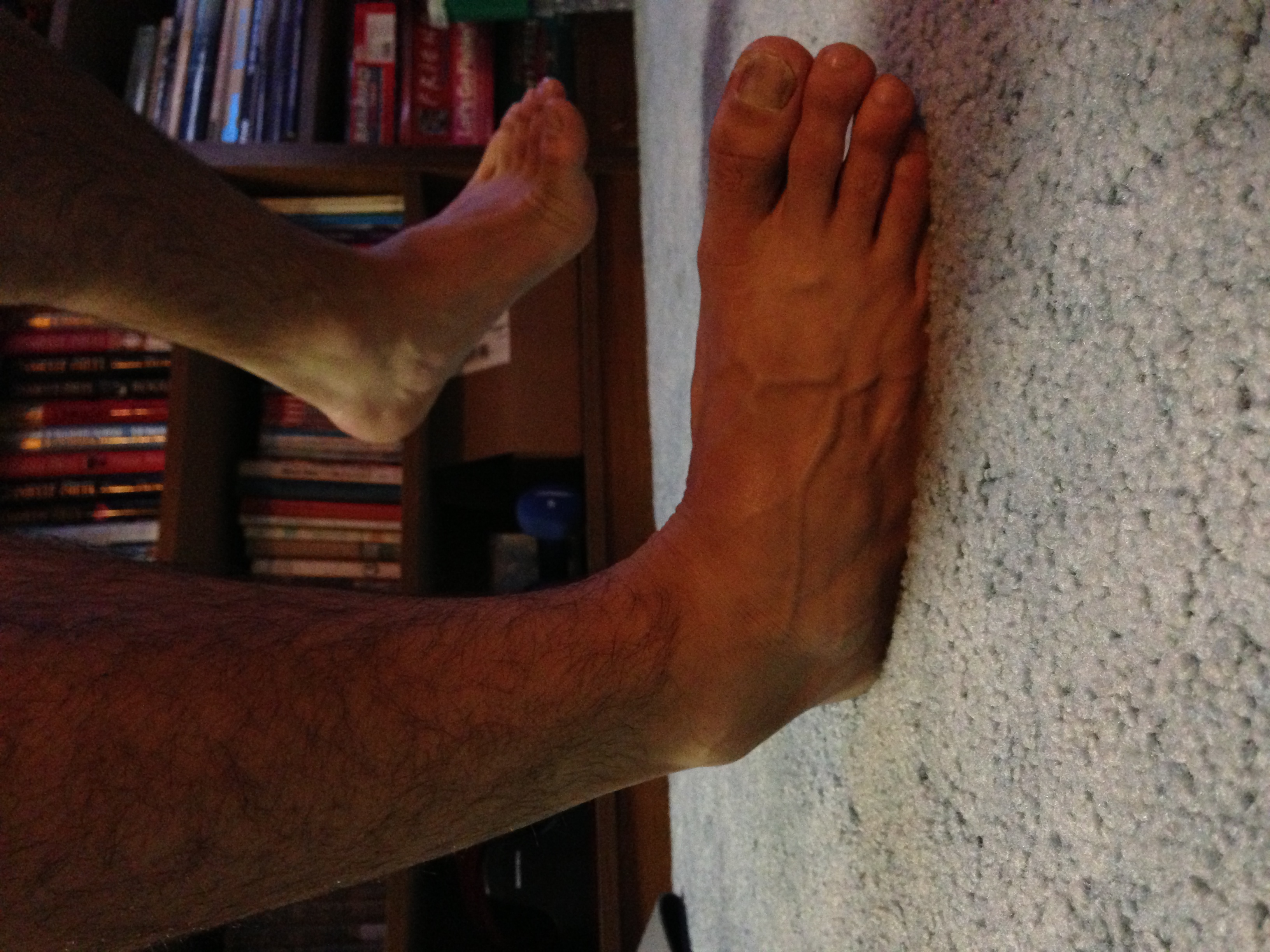
The lateral collateral ligament of ankle joint (or external lateral ligament of the ankle-joint) are ligaments of the ankle which attach to the fibula. Structure Its components are: * anterior talofibular ligament The anterior talofibular ligament attaches the anterior margin of the lateral malleolus to the adjacent region of the talus bone. The most common ligament involved in ankle sprain is the anterior talofibular ligament. * posterior talofibular ligament The posterior talofibular ligament runs horizontally between the neck of the talus and the medial side of lateral malleolus * calcaneofibular ligament The calcaneofibular ligament is attached on the posteromedial side of lateral malleolus and descends posteroinferiorly below to a lateral side of the calcaneus. References See also * Sprained ankle A sprained ankle, also known as a twisted ankle or rolled ankle, is an injury where sprain occurs on one or more ligaments of the ankle. It is the most common injury to occur ... [...More Info...] [...Related Items...] OR: [Wikipedia] [Google] [Baidu] |
Talus Bone
The talus (; Latin for ankle or ankle bone), talus bone, astragalus (), or ankle bone is one of the group of foot bones known as the tarsus. The tarsus forms the lower part of the ankle joint. It transmits the entire weight of the body from the lower legs to the foot.Platzer (2004), p 216 The talus has joints with the two bones of the lower leg, the tibia and thinner fibula. These leg bones have two prominences (the lateral and medial malleoli) that articulate with the talus. At the foot end, within the tarsus, the talus articulates with the calcaneus (heel bone) below, and with the curved navicular bone in front; together, these foot articulations form the ball-and-socket-shaped talocalcaneonavicular joint. The talus is the second largest of the tarsal bones; it is also one of the bones in the human body with the highest percentage of its surface area covered by articular cartilage. It is also unusual in that it has a retrograde blood supply, i.e. arterial blood enters the ... [...More Info...] [...Related Items...] OR: [Wikipedia] [Google] [Baidu] |
Calcaneus
In humans and many other primates, the calcaneus (; from the Latin ''calcaneus'' or ''calcaneum'', meaning heel) or heel bone is a bone of the tarsus of the foot which constitutes the heel. In some other animals, it is the point of the hock. Structure In humans, the calcaneus is the largest of the tarsal bones and the largest bone of the foot. Its long axis is pointed forwards and laterally. The talus bone, calcaneus, and navicular bone are considered the proximal row of tarsal bones. In the calcaneus, several important structures can be distinguished:Platzer (2004), p 216 There is a large calcaneal tuberosity located posteriorly on plantar surface with medial and lateral tubercles on its surface. Besides, there is another peroneal tubecle on its lateral surface. On its lower edge on either side are its lateral and medial processes (serving as the origins of the abductor hallucis and abductor digiti minimi). The Achilles tendon is inserted into a roughened area on its superio ... [...More Info...] [...Related Items...] OR: [Wikipedia] [Google] [Baidu] |
Fibula
The fibula or calf bone is a leg bone on the lateral side of the tibia, to which it is connected above and below. It is the smaller of the two bones and, in proportion to its length, the most slender of all the long bones. Its upper extremity is small, placed toward the back of the head of the tibia, below the knee joint and excluded from the formation of this joint. Its lower extremity inclines a little forward, so as to be on a plane anterior to that of the upper end; it projects below the tibia and forms the lateral part of the ankle joint. Structure The bone has the following components: * Lateral malleolus * Interosseous membrane connecting the fibula to the tibia, forming a syndesmosis joint * The superior tibiofibular articulation is an arthrodial joint between the lateral condyle of the tibia and the head of the fibula. * The inferior tibiofibular articulation (tibiofibular syndesmosis) is formed by the rough, convex surface of the medial side of the lower end of the f ... [...More Info...] [...Related Items...] OR: [Wikipedia] [Google] [Baidu] |
Lateral Malleolus
A malleolus is the bony prominence on each side of the human ankle. Each leg is supported by two bones, the tibia on the inner side (medial) of the leg and the fibula on the outer side (lateral) of the leg. The medial malleolus is the prominence on the inner side of the ankle, formed by the lower end of the tibia. The lateral malleolus is the prominence on the outer side of the ankle, formed by the lower end of the fibula. The word ''malleolus'' (), plural ''malleoli'' (), comes from Latin and means "small hammer". (It is cognate with '' mallet''.) Medial malleolus The medial malleolus is found at the foot end of the tibia. The medial surface of the lower extremity of tibia is prolonged downward to form a strong pyramidal process, flattened from without inward - the medial malleolus. * The ''medial surface'' of this process is convex and subcutaneous. * The ''lateral'' or ''articular surface'' is smooth and slightly concave, and articulates with the talus. * The ''anterior ... [...More Info...] [...Related Items...] OR: [Wikipedia] [Google] [Baidu] |
Ankle
The ankle, or the talocrural region, or the jumping bone (informal) is the area where the foot and the leg meet. The ankle includes three joints: the ankle joint proper or talocrural joint, the subtalar joint, and the inferior tibiofibular joint. The movements produced at this joint are dorsiflexion and plantarflexion of the foot. In common usage, the term ankle refers exclusively to the ankle region. In medical terminology, "ankle" (without qualifiers) can refer broadly to the region or specifically to the talocrural joint. The main bones of the ankle region are the talus (in the foot), and the tibia and fibula (in the leg). The talocrural joint is a synovial hinge joint that connects the distal ends of the tibia and fibula in the lower limb with the proximal end of the talus. The articulation between the tibia and the talus bears more weight than that between the smaller fibula and the talus. Structure Region The ankle region is found at the junction of the leg and the f ... [...More Info...] [...Related Items...] OR: [Wikipedia] [Google] [Baidu] |
Anterior Talofibular Ligament
The anterior talofibular ligament is a ligament in the ankle. It passes from the anterior margin of the fibular malleolus, anteriorly and laterally, to the talus bone, in front of its lateral articular facet. It is one of the lateral ligaments of the ankle and prevents the foot from sliding forward in relation to the shin. It is the most commonly injured ligament in a sprained ankle—from an inversion injury—and will allow a positive anterior drawer test of the ankle if completely torn. See also * Sprained ankle * Posterior talofibular ligament The posterior talofibular ligament is a ligament that connects the fibula to the talus bone. It runs almost horizontally from the malleolar fossa of the lateral malleolus of the fibula The fibula or calf bone is a leg bone on the lateral side ... References Further reading * External links * - "Lateral view of the ligaments of the ankle." * () Ligaments of the lower limb {{ligament-stub ... [...More Info...] [...Related Items...] OR: [Wikipedia] [Google] [Baidu] |
Posterior Talofibular Ligament
The posterior talofibular ligament is a ligament that connects the fibula to the talus bone. It runs almost horizontally from the malleolar fossa of the lateral malleolus of the fibula to the lateral tubercle on the posterior surface of the talus. This insertion lies immediately lateral to the groove for the tendon of the flexor hallucis longus The flexor hallucis longus muscle (FHL) is one of the three deep muscles of the posterior compartment of the leg that attaches to the plantar surface of the distal phalanx of the great toe. The other deep muscles are the flexor digitorum longus an .... References External links * () Ligaments of the lower limb {{ligament-stub ... [...More Info...] [...Related Items...] OR: [Wikipedia] [Google] [Baidu] |
Calcaneofibular Ligament
The calcaneofibular ligament is a narrow, rounded cord, running from the tip of the lateral malleolus of the fibula downward and slightly backward to a tubercle on the lateral surface of the calcaneus. It is part of the lateral collateral ligament, which opposes the hyperinversion of the subtalar joint, as in a common type of ankle sprain. It is covered by the tendons of the fibularis longus and brevis muscles. Clinical significance The calcaneofibular ligament is commonly sprained ligament in ankle injuries. It may be injured individually, or in combination with other ligaments such as the anterior talofibular ligament and the posterior talofibular ligament The posterior talofibular ligament is a ligament that connects the fibula to the talus bone. It runs almost horizontally from the malleolar fossa of the lateral malleolus of the fibula The fibula or calf bone is a leg bone on the lateral side .... References Further reading * External links * * —Calcaneofibu ... [...More Info...] [...Related Items...] OR: [Wikipedia] [Google] [Baidu] |
Sprained Ankle
A sprained ankle, also known as a twisted ankle or rolled ankle, is an injury where sprain occurs on one or more ligaments of the ankle. It is the most common injury to occur in ball sports, such as basketball, volleyball, football, and racquet sports. Signs and symptoms Knowing the symptoms that can be experienced with a sprain is important in determining that the injury is not really a break in the bone. When a sprain occurs, hematoma occurs within the tissue that surrounds the joint, causing a bruise. White blood cells responsible for inflammation migrate to the area, and blood flow increases as well. Along with this inflammation, swelling and pain is experienced. The nerves in the area become more sensitive when the injury is suffered, so pain is felt as throbbing and will worsen if there is pressure placed on the area. Warmth and redness are also seen as blood flow is increased. Also there is a decreased ability to move the joint. Image:Freshspraininbrace.JPG, Right foot, ... [...More Info...] [...Related Items...] OR: [Wikipedia] [Google] [Baidu] |


.jpg)
