|
Laser Capture Microdissection
Laser capture microdissection (LCM), also called microdissection, laser microdissection (LMD), or laser-assisted microdissection (LMD or LAM), is a method for isolating specific cells of interest from microscopic regions of tissue/cells/organisms (dissection on a microscopic scale with the help of a laser). Principle Laser-capture microdissection (LCM) is a method to procure subpopulations of tissue cells under direct microscopic visualization. LCM technology can harvest the cells of interest directly or can isolate specific cells by cutting away unwanted cells to give histologically pure enriched cell populations. A variety of downstream applications exist: DNA genotyping and loss of heterozygosity (LOH) analysis, RNA transcript profiling, cDNA library generation, proteomics discovery and signal-pathway profiling. The total time required to carry out this protocol is typically 1–1.5 h. Extraction A laser is coupled into a microscope and focuses onto the tissue on the slide. ... [...More Info...] [...Related Items...] OR: [Wikipedia] [Google] [Baidu] |
Laser Capture Microdissection Transfer Of Pure Breast Duct Epithelial Cells
A laser is a device that emits light through a process of optical amplification based on the stimulated emission of electromagnetic radiation. The word "laser" is an acronym for "light amplification by stimulated emission of radiation". The first laser was built in 1960 by Theodore H. Maiman at Hughes Research Laboratories, based on theoretical work by Charles Hard Townes and Arthur Leonard Schawlow. A laser differs from other sources of light in that it emits light which is coherence (physics), ''coherent''. Spatial coherence allows a laser to be focused to a tight spot, enabling applications such as laser cutting and Photolithography#Light sources, lithography. Spatial coherence also allows a laser beam to stay narrow over great distances (collimated light, collimation), enabling applications such as laser pointers and lidar (light detection and ranging). Lasers can also have high temporal coherence, which allows them to emit light with a very narrow frequency spectrum, spectru ... [...More Info...] [...Related Items...] OR: [Wikipedia] [Google] [Baidu] |
Microscope
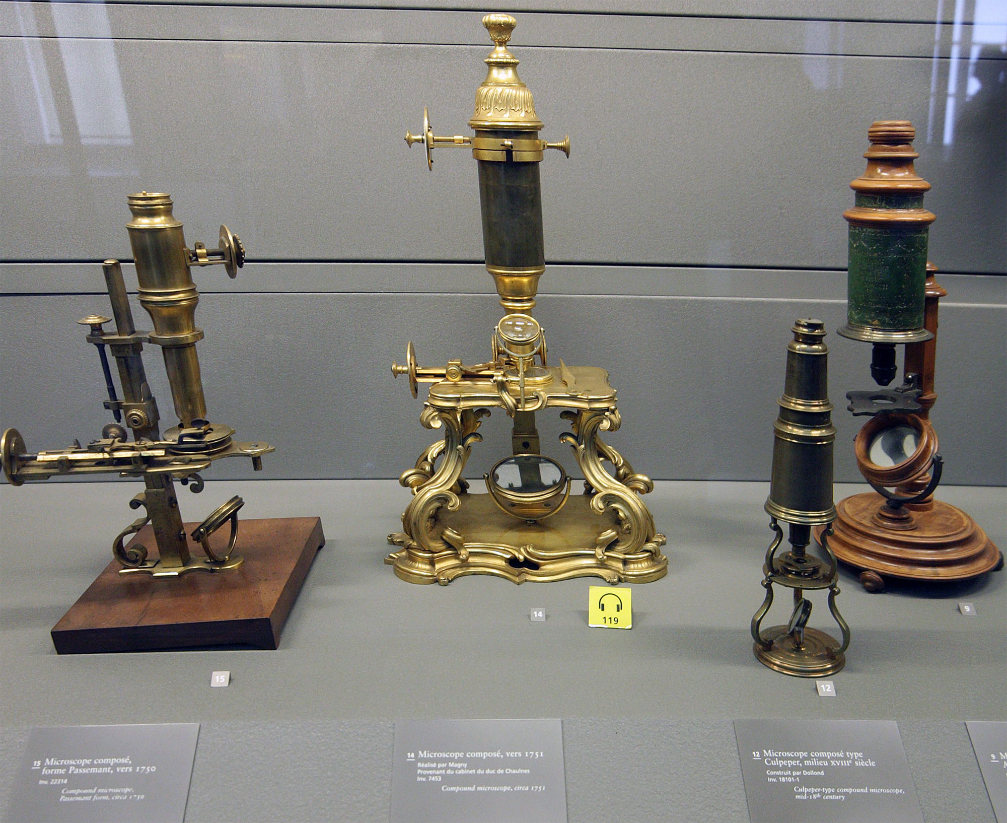
A microscope () is a laboratory instrument used to examine objects that are too small to be seen by the naked eye. Microscopy is the science of investigating small objects and structures using a microscope. Microscopic means being invisible to the eye unless aided by a microscope. There are many types of microscopes, and they may be grouped in different ways. One way is to describe the method an instrument uses to interact with a sample and produce images, either by sending a beam of light or electrons through a sample in its optical path, by detecting photon emissions from a sample, or by scanning across and a short distance from the surface of a sample using a probe. The most common microscope (and the first to be invented) is the optical microscope, which uses lenses to refract visible light that passed through a thinly sectioned sample to produce an observable image. Other major types of microscopes are the fluorescence microscope, electron microscope (both the ... [...More Info...] [...Related Items...] OR: [Wikipedia] [Google] [Baidu] |
Cell Imaging
Cell most often refers to: * Cell (biology), the functional basic unit of life Cell may also refer to: Locations * Monastic cell, a small room, hut, or cave in which a religious recluse lives, alternatively the small precursor of a monastery with only a few monks or nuns * Prison cell, a room used to hold people in prisons Groups of people * Cell, a group of people in a cell group, a form of Christian church organization * Cell, a unit of a clandestine cell system, a penetration-resistant form of a secret or outlawed organization * Cellular organizational structure, such as in business management Science, mathematics, and technology Computing and telecommunications * Cell (EDA), a term used in an electronic circuit design schematics * Cell (microprocessor), a microprocessor architecture developed by Sony, Toshiba, and IBM * Memory cell (computing), the basic unit of (volatile or non-volatile) computer memory * Cell, a unit in a database table or spreadsheet, formed by the ... [...More Info...] [...Related Items...] OR: [Wikipedia] [Google] [Baidu] |
Complete Blood Count
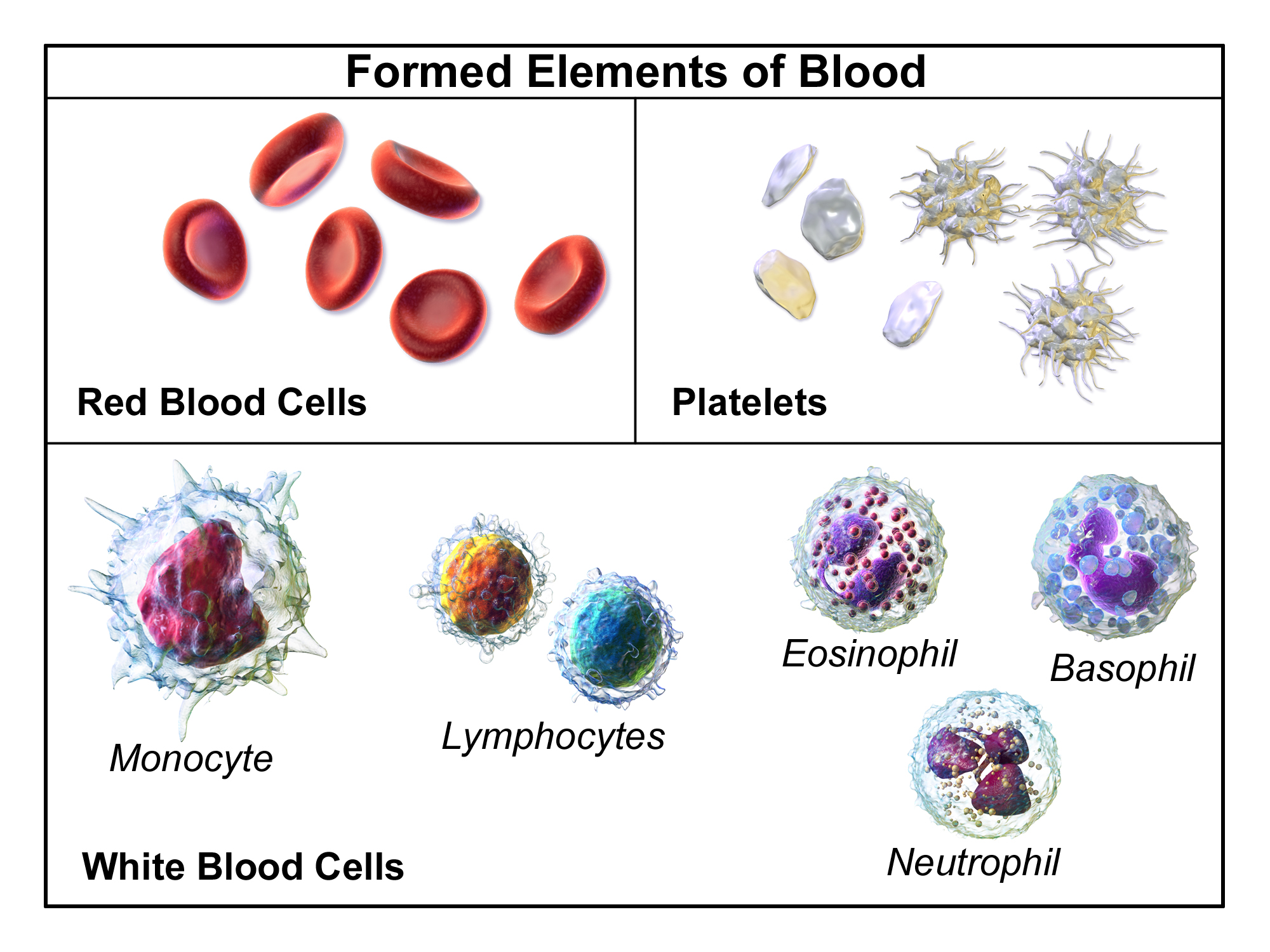
A complete blood count (CBC), also known as a full blood count (FBC), is a set of medical laboratory tests that provide information about the cells in a person's blood. The CBC indicates the counts of white blood cells, red blood cells and platelets, the concentration of hemoglobin, and the hematocrit (the volume percentage of red blood cells). The red blood cell indices, which indicate the average size and hemoglobin content of red blood cells, are also reported, and a white blood cell differential, which counts the different types of white blood cells, may be included. The CBC is often carried out as part of a medical assessment and can be used to monitor health or diagnose diseases. The results are interpreted by comparing them to reference ranges, which vary with sex and age. Conditions like anemia and thrombocytopenia are defined by abnormal complete blood count results. The red blood cell indices can provide information about the cause of a person's anemia such as iron ... [...More Info...] [...Related Items...] OR: [Wikipedia] [Google] [Baidu] |
Amyloid Plaques
Amyloid plaques (also known as neuritic plaques, amyloid beta plaques or senile plaques) are extracellular deposits of the amyloid beta (Aβ) protein mainly in the grey matter of the brain. Degenerative neuronal elements and an abundance of microglia and astrocytes can be associated with amyloid plaques. Some plaques occur in the brain as a result of aging, but large numbers of plaques and neurofibrillary tangles are characteristic features of Alzheimer's disease. Abnormal neurites in amyloid plaques are tortuous, often swollen axons and dendrites. The neurites contain a variety of organelles and cellular debris, and many of them include characteristic paired helical filaments, the ultrastructural component of neurofibrillary tangles. The plaques are highly variable in shape and size; in tissue sections immunostained for Aβ, they comprise a log-normal size distribution curve with an average plaque area of 400-450 square micrometers (µm²). The smallest plaques (less than ... [...More Info...] [...Related Items...] OR: [Wikipedia] [Google] [Baidu] |
Protein
Proteins are large biomolecules and macromolecules that comprise one or more long chains of amino acid residues. Proteins perform a vast array of functions within organisms, including catalysing metabolic reactions, DNA replication, responding to stimuli, providing structure to cells and organisms, and transporting molecules from one location to another. Proteins differ from one another primarily in their sequence of amino acids, which is dictated by the nucleotide sequence of their genes, and which usually results in protein folding into a specific 3D structure that determines its activity. A linear chain of amino acid residues is called a polypeptide. A protein contains at least one long polypeptide. Short polypeptides, containing less than 20–30 residues, are rarely considered to be proteins and are commonly called peptides. The individual amino acid residues are bonded together by peptide bonds and adjacent amino acid residues. The sequence of amino acid ... [...More Info...] [...Related Items...] OR: [Wikipedia] [Google] [Baidu] |
UV-A
Ultraviolet (UV) is a form of electromagnetic radiation with wavelength from 10 nm (with a corresponding frequency around 30 PHz) to 400 nm (750 THz), shorter than that of visible light, but longer than X-rays. UV radiation is present in sunlight, and constitutes about 10% of the total electromagnetic radiation output from the Sun. It is also produced by electric arcs and specialized lights, such as mercury-vapor lamps, tanning lamps, and black lights. Although long-wavelength ultraviolet is not considered an ionizing radiation because its photons lack the energy to ionize atoms, it can cause chemical reactions and causes many substances to glow or fluoresce. Consequently, the chemical and biological effects of UV are greater than simple heating effects, and many practical applications of UV radiation derive from its interactions with organic molecules. Short-wave ultraviolet light damages DNA and sterilizes surfaces with which it comes into contact. For huma ... [...More Info...] [...Related Items...] OR: [Wikipedia] [Google] [Baidu] |
Solid-state Laser
A solid-state laser is a laser that uses a gain medium that is a solid, rather than a liquid as in dye lasers or a gas as in gas lasers. Semiconductor-based lasers are also in the solid state, but are generally considered as a separate class from solid-state lasers, called laser diodes. Solid-state media Generally, the active medium of a solid-state laser consists of a glass or crystalline "host" material, to which is added a "dopant" such as neodymium, chromium, erbium, thulium or ytterbium.Z. Su, J. D. Bradley, N. Li, E. S. Magden, Purnawirman, D. Coleman, N. Fahrenkopf, C. Baiocco, T. Adam, G. Leake, D. Coolbaugh, D. Vermeulen, and M. R. Watts (2016"Ultra-Compact CMOS-Compatible Ytterbium Microlaser" ''Integrated Photonics Research, Silicon and Nanophotonics 2016'', IW1A.3. Many of the common dopants are rare-earth elements, because the excited states of such ions are not strongly coupled with the thermal vibrations of their crystal lattices (phonons), and their operati ... [...More Info...] [...Related Items...] OR: [Wikipedia] [Google] [Baidu] |
ION LMD
ION LMD system is one of the laser microdissection systems and a name of device that follows Gravity-Assisted Microdissection method, also known as GAM method. This non-contact laser microdissection system makes cell isolation for further genetic analysis possible. It is the first developed laser microdissection system in Asia. History At first, proto type of ION LMD system was developed in 2004. The first generation of ION LMD was developed in 2005 and then the second generation(so-called G2) was developed in 2008. At last, the third generation(so-called ION LMD Pro) was developed in 2012. Manufacturer JungWoo F&B was founded in 1994, and offers various factory automation products for clients in semiconductor, consumer electronics, LCD, automotive manufacturing and ship-building industries. In 2003, the company entered the bio-mechanics business for the medical laboratory market and developed an ION LMD system which is utilized in cancer research. Awards This ION LMD ... [...More Info...] [...Related Items...] OR: [Wikipedia] [Google] [Baidu] |
Phase Contrast Microscopy
__NOTOC__ Phase-contrast microscopy (PCM) is an optical microscopy technique that converts phase shifts in light passing through a transparent specimen to brightness changes in the image. Phase shifts themselves are invisible, but become visible when shown as brightness variations. When light waves travel through a medium other than a vacuum, interaction with the medium causes the wave amplitude and phase to change in a manner dependent on properties of the medium. Changes in amplitude (brightness) arise from the scattering and absorption of light, which is often wavelength-dependent and may give rise to colors. Photographic equipment and the human eye are only sensitive to amplitude variations. Without special arrangements, phase changes are therefore invisible. Yet, phase changes often convey important information. Phase-contrast microscopy is particularly important in biology. It reveals many cellular structures that are invisible with a bright-field microscope, as exemp ... [...More Info...] [...Related Items...] OR: [Wikipedia] [Google] [Baidu] |
Differential Interference Contrast Microscopy
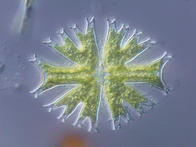
Differential interference contrast (DIC) microscopy, also known as Nomarski interference contrast (NIC) or Nomarski microscopy, is an optical microscopy technique used to enhance the contrast in unstained, transparent samples. DIC works on the principle of interferometry to gain information about the optical path length of the sample, to see otherwise invisible features. A relatively complex optical system produces an image with the object appearing black to white on a grey background. This image is similar to that obtained by phase contrast microscopy but without the bright diffraction halo. The technique was developed by Polish physicist Georges Nomarski in 1952. DIC works by separating a polarized light source into two orthogonally polarized mutually coherent parts which are spatially displaced (sheared) at the sample plane, and recombined before observation. The interference of the two parts at recombination is sensitive to their optical path difference (i.e. the product ... [...More Info...] [...Related Items...] OR: [Wikipedia] [Google] [Baidu] |
Bright Field Microscopy
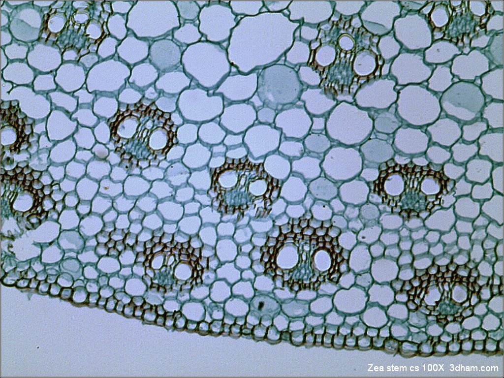
Bright-field microscopy (BF) is the simplest of all the optical microscopy illumination techniques. Sample illumination is transmitted (i.e., illuminated from below and observed from above) white light, and contrast in the sample is caused by attenuation of the transmitted light in dense areas of the sample. Bright-field microscopy is the simplest of a range of techniques used for illumination of samples in light microscopes, and its simplicity makes it a popular technique. The typical appearance of a bright-field microscopy image is a dark sample on a bright background, hence the name. Light path The light path of a bright-field microscope is extremely simple, no additional components are required beyond the normal light-microscope setup. The light path therefore consists of: * a transillumination light source, commonly a halogen lamp in the microscope stand; * a condenser lens, which focuses light from the light source onto the sample; * an objective lens, which collects light ... [...More Info...] [...Related Items...] OR: [Wikipedia] [Google] [Baidu] |


