|
Lacrimal Sac
The lacrimal sac or lachrymal sac is the upper dilated end of the nasolacrimal duct, and is lodged in a deep groove formed by the lacrimal bone and frontal process of the maxilla. It connects the lacrimal canaliculi, which drain tears from the eye's surface, and the nasolacrimal duct, which conveys this fluid into the nasal cavity. Lacrimal sac occlusion leads to dacryocystitis. Structure It is oval in form and measures from 12 to 15 mm. in length; its upper end is closed and rounded; its lower is continued into the nasolacrimal duct. Its superficial surface is covered by a fibrous expansion derived from the medial palpebral ligament, and its deep surface is crossed by the lacrimal part of the orbicularis oculi, which is attached to the crest on the lacrimal bone. Histology Like the nasolacrimal duct, the sac is lined by stratified columnar epithelium with mucus-secreting goblet cells, with surrounding connective tissue. The Lacrimal Sac also drains the eye of debris and ... [...More Info...] [...Related Items...] OR: [Wikipedia] [Google] [Baidu] |
Lacrimal Apparatus
The lacrimal apparatus is the physiological system containing the Orbit (anatomy), orbital structures for tears, tear production and drainage.Cassin, B. and Solomon, S. ''Dictionary of Eye Terminology''. Gainesville, Florida: Triad Publishing Company, 1990. It consists of: * The lacrimal gland, which secretes the tears, and its excretory ducts, which convey the fluid to the surface of the human eye; it is a j-shaped serous gland located in lacrimal fossa. * The lacrimal canaliculi, the lacrimal sac, and the nasolacrimal duct, by which the fluid is conveyed into the cavity of the Human nose, nose, emptying anterioinferiorly to the inferior nasal conchae from the nasolacrimal duct; * The innervation of the lacrimal apparatus involves both the a Sympathetic nervous system, sympathetic supply through the Internal carotid plexus, carotid plexus of nerves around the internal carotid artery, and parasympathetic nervous system, parasympathetically from the lacrimal nucleus of the facial n ... [...More Info...] [...Related Items...] OR: [Wikipedia] [Google] [Baidu] |
Angular Artery
The angular artery is an artery of the face. It is the terminal part of the facial artery. It ascends to the medial angle of the eye's Orbit (anatomy), orbit. It is accompanied by the angular vein. It ends by anastomosing with the dorsal nasal branch of the ophthalmic artery. It supplies the lacrimal sac, the orbicularis oculi muscle, and the outer side of the nose. Structure The angular artery is the terminal part of the facial artery. It ascends to the medial angle of the eye's Orbit (anatomy), orbit (the medial canthus). It is embedded in the fibers of the angular head of the levator labii superioris muscle. It is accompanied by the angular vein. On the cheek, it distributes branches which anastomose with the infraorbital artery. It ends by anastomosing with the dorsal nasal branch of the ophthalmic artery. Function The angular artery supplies the lacrimal sac, most of the outer side of the nose, part of the lower eyelid, and the orbicularis oculi muscle. Clinical signif ... [...More Info...] [...Related Items...] OR: [Wikipedia] [Google] [Baidu] |
Nasolacrimal Duct
The nasolacrimal duct (also called the tear duct) carries tears from the lacrimal sac of the eye into the nasal cavity. The duct begins in the eye socket between the maxillary and lacrimal bones, from where it passes downwards and backwards. The opening of the nasolacrimal duct into the inferior nasal meatus of the nasal cavity is partially covered by a mucosal fold ( valve of Hasner or ''plica lacrimalis''). Excess tears flow through the nasolacrimal duct which drains into the inferior nasal meatus. This is the reason the nose starts to run when a person is crying or has watery eyes from an allergy, and why one can sometimes taste eye drops. This is for the same reason when applying some eye drops it is often advised to close the nasolacrimal duct by pressing it with a finger to prevent the medicine from escaping the eye and having unwanted side effects elsewhere in the body as it will proceed through the canal to the Nasal Cavity. Like the lacrimal sac, the duct is lined by st ... [...More Info...] [...Related Items...] OR: [Wikipedia] [Google] [Baidu] |
Lacrimal Bone
The lacrimal bone is a small and fragile bone of the facial skeleton; it is roughly the size of the little fingernail. It is situated at the front part of the medial wall of the orbit. It has two surfaces and four borders. Several bony landmarks of the lacrimal bone function in the process of lacrimation or crying. Specifically, the lacrimal bone helps form the nasolacrimal canal necessary for tear translocation. A depression on the anterior inferior portion of the bone, the lacrimal fossa, houses the membranous lacrimal sac. Tears or lacrimal fluid, from the lacrimal glands, collect in this sac during excessive lacrimation. The fluid then flows through the nasolacrimal duct and into the nasopharynx. This drainage results in what is commonly referred to a runny nose during excessive crying or tear production. Injury or fracture of the lacrimal bone can result in posttraumatic obstruction of the lacrimal pathways. Structure Lateral or orbital surface The lateral or orbital surface i ... [...More Info...] [...Related Items...] OR: [Wikipedia] [Google] [Baidu] |
Frontal Process Of Maxilla
The frontal process of maxilla is a strong plate, which projects upward, medialward, and backward from the maxilla, forming part of the lateral boundary of the nose. Its ''lateral surface'' is smooth, continuous with the anterior surface of the body, and gives attachment to the quadratus labii superioris, the orbicularis oculi, and the medial palpebral ligament. Its ''medial surface'' forms part of the lateral wall of the nasal cavity; at its upper part is a rough, uneven area, which articulates with the ethmoid, closing in the anterior ethmoidal cells; below this is an oblique ridge, the ethmoidal crest, the posterior end of which articulates with the middle nasal concha, while the anterior part is termed the agger nasi; the crest forms the upper limit of the atrium of the middle meatus. The ''upper border'' articulates with the frontal bone and the ''anterior'' with the nasal; the ''posterior border'' is thick, and hollowed into a groove, which is continuous below with the lac ... [...More Info...] [...Related Items...] OR: [Wikipedia] [Google] [Baidu] |
Lacrimal Canaliculi
The lacrimal canaliculi, (sing. canaliculus), are the small channels in each eyelid that drain lacrimal fluid, from the lacrimal puncta to the lacrimal sac. This forms part of the lacrimal apparatus that drains lacrimal fluid from the surface of the eye to the nasal cavity. Structure There is a single lacrimal canaliculus in each eyelid, a superior lacrimal canaliculus in the upper eyelid and an inferior lacrimal canaliculus in the lower eyelid. The canaliculi travel vertically and then turn medially to travel towards the lacrimal sac. At the bend, the canaliculus is dilated and called the ampulla. Usually, the superior and inferior lacrimal canaliculi join to form a common passage that enters the lateral wall of the lacrimal sac. Superior lacrimal canaliculus The superior lacrimal canaliculus is located in the upper eyelid. It first ascends, then bends medially towards the lacrimal sac. It drains lacrimal fluid from the superior lacrimal punctum. It is smaller and shorter th ... [...More Info...] [...Related Items...] OR: [Wikipedia] [Google] [Baidu] |
Dacryocystitis
Dacryocystitis is an infection of the lacrimal sac, secondary to obstruction of the nasolacrimal duct at the junction of lacrimal sac. The term derives from the Greek ''dákryon'' ( tear), ''cysta'' (sac), and ''-itis'' (inflammation). It causes pain, redness, and swelling over the inner aspect of the lower eyelid and epiphora. When nasolacrimal duct obstruction is secondary to a congenital barrier it is referred to as dacryocystocele. It is most commonly caused by '' Staphylococcus aureus'' and ''Streptococcus pneumoniae''. The most common complication is corneal ulceration, frequently in association with ''S. pneumoniae''. The mainstays of treatment are oral antibiotics, warm compresses, and relief of nasolacrimal duct obstruction by dacryocystorhinostomy. Signs and symptoms *Pain, swelling, redness over the lacrimal sac at medial canthus *Tearing, crusting, fever *Digital pressure over the lacrimal sac may extrude pus through the punctum *In chronic cases, tearing may be th ... [...More Info...] [...Related Items...] OR: [Wikipedia] [Google] [Baidu] |
Medial Palpebral Ligament
The medial palpebral ligament (medial canthal tendon) is a ligament of the face. It attaches to the Frontal process of maxilla, frontal process of the maxilla, the lacrimal groove, and the Tarsus (eyelids), tarsus of each eyelid. It has a superficial (anterior) and a deep (posterior) layer, with many surrounding attachments. It connects the Canthus, medial canthus of each eyelid to the medial part of the Orbit (anatomy), orbit. It is a useful point of fixation during eyelid reconstructive surgery. Structure The anterior attachment of the medial palpebral ligament is to the frontal process of maxilla, frontal process of the maxilla in front of the lacrimal groove (near the nasal bone and the frontal bone), and its posterior attachment is the lacrimal bone. Crossing the lacrimal sac, it divides into two parts, upper and lower, each attached to the medial end of the corresponding tarsus (eyelids), tarsus of each eyelid. As the ligament crosses the lacrimal sac, a strong aponeuroti ... [...More Info...] [...Related Items...] OR: [Wikipedia] [Google] [Baidu] |
Orbicularis Oculi
The orbicularis oculi is a muscle in the face that closes the eyelids. It arises from the nasal part of the frontal bone, from the frontal process of the maxilla in front of the lacrimal groove, and from the anterior surface and borders of a short fibrous band, the medial palpebral ligament. From this origin, the fibers are directed laterally, forming a broad and thin layer, which occupies the eyelids or palpebræ, surrounds the circumference of the orbit, and spreads over the temple, and downward on the cheek. Structure There are at least 3 clearly defined sections of the orbicularis muscle. However, it is not clear whether the lacrimal section is a separate section, or whether it is just an extension of the preseptal and pretarsal sections. Orbital orbicularis The orbital portion is thicker and of a reddish color; its fibers form a complete ellipse without interruption at the lateral palpebral commissure; the upper fibers of this portion blend with the frontalis and corrugator ... [...More Info...] [...Related Items...] OR: [Wikipedia] [Google] [Baidu] |
Stratified Columnar Epithelium
Stratified columnar epithelium is a rare type of epithelial tissue composed of column-shaped cells arranged in multiple layers. It is found in the conjunctiva, pharynx, anus, and male urethra. It also occurs in embryo. Location Stratified columnar epithelia are found in a variety of locations, including: * parts of the conjunctiva of the eye * parts of the pharynx * anus * male urethra and vas deferens * excretory duct of mammary gland and major salivary glands Embryology Stratified columnar epithelium is initially present in parts of the gastrointestinal tract in utero ''In Utero'' is the third and final studio album by American rock band Nirvana. It was released on September 21, 1993, by DGC Records. After breaking into the mainstream with their second album, ''Nevermind'' (1991), Nirvana hired Steve Albini t ..., before being replaced with other types of epithelium. For example, by 8 weeks, it covers the lining of the stomach. By 17 weeks, it is replaced by simple c ... [...More Info...] [...Related Items...] OR: [Wikipedia] [Google] [Baidu] |
Goblet Cells
Goblet cells are simple columnar epithelial cells that secrete gel-forming mucins, like mucin 5AC. The goblet cells mainly use the merocrine method of secretion, secreting vesicles into a duct, but may use apocrine methods, budding off their secretions, when under stress. The term ''goblet'' refers to the cell's goblet-like shape. The apical portion is shaped like a cup, as it is distended by abundant mucus laden granules; its basal portion lacks these granules and is shaped like a stem. The goblet cell is highly polarized with the nucleus and other organelles concentrated at the base of the cell and secretory granules containing mucin, at the apical surface. The apical plasma membrane projects short microvilli to give an increased surface area for secretion. Goblet cells are typically found in the respiratory, reproductive and gastrointestinal tracts and are surrounded by other columnar cells. Biased differentiation of airway basal cells in the respiratory epithelium, into gob ... [...More Info...] [...Related Items...] OR: [Wikipedia] [Google] [Baidu] |
Radiocontrast
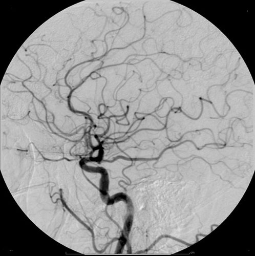
Radiocontrast agents are substances used to enhance the visibility of internal structures in X-ray-based imaging techniques such as computed tomography (contrast CT), projectional radiography, and fluoroscopy. Radiocontrast agents are typically iodine, or more rarely barium sulfate. The contrast agents absorb external X-rays, resulting in decreased exposure on the X-ray detector. This is different from radiopharmaceuticals used in nuclear medicine which emit radiation. Magnetic resonance imaging (MRI) functions through different principles and thus MRI contrast agents have a different mode of action. These compounds work by altering the magnetic properties of nearby hydrogen nuclei. Types and uses Radiocontrast agents used in X-ray examinations can be grouped in positive (iodinated agents, barium sulfate), and negative agents (air, carbon dioxide, methylcellulose). Iodine (circulatory system) Iodinated contrast contains iodine. It is the main type of radiocontrast used for intr ... [...More Info...] [...Related Items...] OR: [Wikipedia] [Google] [Baidu] |
_and_foveolar_cells_in_incomplete_Barrett's_esophagus.jpg)
