|
Lacrimal Caruncle
The lacrimal caruncle, or caruncula lacrimalis, is the small, pink, globular nodule at the inner corner (the medial canthus) of the eye. It consists of tissue types of neighbouring eye structures. It may suffer from lesions and allergic inflammation. Structure The lacrimal caruncle is found at the medial canthus of the eye. It consists of skin, hair follicles, sebaceous glands, sweat glands, accessory lacrimal tissue and other tissues that are present in the skin and accessory lacrimal glands. Its non-keratinized epithelium resembles the conjunctival epithelium. Clinical significance Lesions The lacrimal caruncle may have a lesion. This can have any one of a number of causes, which may be difficult to diagnose. Cancer is a rare cause. These lesions include papillomas and oncocytomas. Allergies With ocular allergies, the lacrimal caruncle and the plica semilunaris of the conjunctiva may be inflamed and pruritic (itchy) due to histamine Histamine is an organic nitro ... [...More Info...] [...Related Items...] OR: [Wikipedia] [Google] [Baidu] |
Canthus
The canthus (pl. canthi, palpebral commissures) is either corner of the eye where the upper and lower eyelids meet. More specifically, the inner and outer canthi are, respectively, the medial and lateral ends/angles of the palpebral fissure. The bicanthal plane is the transversal plane linking both canthi and defines the upper boundary of the midface. Etymology The word ' is the Latinized form of the Ancient Greek ('), meaning 'corner of the eye'. Population distribution The eyes of those of East Asian and some Southeast Asian people tend to have the inner canthus veiled by the epicanthus. In the Caucasian or double eyelid, the inner corner tends to be exposed completely. Commissures * The ''lateral palpebral commissure'' (commissura palpebrarum lateralis; external canthus) is more acute than the medial, and the eyelids here lie in close contact with the bulb of the eye. * The ''medial palpebral commissure'' (commissura palpebrarum medialis; internal canthus) is prolon ... [...More Info...] [...Related Items...] OR: [Wikipedia] [Google] [Baidu] |
Human Eye
The human eye is a sensory organ, part of the sensory nervous system, that reacts to visible light and allows humans to use visual information for various purposes including seeing things, keeping balance, and maintaining circadian rhythm. The eye can be considered as a living optical device. It is approximately spherical in shape, with its outer layers, such as the outermost, white part of the eye (the sclera) and one of its inner layers (the pigmented choroid) keeping the eye essentially light tight except on the eye's optic axis. In order, along the optic axis, the optical components consist of a first lens (the cornea—the clear part of the eye) that accomplishes most of the focussing of light from the outside world; then an aperture (the pupil) in a diaphragm (the iris—the coloured part of the eye) that controls the amount of light entering the interior of the eye; then another lens (the crystalline lens) that accomplishes the remaining focussing of light in ... [...More Info...] [...Related Items...] OR: [Wikipedia] [Google] [Baidu] |
Allergy
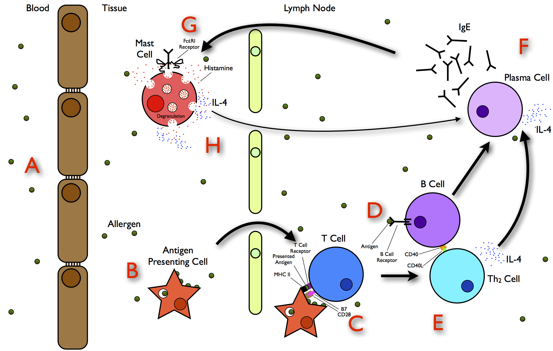
Allergies, also known as allergic diseases, refer a number of conditions caused by the hypersensitivity of the immune system to typically harmless substances in the environment. These diseases include Allergic rhinitis, hay fever, Food allergy, food allergies, atopic dermatitis, allergic asthma, and anaphylaxis. Symptoms may include allergic conjunctivitis, red eyes, an itchy rash, sneeze, sneezing, coughing, a rhinorrhea, runny nose, shortness of breath, or swelling. Note: food intolerances and food poisoning are separate conditions. Common allergens include pollen and certain foods. Metals and other substances may also cause such problems. Food, insect stings, and medications are common causes of severe reactions. Their development is due to both genetic and environmental factors. The underlying mechanism involves immunoglobulin E antibodies (IgE), part of the body's immune system, binding to an allergen and then to FcεRI, a receptor on mast cells or basophils where it tri ... [...More Info...] [...Related Items...] OR: [Wikipedia] [Google] [Baidu] |
Skin
Skin is the layer of usually soft, flexible outer tissue covering the body of a vertebrate animal, with three main functions: protection, regulation, and sensation. Other cuticle, animal coverings, such as the arthropod exoskeleton, have different cellular differentiation, developmental origin, structure and chemical composition. The adjective cutaneous means "of the skin" (from Latin ''cutis'' 'skin'). In mammals, the skin is an organ (anatomy), organ of the integumentary system made up of multiple layers of ectodermal tissue (biology), tissue and guards the underlying muscles, bones, ligaments, and internal organs. Skin of a different nature exists in amphibians, reptiles, and birds. Skin (including cutaneous and subcutaneous tissues) plays crucial roles in formation, structure, and function of extraskeletal apparatus such as horns of bovids (e.g., cattle) and rhinos, cervids' antlers, giraffids' ossicones, armadillos' osteoderm, and os penis/os clitoris. All mammals have som ... [...More Info...] [...Related Items...] OR: [Wikipedia] [Google] [Baidu] |
Hair Follicle
The hair follicle is an organ found in mammalian skin. It resides in the dermal layer of the skin and is made up of 20 different cell types, each with distinct functions. The hair follicle regulates hair growth via a complex interaction between hormones, neuropeptides, and immune cells. This complex interaction induces the hair follicle to produce different types of hair as seen on different parts of the body. For example, terminal hairs grow on the scalp and lanugo hairs are seen covering the bodies of fetuses in the uterus and in some newborn babies. The process of hair growth occurs in distinct sequential stages. The first stage is called ''anagen'' and is the active growth phase, ''telogen'' is the resting stage, ''catagen'' is the regression of the hair follicle phase, ''exogen'' is the active shedding of hair phase and lastly ''kenogen'' is the phase between the empty hair follicle and the growth of new hair. The function of hair in humans has long been a subject ... [...More Info...] [...Related Items...] OR: [Wikipedia] [Google] [Baidu] |
Sebaceous Gland
A sebaceous gland is a microscopic exocrine gland in the skin that opens into a hair follicle to secrete an oily or waxy matter, called sebum, which lubricates the hair and skin of mammals. In humans, sebaceous glands occur in the greatest number on the face and scalp, but also on all parts of the skin except the palms of the hands and soles of the feet. In the eyelids, meibomian glands, also called tarsal glands, are a type of sebaceous gland that secrete a special type of sebum into tears. Surrounding the female nipple, areolar glands are specialized sebaceous glands for lubricating the nipple. Fordyce spots are benign, visible, sebaceous glands found usually on the lips, gums and inner cheeks, and genitals. Structure Location Sebaceous glands are found throughout all areas of the skin, except the palms of the hands and soles of the feet. There are two types of sebaceous glands, those connected to hair follicles and those that exist independently. Sebaceous gland ... [...More Info...] [...Related Items...] OR: [Wikipedia] [Google] [Baidu] |
Sweat Gland
Sweat glands, also known as sudoriferous or sudoriparous glands, , are small tubular structures of the skin that produce sweat. Sweat glands are a type of exocrine gland, which are glands that produce and secrete substances onto an epithelial surface by way of a duct. There are two main types of sweat glands that differ in their structure, function, secretory product, mechanism of excretion, anatomic distribution, and distribution across species: * Eccrine sweat glands are distributed almost all over the human body, in varying densities, with the highest density in palms and soles, then on the head, but much less on the trunk and the extremities. Its water-based secretion represents a primary form of cooling in humans. * Apocrine sweat glands are mostly limited to the axillae (armpits) and perineal area in humans. They are not significant for cooling in humans, but are the sole effective sweat glands in hoofed animals, such as the camels, donkeys, horses, and cattle. Ce ... [...More Info...] [...Related Items...] OR: [Wikipedia] [Google] [Baidu] |
Accessory Lacrimal Glands
Krause's glands, Wolfring's glands (or Ciaccio's glands) and Popov's gland are the accessory lacrimal glands of the lacrimal system of human eye. These glands are structurally and histologically similar to the main lacrimal gland. Glands of Krause are located in the stroma of the conjunctival fornix, and the glands of Wolfring are located along the orbital border of the tarsal plate. These glands are oval and display numerous acini. The acini are surrounded, sometimes incompletely, by a row of myoepithelial cells. Animal studies suggest that the ducts of Wolfring glands have a tortuous course and open onto the palpebral conjunctiva. Like the main lacrimal gland, the accessory lacrimal glands are also densely innervated, but they lack parasympathetic innervation. These glands are exocrine glands, responsible for the basal (unstimulated) secretion of the middle aqueous layer of the tear film. 20 to 40 glands of Krause are found in the upper fornix, and 6-8 glands appear in the ... [...More Info...] [...Related Items...] OR: [Wikipedia] [Google] [Baidu] |
Epithelium
Epithelium or epithelial tissue is one of the four basic types of animal tissue, along with connective tissue, muscle tissue and nervous tissue. It is a thin, continuous, protective layer of compactly packed cells with a little intercellular matrix. Epithelial tissues line the outer surfaces of organs and blood vessels throughout the body, as well as the inner surfaces of cavities in many internal organs. An example is the epidermis, the outermost layer of the skin. There are three principal shapes of epithelial cell: squamous (scaly), columnar, and cuboidal. These can be arranged in a singular layer of cells as simple epithelium, either squamous, columnar, or cuboidal, or in layers of two or more cells deep as stratified (layered), or ''compound'', either squamous, columnar or cuboidal. In some tissues, a layer of columnar cells may appear to be stratified due to the placement of the nuclei. This sort of tissue is called pseudostratified. All glands are made up of epi ... [...More Info...] [...Related Items...] OR: [Wikipedia] [Google] [Baidu] |
Conjunctiva
The conjunctiva is a thin mucous membrane that lines the inside of the eyelids and covers the sclera (the white of the eye). It is composed of non-keratinized, stratified squamous epithelium with goblet cells, stratified columnar epithelium and stratified cuboidal epithelium (depending on the zone). The conjunctiva is highly vascularised, with many microvessels easily accessible for imaging studies. Structure The conjunctiva is typically divided into three parts: Blood supply Blood to the bulbar conjunctiva is primarily derived from the ophthalmic artery. The blood supply to the palpebral conjunctiva (the eyelid) is derived from the external carotid artery. However, the circulations of the bulbar conjunctiva and palpebral conjunctiva are linked, so both bulbar conjunctival and palpebral conjunctival vessels are supplied by both the ophthalmic artery and the external carotid artery, to varying extents. Nerve supply Sensory innervation of the conjunctiva is divided i ... [...More Info...] [...Related Items...] OR: [Wikipedia] [Google] [Baidu] |
The American Journal Of Dermatopathology
The International Society of Dermatopathology is a non-profit, international society for the discipline of dermatopathology which was founded in 1979. It is based in Half Moon Bay Half Moon Bay is a coastal city in San Mateo County, California, United States, approximately south of San Francisco. Its population was 11,795 as of the 2020 census. Immediately at the north of Half Moon Bay is Pillar Point Harbor and the un ..., California, US. It publishes a peer-reviewed journal, ''The American Journal of Dermatopathology'' (AJDP). It was founded by Helmut Kerl, MD; Gérald E. Piérard, MD; Jorge Sánchez, MD; and the late A. Bernard Ackerman, MD. References External links Official website Dermatology organizations Pathology organizations International medical associations 1979 establishments in California Non-profit organizations based in California {{med-journal-stub ... [...More Info...] [...Related Items...] OR: [Wikipedia] [Google] [Baidu] |
Papilloma
A papilloma (plural papillomas or papillomata) ('' papillo-'' + ''-oma'') is a benign epithelial tumor growing exophytically (outwardly projecting) in nipple-like and often finger-like fronds. In this context, papilla refers to the projection created by the tumor, not a tumor on an already existing papilla (such as the nipple). When used without context, it frequently refers to infections ( squamous cell papilloma) caused by human papillomavirus (HPV), such as warts. Human papillomavirus infection is a major cause of cervical cancer, vulvar cancer, vaginal cancer, penis cancer, anal cancer, and HPV-positive oropharyngeal cancers. Most viral warts are caused by human papillomavirus infection (HPV), of which there are nearly 200 distinct human papillomaviruses (HPVs), and many HPV types are carcinogenic. There are, however, a number of other conditions that cause papilloma, as well as many cases in which there is no known cause. Signs and symptoms A benign papillomatous tumor ... [...More Info...] [...Related Items...] OR: [Wikipedia] [Google] [Baidu] |


