|
LRP6
Low-density lipoprotein receptor-related protein 6 is a protein that in humans is encoded by the ''LRP6'' gene. LRP6 is a key component of the LRP5/LRP6/Frizzled co-receptor group that is involved in canonical Wnt pathway. Structure LRP6 is a transmembrane low-density lipoprotein receptor that shares a similar structure with LRP5. In each protein, about 85% of its 1600-amino-acid length is extracellular. Each has four β-propeller motifs at the amino terminal end that alternate with four epidermal growth factor (EGF)-like repeats. Most extracellular ligands bind to LRP5 and LRP6 at the β-propellers. Each protein has a single-pass, 22-amino-acid segment that crosses the cell membrane and a 207-amino-acid segment that is internal to the cell. Function LRP6 acts as a co-receptor with LRP5 and the Frizzled protein family members for transducing signals by Wnt proteins through the canonical Wnt pathway. Interactions Canonical WNT signals are transduced through Frizzled receptor an ... [...More Info...] [...Related Items...] OR: [Wikipedia] [Google] [Baidu] |
LRP5
Low-density lipoprotein receptor-related protein 5 is a protein that in humans is encoded by the ''LRP5'' gene. LRP5 is a key component of the LRP5/LRP6/Frizzled co-receptor group that is involved in canonical Wnt pathway. Mutations in LRP5 can lead to considerable changes in bone mass. A loss-of-function mutation causes osteoporosis pseudoglioma syndrome with a decrease in bone mass, while a gain-of-function mutation causes drastic increases in bone mass. Structure LRP5 is a transmembrane low-density lipoprotein receptor that shares a similar structure with LRP6. In each protein, about 85% of its 1600-amino-acid length is extracellular. Each has four β-propeller motifs at the amino terminal end that alternate with four epidermal growth factor (EGF)-like repeats. Most extracellular ligands bind to LRP5 and LRP6 at the β-propellers. Each protein has a single-pass, 22-amino-acid segment that crosses the cell membrane and a 207-amino-acid segment that is internal to the cell. ... [...More Info...] [...Related Items...] OR: [Wikipedia] [Google] [Baidu] |
DKK1
Dickkopf-related protein 1 is a protein that in humans is encoded by the ''DKK1'' gene. Function This gene encodes a protein that is a member of the dickkopf family. It is a secreted protein with two cysteine rich regions and is involved in embryonic development through its inhibition of the Wnt signaling pathway. Dickkopf WNT signaling pathway inhibitor 1 (Dkk1) is a protein-coding gene that acts from the anterior visceral endoderm. The dickkopf protein encoded by DKK1 is an antagonist of the Wnt/β-catenin signalling pathway that acts by isolating the LRP6 co-receptor so that it cannot aid in activating the WNT signaling pathway. DKK1 was also demonstrated to antagonize the Wnt/β-catenin pathway via a reduction in β-catenin and an increase in OCT4 expression. This inhibition plays a key role in heart, head and forelimb development during anterior morphogenesis of the embryo. Interactions DKK1 has been shown to Protein-protein interaction, interact with LRP6 and is a hi ... [...More Info...] [...Related Items...] OR: [Wikipedia] [Google] [Baidu] |
Wnt Signaling Pathway
The Wnt signaling pathways are a group of signal transduction pathways which begin with proteins that pass signals into a cell through cell surface receptors. The name Wnt is a portmanteau created from the names Wingless and Int-1. Wnt signaling pathways use either nearby cell-cell communication (paracrine) or same-cell communication (autocrine). They are highly evolutionarily conserved in animals, which means they are similar across animal species from fruit flies to humans. Three Wnt signaling pathways have been characterized: the canonical Wnt pathway, the noncanonical planar cell polarity pathway, and the noncanonical Wnt/calcium pathway. All three pathways are activated by the binding of a Wnt-protein ligand to a Frizzled family receptor, which passes the biological signal to the Dishevelled protein inside the cell. The canonical Wnt pathway leads to regulation of gene transcription, and is thought to be negatively regulated in part by the SPATS1 gene. The noncanonical plana ... [...More Info...] [...Related Items...] OR: [Wikipedia] [Google] [Baidu] |
Sclerostin
Sclerostin is a protein that in humans is encoded by the ''SOST'' gene. Sclerostin is a secreted glycoprotein with a C-terminal cysteine knot-like (CTCK) domain and sequence similarity to the DAN (differential screening-selected gene aberrative in neuroblastoma) family of bone morphogenetic protein (BMP) antagonists. Sclerostin is produced primarily by the osteocyte but is also expressed in other tissues, and has anti-anabolic effects on bone formation. Structure The sclerostin protein, with a length of 213 residues, has a secondary structure that has been determined by protein NMR to be 28% beta sheet (6 strands; 32 residues). Function Sclerostin, the product of the SOST gene, located on chromosome 17q12–q21 in humans, was originally believed to be a non-classical bone morphogenetic protein (BMP) antagonist. More recently, sclerostin has been identified as binding to LRP5/ 6 receptors and inhibiting the Wnt signaling pathway. The inhibition of the Wnt pathway leads to ... [...More Info...] [...Related Items...] OR: [Wikipedia] [Google] [Baidu] |
Dickkopf
Dickkopf (DKK) is a family of proteins consisting of five members as of 2020. The most well-studied is DKK1, Dickkopf-related protein 1 (DKK1). DKK proteins inhibit the Wnt signaling pathway coreceptors LRP5 and LRP6. They bind with high affinity as Ligand (biochemistry), ligands to KREMEN1 and KREMEN2, which are Transmembrane protein, transmembrane proteins. DKK proteins have important roles in the development of vertebrates. Structure DKK proteins are Glycoprotein, glycoproteins consisting of 255–350 Amino acid, amino acids. DKK1, DKK2, and DKK4 have similar molecular weights, at 24–29 kDalton (unit), Da (kilodaltons). DKK3 is heaviest, at 38 kDa. In addition to having similar weights, DKK1, -2, and -4 have high structural similarity, with two shared cysteine-rich domains. DKK3 differs from -1, -2, and -4 by the presence of a Soggy domain at its N-terminus. Proteins Four DKK proteins and one DKK-like protein occur in humans and other vertebrates, with five pro ... [...More Info...] [...Related Items...] OR: [Wikipedia] [Google] [Baidu] |
Protein
Proteins are large biomolecules and macromolecules that comprise one or more long chains of amino acid residues. Proteins perform a vast array of functions within organisms, including catalysing metabolic reactions, DNA replication, responding to stimuli, providing structure to cells and organisms, and transporting molecules from one location to another. Proteins differ from one another primarily in their sequence of amino acids, which is dictated by the nucleotide sequence of their genes, and which usually results in protein folding into a specific 3D structure that determines its activity. A linear chain of amino acid residues is called a polypeptide. A protein contains at least one long polypeptide. Short polypeptides, containing less than 20–30 residues, are rarely considered to be proteins and are commonly called peptides. The individual amino acid residues are bonded together by peptide bonds and adjacent amino acid residues. The sequence of amino acid residue ... [...More Info...] [...Related Items...] OR: [Wikipedia] [Google] [Baidu] |
GSK3B
Glycogen synthase kinase-3 beta, (GSK-3 beta), is an enzyme that in humans is encoded by the ''GSK3B'' gene. In mice, the enzyme is encoded by the Gsk3b gene. Abnormal regulation and expression of GSK-3 beta is associated with an increased susceptibility towards bipolar disorder. Function Glycogen synthase kinase-3 (GSK-3) is a proline-directed serine-threonine kinase that was initially identified as a phosphorylating and an inactivating agent of glycogen synthase. Two isoforms, alpha (GSK3A) and beta, show a high degree of amino acid homology. GSK3B is involved in energy metabolism, neuronal cell development, and body pattern formation. It might be a new therapeutic target for ischemic stroke. Disease relevance Homozygous disruption of the Gsk3b locus in mice results in embryonic lethality during mid-gestation. This lethality phenotype could be rescued by inhibition of tumor necrosis factor. Two SNPs at this gene, rs334558 (-50T/C) and rs3755557 (-1727A/T), are associat ... [...More Info...] [...Related Items...] OR: [Wikipedia] [Google] [Baidu] |
Anabolic
Anabolism () is the set of metabolic pathways that construct molecules from smaller units. These reactions require energy, known also as an endergonic process. Anabolism is the building-up aspect of metabolism, whereas catabolism is the breaking-down aspect. Anabolism is usually synonymous with biosynthesis. Pathway Polymerization, an anabolic pathway used to build macromolecules such as nucleic acids, proteins, and polysaccharides, uses condensation reactions to join monomers. Macromolecules are created from smaller molecules using enzymes and cofactors. Energy source Anabolism is powered by catabolism, where large molecules are broken down into smaller parts and then used up in cellular respiration. Many anabolic processes are powered by the cleavage of adenosine triphosphate (ATP). Anabolism usually involves reduction and decreases entropy, making it unfavorable without energy input. The starting materials, called the precursor molecules, are joined using the chemical ene ... [...More Info...] [...Related Items...] OR: [Wikipedia] [Google] [Baidu] |
Catenin
Catenins are a family of proteins found in complexes with cadherin cell adhesion molecules of animal cells. The first two catenins that were identified became known as α-catenin and β-catenin. α-Catenin can bind to β-catenin and can also bind filamentous actin (F-actin). β-Catenin binds directly to the cytoplasmic tail of classical cadherins. Additional catenins such as γ-catenin and δ-catenin have been identified. The name "catenin" was originally selected ('catena' means 'chain' in Latin) because it was suspected that catenins might link cadherins to the cytoskeleton. Types * α-catenin * β-catenin *γ-catenin * δ-catenin All but α-catenin contain armadillo repeats. They exhibit a high degree of protein dynamics, alone or in complex. Function Several types of catenins work with N-cadherins to play an important role in learning and memory. Cell-cell adhesion complexes are required for simple epithelia in higher organisms to maintain structure, function and pola ... [...More Info...] [...Related Items...] OR: [Wikipedia] [Google] [Baidu] |
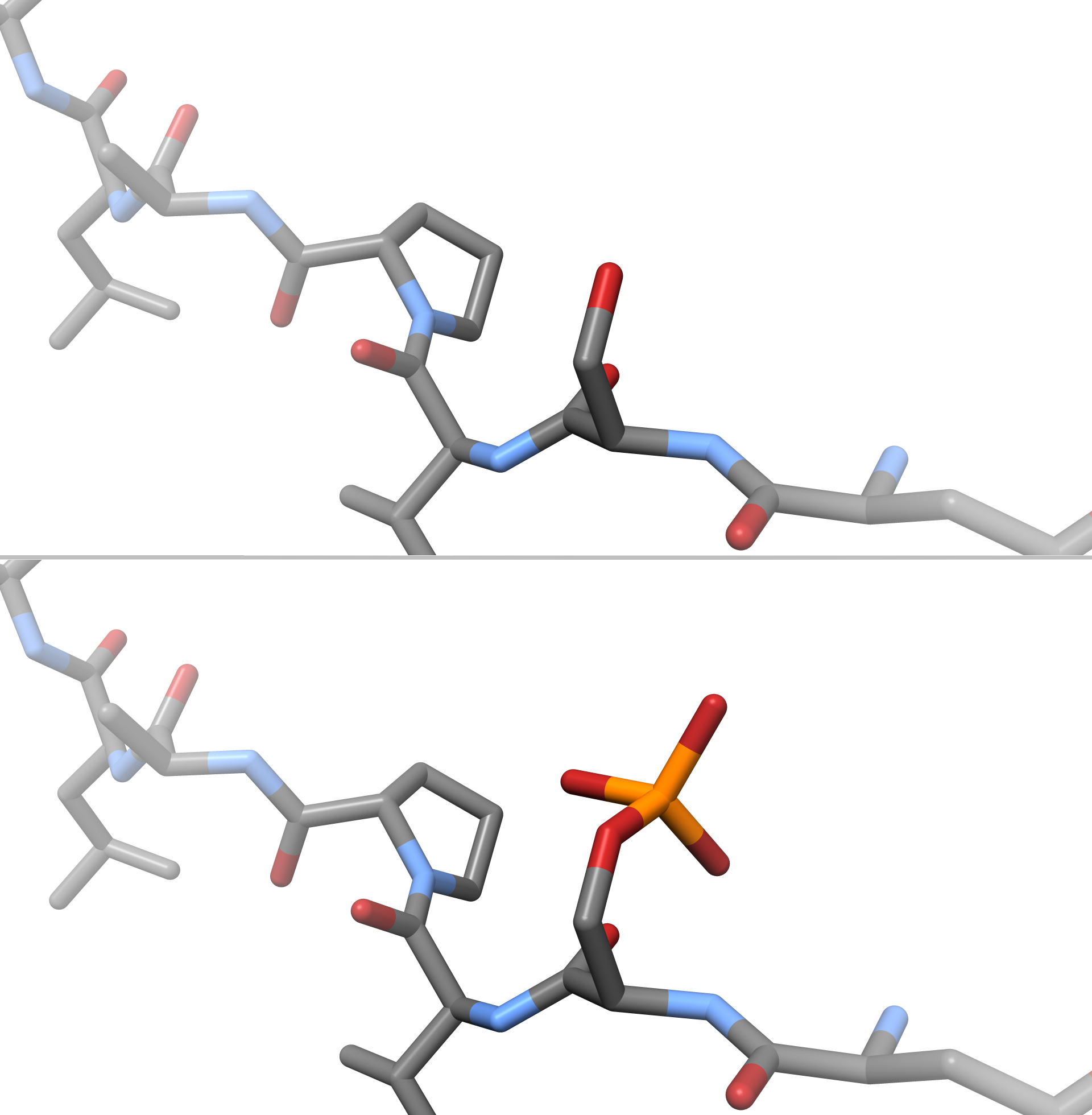
Phosphorylation
In chemistry, phosphorylation is the attachment of a phosphate group to a molecule or an ion. This process and its inverse, dephosphorylation, are common in biology and could be driven by natural selection. Text was copied from this source, which is available under a Creative Commons Attribution 4.0 International License. Protein phosphorylation often activates (or deactivates) many enzymes. Glucose Phosphorylation of sugars is often the first stage in their catabolism. Phosphorylation allows cells to accumulate sugars because the phosphate group prevents the molecules from diffusing back across their transporter. Phosphorylation of glucose is a key reaction in sugar metabolism. The chemical equation for the conversion of D-glucose to D-glucose-6-phosphate in the first step of glycolysis is given by :D-glucose + ATP → D-glucose-6-phosphate + ADP : ΔG° = −16.7 kJ/mol (° indicates measurement at standard condition) Hepatic cells are freely permeable to glucose, and ... [...More Info...] [...Related Items...] OR: [Wikipedia] [Google] [Baidu] |
Amino Terminal
The N-terminus (also known as the amino-terminus, NH2-terminus, N-terminal end or amine-terminus) is the start of a protein or polypeptide, referring to the free amine group (-NH2) located at the end of a polypeptide. Within a peptide, the amine group is bonded to the carboxylic group of another amino acid, making it a chain. That leaves a free carboxylic group at one end of the peptide, called the C-terminus, and a free amine group on the other end called the N-terminus. By convention, peptide sequences are written N-terminus to C-terminus, left to right (in LTR writing systems). This correlates the translation direction to the text direction, because when a protein is translated from messenger RNA, it is created from the N-terminus to the C-terminus, as amino acids are added to the carboxyl end of the protein. Chemistry Each amino acid has an amine group and a carboxylic group. Amino acids link to one another by peptide bonds which form through a dehydration reaction that jo ... [...More Info...] [...Related Items...] OR: [Wikipedia] [Google] [Baidu] |
Epidermal Growth Factor
Epidermal growth factor (EGF) is a protein that stimulates cell growth and differentiation by binding to its receptor, EGFR. Human EGF is 6-k Da and has 53 amino acid residues and three intramolecular disulfide bonds. EGF was originally described as a secreted peptide found in the submaxillary glands of mice and in human urine. EGF has since been found in many human tissues, including platelets, submandibular gland (submaxillary gland), and parotid gland. Initially, human EGF was known as urogastrone. Structure In humans, EGF has 53 amino acids (sequence NSDSECPLSHDGYCLHDGVCMYIEALDKYACNCVVGYIGERCQYRDLKWWELR), with a molecular mass of around 6 kDa. It contains three disulfide bridges (Cys6-Cys20, Cys14-Cys31, Cys33-Cys42). Function EGF, via binding to its cognate receptor, results in cellular proliferation, differentiation, and survival. Salivary EGF, which seems to be regulated by dietary inorganic iodine, also plays an important physiological role in the maintenance of ... [...More Info...] [...Related Items...] OR: [Wikipedia] [Google] [Baidu] |


