|
L1 Family
The L1 family is a family of cell adhesion molecules that includes four different L1-like proteins. They are members of the immunoglobulin superfamily (IgSF CAM). The members of the L1-family in humans are called L1 or L1cam, CHL1 (close homologue of L1), Neurofascin and NRCAM (NgCAM related cell adhesion molecule). L1 family members are found on neurons, especially on their axons. Sometimes they are found on glia, such as Schwann cells, radial glia and Bergmann glia cells and, as such, are important for neural cell migration during development. L1 family members are expressed throughout the vertebrate and invertebrate kingdoms. L1 family members are able to bind to a number of other proteins. As cell adhesion molecules, they often bind "homophilically" to themselves; for example L1 on one cell binding to L1 on an adjacent cell. L1 family members also bind "heterophilically" to members of the contactin or CNTN1 family. L1 family members bind to many cytoplasmic pro ... [...More Info...] [...Related Items...] OR: [Wikipedia] [Google] [Baidu] |
Cell Adhesion Molecule
Cell adhesion molecules (CAMs) are a subset of cell surface proteins that are involved in the binding of cells with other cells or with the extracellular matrix (ECM), in a process called cell adhesion. In essence, CAMs help cells stick to each other and to their surroundings. CAMs are crucial components in maintaining tissue structure and function. In fully developed animals, these molecules play an integral role in generating force and movement and consequently ensuring that organs are able to execute their functions normally. In addition to serving as "molecular glue", CAMs play important roles in the cellular mechanisms of growth, contact inhibition, and apoptosis. Aberrant expression of CAMs may result in a wide range of pathologies, ranging from frostbite to cancer. Structure CAMs are typically single-pass transmembrane receptors and are composed of three conserved domains: an intracellular domain that interacts with the cytoskeleton, a transmembrane domain, and an ext ... [...More Info...] [...Related Items...] OR: [Wikipedia] [Google] [Baidu] |
L1 (protein)
L1, also known as L1CAM, is a transmembrane protein member of the L1 protein family, encoded by the L1CAM gene. This protein, of 200-220 kDa, is a neuronal cell adhesion molecule with a strong implication in cell migration, adhesion, neurite outgrowth, myelination and neuronal differentiation. It also plays a key role in treatment-resistant cancers due to its function. It was first identified in 1984 by M. Schachner who found the protein in post-mitotic mice neurons. Mutations in the L1 protein are the cause of L1 syndrome, sometimes known by the acronym CRASH (corpus callosum hypoplasia, retardation, aphasia, spastic paraplegia and hydrocephalus). Tissue and cellular distribution L1 protein is located all over the nervous system on the surface of neurons. It is placed along the cellular membrane so that one end of the protein remains inside the nerve cell while the other end stays on the outer surface of the neurone. This position allows the protein to activate chemical si ... [...More Info...] [...Related Items...] OR: [Wikipedia] [Google] [Baidu] |
IgSF CAM
IgSF CAMs (Immunoglobulin-like Cell Adhesion Molecules) are cell adhesion molecules that belong to Immunoglobulin superfamily. It is regarded as the most diverse superfamily of CAMs. This family is characterized by their extracellular domains containing Ig-like domains. The Ig domains are then followed by Fibronectin type III domain The Fibronectin type III domain is an evolutionarily conserved protein domain that is widely found in animal proteins. The fibronectin protein in which this domain was first identified contains 16 copies of this domain. The domain is about 100 am ... repeats and IgSFs are anchored to the membrane by a Glycosylphosphatidylinositol, GPI moiety. This family is involved in both homophilic or heterophilic binding and has the ability to bind integrins or different IgSF CAMs. Examples Here is a list of some molecules of this family: *NCAMs Neural Cell Adhesion Molecules *ICAM-1 Intercellular Cell Adhesion Molecule *VCAM-1 Vascular Cell Adhesion Molecule * ... [...More Info...] [...Related Items...] OR: [Wikipedia] [Google] [Baidu] |
CHL1
Neural cell adhesion molecule L1-like protein also known as close homolog of L1 (CHL1) is a protein that in humans is encoded by the ''CHL1'' gene. CHL1 is a cell adhesion molecule Cell adhesion molecules (CAMs) are a subset of cell surface proteins that are involved in the binding of cells with other cells or with the extracellular matrix (ECM), in a process called cell adhesion. In essence, CAMs help cells stick to each ... closely related to the L1. In melanocytic cells CHL1 gene expression may be regulated by MITF, and can act as a helicase protein during the interphase stage of mitosis. The protein, however, has dynamic localisation, meaning that it has not only multiple roles in the cell, but also various locations. References Further reading * * * * * * * * * * * * * External links * * {{membrane-protein-stub ... [...More Info...] [...Related Items...] OR: [Wikipedia] [Google] [Baidu] |
Neurofascin
Neurofascin is a protein that in humans is encoded by the ''NFASC'' gene. Function Neurofascin is an L1 family immunoglobulin cell adhesion molecule (see L1CAM) involved in axon subcellular targeting and synapse formation during neural development The development of the nervous system, or neural development (neurodevelopment), refers to the processes that generate, shape, and reshape the nervous system of animals, from the earliest stages of embryonic development to adulthood. The fiel .... Clinical importance A homozygous mutation causing loss of Nfasc155 causes severe congenital hypotonia, contractures of fingers and toes and no reaction to touch or pain. References Further reading * * * * * * * * * * * * * Human proteins {{gene-1-stub ... [...More Info...] [...Related Items...] OR: [Wikipedia] [Google] [Baidu] |
Radial Glia
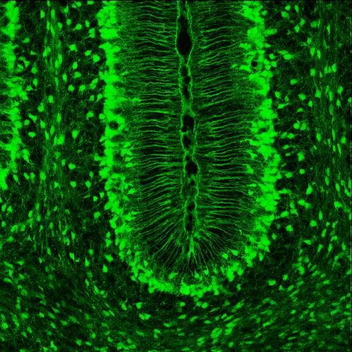
Radial glial cells, or radial glial progenitor cells (RGPs), are bipolar-shaped progenitor cells that are responsible for producing all of the neurons in the cerebral cortex. RGPs also produce certain lineages of glia, including astrocytes and oligodendrocytes. Their cell bodies ( somata) reside in the embryonic ventricular zone, which lies next to the developing ventricular system. During development, newborn neurons use radial glia as scaffolds, traveling along the radial glial fibers in order to reach their final destinations. Despite the various possible fates of the radial glial population, it has been demonstrated through clonal analysis that most radial glia have restricted, unipotent or multipotent, fates. Radial glia can be found during the neurogenic phase in all vertebrates (studied to date). The term "radial glia" refers to the morphological characteristics of these cells that were first observed: namely, their radial processes and their similarity to astrocytes, a ... [...More Info...] [...Related Items...] OR: [Wikipedia] [Google] [Baidu] |
Bergmann Glia
Radial glial cells, or radial glial progenitor cells (RGPs), are bipolar-shaped progenitor cells that are responsible for producing all of the neurons in the cerebral cortex. RGPs also produce certain lineages of glia, including astrocytes and oligodendrocytes. Their cell bodies ( somata) reside in the embryonic ventricular zone, which lies next to the developing ventricular system. During development, newborn neurons use radial glia as scaffolds, traveling along the radial glial fibers in order to reach their final destinations. Despite the various possible fates of the radial glial population, it has been demonstrated through clonal analysis that most radial glia have restricted, unipotent or multipotent, fates. Radial glia can be found during the neurogenic phase in all vertebrates (studied to date). The term "radial glia" refers to the morphological characteristics of these cells that were first observed: namely, their radial processes and their similarity to astrocytes, ... [...More Info...] [...Related Items...] OR: [Wikipedia] [Google] [Baidu] |
CNTN1
Contactin 1, also known as CNTN1, is a protein which in humans is encoded by the ''CNTN1'' gene. Function The protein encoded by this gene is a member of the immunoglobulin superfamily. It is a glycosylphosphatidylinositol (GPI)-anchored neuronal membrane protein that functions as a cell adhesion molecule. It may play a role in the formation of axon connections in the developing nervous system. Two alternatively spliced transcript variants encoding different isoforms have been described for this gene. Interactions CNTN1 has been shown to interact with PTPRB Receptor-type tyrosine-protein phosphatase beta or VE-PTP is an enzyme specifically expressed in endothelial cells that in humans is encoded by the ''PTPRB'' gene. Function VE-PTP is a member of the classical protein tyrosine phosphatase (P .... References External links * * Further reading * * * * * * * * * * * * * * * * * {{gene-12-stub ... [...More Info...] [...Related Items...] OR: [Wikipedia] [Google] [Baidu] |
Ankyrin
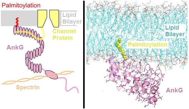
Ankyrins are a family of proteins that mediate the attachment of integral membrane proteins to the spectrin-actin based membrane cytoskeleton. Ankyrins have binding sites for the beta subunit of spectrin and at least 12 families of integral membrane proteins. This linkage is required to maintain the integrity of the plasma membranes and to anchor specific ion channels, ion exchangers and ion transporters in the plasma membrane. The name is derived from the Greek word ἄγκυρα (''ankyra'') for "anchor". Structure Ankyrins contain four functional domains: an N-terminal domain that contains 24 tandem ankyrin repeats, a central domain that binds to spectrin, a death domain that binds to proteins involved in apoptosis, and a C-terminal regulatory domain that is highly variable between different ankyrin proteins. Membrane protein recognition The 24 tandem ankyrin repeats are responsible for the recognition of a wide range of membrane proteins. These 24 repeats contain 3 struc ... [...More Info...] [...Related Items...] OR: [Wikipedia] [Google] [Baidu] |
Src Gene
Tyrosine-protein kinase CSK also known as C-terminal Src kinase is an enzyme that, in humans, is encoded by the CSK gene. This enzyme phosphorylates tyrosine residues located in the C-terminal end of Src-family kinases (SFKs) including SRC, HCK, FYN, LCK, LYN and YES1. Function This Non-receptor tyrosine-protein kinase plays an important role in the regulation of cell growth, differentiation, migration and immune response. CSK acts by suppressing the activity of the Src family of protein kinases by phosphorylation of Src family members at a conserved C-terminal tail site in Src. Upon phosphorylation by other kinases, Src-family members engage in intramolecular interactions between the phosphotyrosine tail and the SH2 domain that result in an inactive conformation. To inhibit SFKs, CSK is then recruited to the plasma membrane via binding to transmembrane proteins or adapter proteins located near the plasma membrane and ultimately suppresses signaling through various surface ... [...More Info...] [...Related Items...] OR: [Wikipedia] [Google] [Baidu] |
Extracellular Signal-regulated Kinases
In molecular biology, extracellular signal-regulated kinases (ERKs) or classical MAP kinases are widely expressed protein kinase intracellular signalling molecules that are involved in functions including the regulation of meiosis, mitosis, and postmitotic functions in differentiated cells. Many different stimuli, including growth factors, cytokines, virus infection, ligands for heterotrimeric G protein-coupled receptors, transforming agents, and carcinogens, activate the ERK pathway. The term, "extracellular signal-regulated kinases", is sometimes used as a synonym for mitogen-activated protein kinase (MAPK), but has more recently been adopted for a specific subset of the mammalian MAPK family. In the MAPK/ERK pathway, Ras activates c-Raf, followed by mitogen-activated protein kinase kinase (abbreviated as MKK, MEK, or MAP2K) and then MAPK1/2 (below). Ras is typically activated by growth hormones through receptor tyrosine kinases and GRB2/SOS, but may also receive o ... [...More Info...] [...Related Items...] OR: [Wikipedia] [Google] [Baidu] |


