|
Klaus Weber
Klaus Weber (5 April 1936 – 8 August 2016) was a German scientist who made many fundamentally important contributions to biochemistry, cell biology, and molecular biology, and was for many years the director of the Laboratory of Biochemistry and Cell Biology at the Max Planck Institute for Biophysical Chemistry in Göttingen, Germany. Biography Weber was born in Łódź, Poland in 1936. After earning an undergraduate degree in 1962 and a graduate degree in 1964 from the University of Freiburg, Weber came to the United States to work as a postdoctoral fellow with James D. Watson at Harvard University. Career After a successful period as a postdoctoral fellow with Watson starting in the spring of 1965, Weber was hired as an assistant professor at Harvard and ran a joint laboratory with Watson and Walter Gilbert. During this period he worked on protein chemistry of RNA phages, but was beginning to shift his focus to animal cells and their viruses, and spent a sabbatical at C ... [...More Info...] [...Related Items...] OR: [Wikipedia] [Google] [Baidu] |
Łódź
Łódź, also rendered in English as Lodz, is a city in central Poland and a former industrial centre. It is the capital of Łódź Voivodeship, and is located approximately south-west of Warsaw. The city's coat of arms is an example of canting arms, canting, as it depicts a boat ( in Polish language, Polish), which alludes to the city's name. As of 2022, Łódź has a population of 670,642 making it the country's List of cities and towns in Poland, fourth largest city. Łódź was once a small settlement that first appeared in 14th-century records. It was granted city rights, town rights in 1423 by Polish King Władysław II Jagiełło and it remained a private town of the Kuyavian bishops and clergy until the late 18th century. In the Second Partition of Poland in 1793, Łódź was annexed to Kingdom of Prussia, Prussia before becoming part of the Napoleonic Duchy of Warsaw; the city joined Congress Poland, a Russian Empire, Russian client state, at the 1815 Congress of Vien ... [...More Info...] [...Related Items...] OR: [Wikipedia] [Google] [Baidu] |
SDS-PAGE
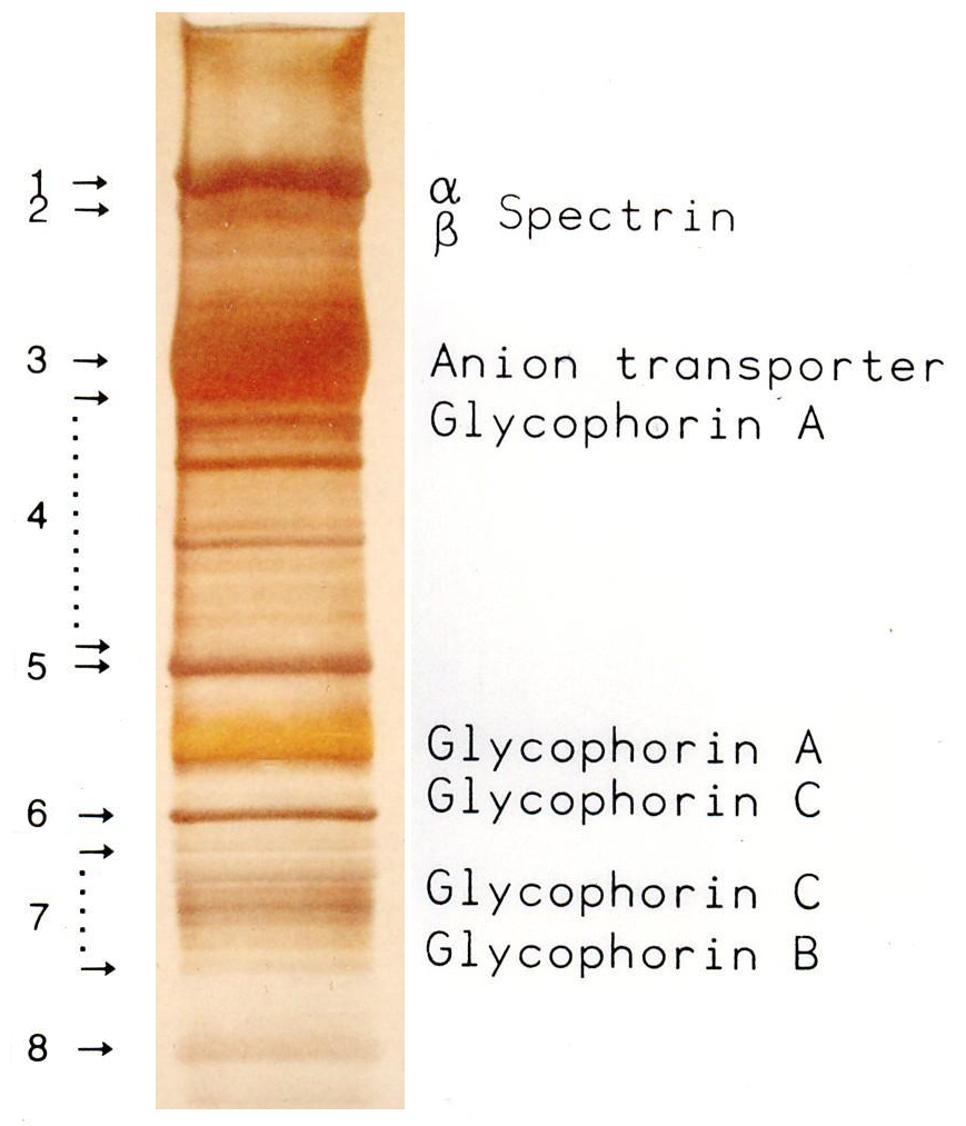
SDS-PAGE (sodium dodecyl sulfate–polyacrylamide gel electrophoresis) is a discontinuous electrophoretic system developed by Ulrich K. Laemmli which is commonly used as a method to separate proteins with molecular masses between 5 and 250 kDa. The combined use of sodium dodecyl sulfate (SDS, also known as sodium lauryl sulfate) and polyacrylamide gel allows to eliminate the influence of structure and charge, and proteins are separated solely on the basis of differences in their molecular weight. Properties SDS-PAGE is an electrophoresis method that allows protein separation by mass. The medium (also referred to as ′matrix′) is a polyacrylamide-based discontinuous gel. The polyacrylamide-gel is typically sandwiched between two glass plates in a slab gel. Although tube gels (in glass cylinders) were used historically, they were rapidly made obsolete with the invention of the more convenient slab gels. In addition, SDS (sodium dodecyl sulfate) is used. About 1.4 grams of S ... [...More Info...] [...Related Items...] OR: [Wikipedia] [Google] [Baidu] |
Thomas Tuschl
Thomas Tuschl (born June 1, 1966) is a German biochemist and molecular biologist, known for his research on RNA. Biography Tuschl was born in Altdorf bei Nürnberg. After graduating in Chemistry from Regensburg University, Tuschl received his PhD in 1995 from the Max Planck Institute for Experimental Medicine in Göttingen. He spent four years as a post-doctoral fellow at the Whitehead Institute of the Massachusetts Institute of Technology (MIT) in Cambridge, USA. In 1999 he returned to Göttingen, to continue his research at the Max Planck Institute for Biophysical Chemistry. There he received international recognition in Genetics for his studies of RNA interference in collaboration with the laboratory of Klaus Weber. This enables "switching off" certain genes by introducing synthetic short RNA into the cell. The mRNA is destroyed and the gene in deactivated. Possible future applications of this method include treatment of tumors or genetic disorders. The function of certain ge ... [...More Info...] [...Related Items...] OR: [Wikipedia] [Google] [Baidu] |
RNA Interference
RNA interference (RNAi) is a biological process in which RNA molecules are involved in sequence-specific suppression of gene expression by double-stranded RNA, through translational or transcriptional repression. Historically, RNAi was known by other names, including ''co-suppression'', ''post-transcriptional gene silencing'' (PTGS), and ''quelling''. The detailed study of each of these seemingly different processes elucidated that the identity of these phenomena were all actually RNAi. Andrew Fire and Craig C. Mello shared the 2006 Nobel Prize in Physiology or Medicine for their work on RNAi in the nematode worm '' Caenorhabditis elegans'', which they published in 1998. Since the discovery of RNAi and its regulatory potentials, it has become evident that RNAi has immense potential in suppression of desired genes. RNAi is now known as precise, efficient, stable and better than antisense therapy for gene suppression. Antisense RNA produced intracellularly by an expression vector m ... [...More Info...] [...Related Items...] OR: [Wikipedia] [Google] [Baidu] |
Fluorescence Microscope
A fluorescence microscope is an optical microscope that uses fluorescence instead of, or in addition to, scattering, reflection, and attenuation or absorption, to study the properties of organic or inorganic substances. "Fluorescence microscope" refers to any microscope that uses fluorescence to generate an image, whether it is a simple set up like an epifluorescence microscope or a more complicated design such as a confocal microscope, which uses optical sectioning to get better resolution of the fluorescence image. Principle The specimen is illuminated with light of a specific wavelength (or wavelengths) which is absorbed by the fluorophores, causing them to emit light of longer wavelengths (i.e., of a different color than the absorbed light). The illumination light is separated from the much weaker emitted fluorescence through the use of a spectral emission filter. Typical components of a fluorescence microscope are a light source (xenon arc lamp or mercury-vapor lamp are ... [...More Info...] [...Related Items...] OR: [Wikipedia] [Google] [Baidu] |
Proc
Proc may refer to: * Proč, a village in eastern Slovakia * '' Proč?'', a 1987 Czech film * procfs or proc filesystem, a special file system (typically mounted to ) in Unix-like operating systems for accessing process information * Protein C (PROC) * Proc, a term in video game terminology * Procedures or process, in the programming language ALGOL 68 * People's Republic of China, the formal name of China China, officially the People's Republic of China (PRC), is a country in East Asia. It is the world's most populous country, with a population exceeding 1.4 billion, slightly ahead of India. China spans the equivalent of five time zones and ... * the official acronym for the Canadian House of Commons Standing Committee on Procedure and House Affairs {{disambiguation ... [...More Info...] [...Related Items...] OR: [Wikipedia] [Google] [Baidu] |
Antibodies
An antibody (Ab), also known as an immunoglobulin (Ig), is a large, Y-shaped protein used by the immune system to identify and neutralize foreign objects such as pathogenic bacteria and viruses. The antibody recognizes a unique molecule of the pathogen, called an antigen. Each tip of the "Y" of an antibody contains a paratope (analogous to a lock) that is specific for one particular epitope (analogous to a key) on an antigen, allowing these two structures to bind together with precision. Using this binding mechanism, an antibody can ''tag'' a microbe or an infected cell for attack by other parts of the immune system, or can neutralize it directly (for example, by blocking a part of a virus that is essential for its invasion). To allow the immune system to recognize millions of different antigens, the antigen-binding sites at both tips of the antibody come in an equally wide variety. In contrast, the remainder of the antibody is relatively constant. It only occurs in a few vari ... [...More Info...] [...Related Items...] OR: [Wikipedia] [Google] [Baidu] |
Intermediate Filaments
Intermediate filaments (IFs) are cytoskeletal structural components found in the cells of vertebrates, and many invertebrates. Homologues of the IF protein have been noted in an invertebrate, the cephalochordate ''Branchiostoma''. Intermediate filaments are composed of a family of related proteins sharing common structural and sequence features. Initially designated 'intermediate' because their average diameter (10 nm) is between those of narrower microfilaments (actin) and wider myosin filaments found in muscle cells, the diameter of intermediate filaments is now commonly compared to actin microfilaments (7 nm) and microtubules (25 nm). Animal intermediate filaments are subcategorized into six types based on similarities in amino acid sequence and protein structure. Most types are cytoplasmic, but one type, Type V is a nuclear lamin. Unlike microtubules, IF distribution in cells show no good correlation with the distribution of either mitochondria or endopla ... [...More Info...] [...Related Items...] OR: [Wikipedia] [Google] [Baidu] |
Microfilaments
Microfilaments, also called actin filaments, are protein filaments in the cytoplasm of eukaryotic cells that form part of the cytoskeleton. They are primarily composed of polymers of actin, but are modified by and interact with numerous other proteins in the cell. Microfilaments are usually about 7 nm in diameter and made up of two strands of actin. Microfilament functions include cytokinesis, amoeboid movement, cell motility, changes in cell shape, endocytosis and exocytosis, cell contractility, and mechanical stability. Microfilaments are flexible and relatively strong, resisting buckling by multi-piconewton compressive forces and filament fracture by nanonewton tensile forces. In inducing cell motility, one end of the actin filament elongates while the other end contracts, presumably by myosin II molecular motors. Additionally, they function as part of actomyosin-driven contractile molecular motors, wherein the thin filaments serve as tensile platforms for myosin's ATP-dep ... [...More Info...] [...Related Items...] OR: [Wikipedia] [Google] [Baidu] |
Microtubules
Microtubules are polymers of tubulin that form part of the cytoskeleton and provide structure and shape to eukaryotic cells. Microtubules can be as long as 50 micrometres, as wide as 23 to 27 nm and have an inner diameter between 11 and 15 nm. They are formed by the polymerization of a dimer of two globular proteins, alpha and beta tubulin into protofilaments that can then associate laterally to form a hollow tube, the microtubule. The most common form of a microtubule consists of 13 protofilaments in the tubular arrangement. Microtubules play an important role in a number of cellular processes. They are involved in maintaining the structure of the cell and, together with microfilaments and intermediate filaments, they form the cytoskeleton. They also make up the internal structure of cilia and flagella. They provide platforms for intracellular transport and are involved in a variety of cellular processes, including the movement of secretory vesicles, organell ... [...More Info...] [...Related Items...] OR: [Wikipedia] [Google] [Baidu] |
Immunofluorescence Microscopy
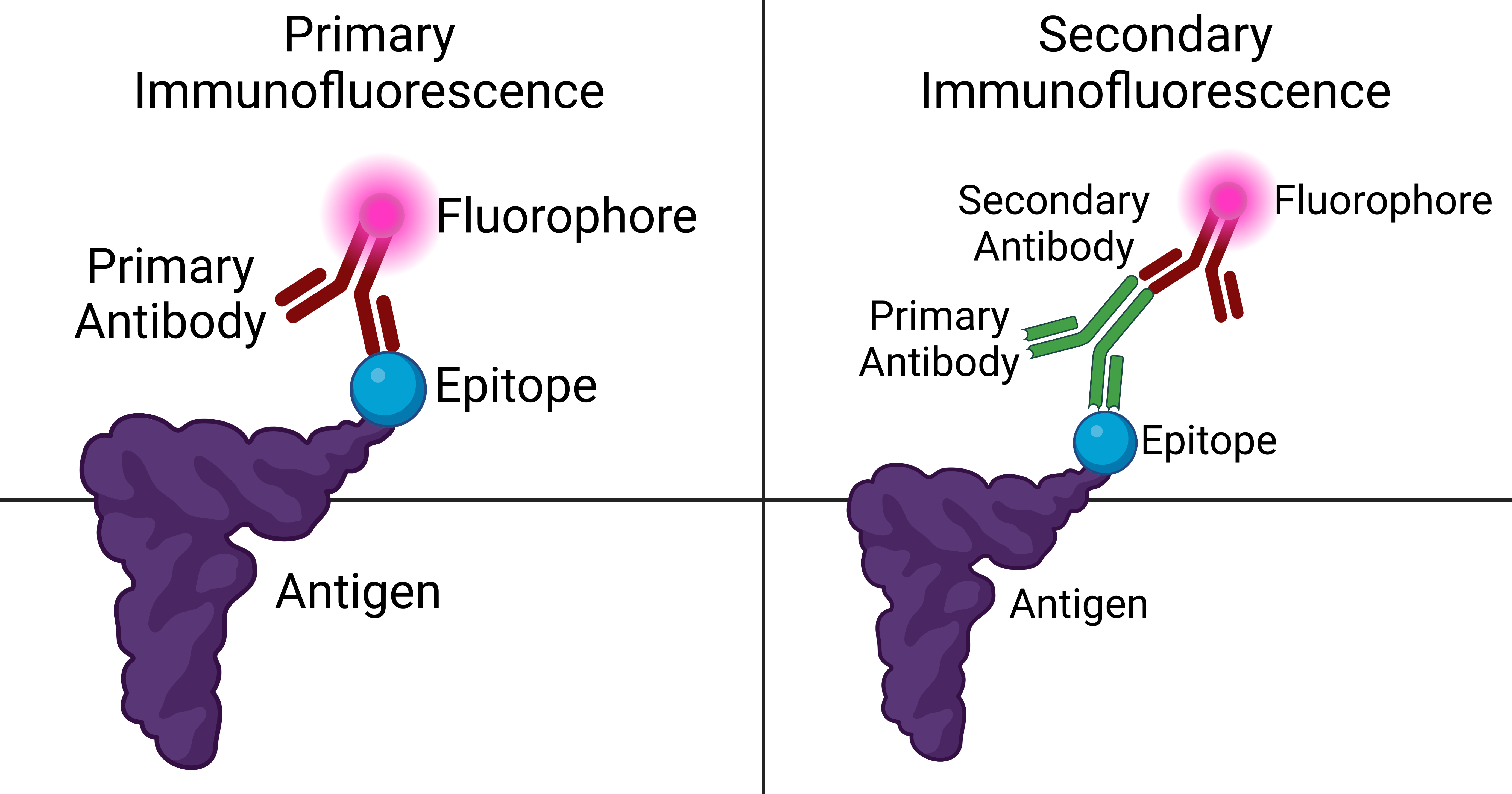
Immunofluorescence is a technique used for light microscopy with a fluorescence microscope and is used primarily on microbiological samples. This technique uses the specificity of antibodies to their antigen to target fluorescent dyes to specific biomolecule targets within a cell, and therefore allows visualization of the distribution of the target molecule through the sample. The specific region an antibody recognizes on an antigen is called an epitope. There have been efforts in epitope mapping since many antibodies can bind the same epitope and levels of binding between antibodies that recognize the same epitope can vary. Additionally, the binding of the fluorophore to the antibody itself cannot interfere with the immunological specificity of the antibody or the binding capacity of its antigen. Immunofluorescence is a widely used example of immunostaining (using antibodies to stain proteins) and is a specific example of immunohistochemistry (the use of the antibody-antigen rela ... [...More Info...] [...Related Items...] OR: [Wikipedia] [Google] [Baidu] |
Regina Nuzzo
Regina Nuzzo is a professor of statistics at Gallaudet University in Washington D.C., a liberal arts school for deaf and hard-of-hearing students. She also writes articles about the importance of statistical and science communication and is an advocate for people with disabilities in the science and technology field. Education Nuzzo graduated from the University of South Florida with a Bachelor's degree in industrial engineering and went on to obtain her Ph.D in statistics from Stanford University in 2004, supervised by Richard A. Olshen. Her dissertation was written on the usage of stochastic models in bio-chemistry. Nuzzo also graduated from the University of California Santa Cruz's science writing program, where she learned how to write effectively for a variety of audiences about science and technology. Career Nuzzo has been a faculty member at Gallaudet University since 2006. She has written multiple articles for publication in major magazines, including WIRED magazine, ... [...More Info...] [...Related Items...] OR: [Wikipedia] [Google] [Baidu] |


