|
Kneecap
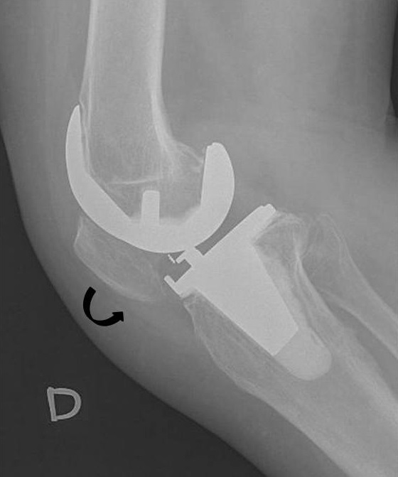
The patella, also known as the kneecap, is a flat, rounded triangular bone which articulates with the femur (thigh bone) and covers and protects the anterior articular surface of the knee joint. The patella is found in many tetrapods, such as mice, cats, birds and dogs, but not in whales, or most reptiles. In humans, the patella is the largest sesamoid bone (i.e., embedded within a tendon or a muscle) in the body. Babies are born with a patella of soft cartilage which begins to ossify into bone at about four years of age. Structure The patella is a sesamoid bone roughly triangular in shape, with the apex of the patella facing downwards. The apex is the most inferior (lowest) part of the patella. It is pointed in shape, and gives attachment to the patellar ligament. The front and back surfaces are joined by a thin margin and towards centre by a thicker margin. The tendon of the quadriceps femoris muscle attaches to the base of the patella., with the vastus intermedius mus ... [...More Info...] [...Related Items...] OR: [Wikipedia] [Google] [Baidu] |
Patella Baja
The patella, also known as the kneecap, is a flat, rounded triangular bone which articulates with the femur (thigh bone) and covers and protects the anterior articular surface of the knee joint. The patella is found in many tetrapods, such as mice, cats, birds and dogs, but not in whales, or most reptiles. In humans, the patella is the largest sesamoid bone (i.e., embedded within a tendon or a muscle) in the body. Babies are born with a patella of soft cartilage which begins to ossify into bone at about four years of age. Structure The patella is a sesamoid bone roughly triangular in shape, with the apex of the patella facing downwards. The apex is the most inferior (lowest) part of the patella. It is pointed in shape, and gives attachment to the patellar ligament. The front and back surfaces are joined by a thin margin and towards centre by a thicker margin. The tendon of the quadriceps femoris muscle attaches to the base of the patella., with the vastus intermedius musc ... [...More Info...] [...Related Items...] OR: [Wikipedia] [Google] [Baidu] |
Knee
In humans and other primates, the knee joins the thigh with the leg and consists of two joints: one between the femur and tibia (tibiofemoral joint), and one between the femur and patella (patellofemoral joint). It is the largest joint in the human body. The knee is a modified hinge joint, which permits flexion and extension as well as slight internal and external rotation. The knee is vulnerable to injury and to the development of osteoarthritis. It is often termed a ''compound joint'' having tibiofemoral and patellofemoral components. (The fibular collateral ligament is often considered with tibiofemoral components.) Structure The knee is a modified hinge joint, a type of synovial joint, which is composed of three functional compartments: the patellofemoral articulation, consisting of the patella, or "kneecap", and the patellar groove on the front of the femur through which it slides; and the medial and lateral tibiofemoral articulations linking the femur, or thigh ... [...More Info...] [...Related Items...] OR: [Wikipedia] [Google] [Baidu] |
Bipartite Patella
Bipartite patella is a condition where the patella, or kneecap, is composed of two separate bones. Instead of fusing together as normally occurs in early childhood, the bones of the patella remain separated. The condition occurs in approximately 12% of the population and is no more likely to occur in males than females. It is often asymptomatic and most commonly diagnosed as an incidental finding Incidental medical findings are previously undiagnosed medical or psychiatric conditions that are discovered unintentionally and during evaluation for a medical or psychiatric condition. Such findings may occur in a variety of settings, including ro ..., with about 2% of cases becoming symptomatic. Saupe introduced a classification system for Bipartite Patella back in 1921. Type 1: Fragment is located at the bottom of the kneecap (5% of cases) Type 2: Fragment is located on the lateral side of the kneecap (20% of cases) Type 3: Fragment is located on the upper lateral border of the kne ... [...More Info...] [...Related Items...] OR: [Wikipedia] [Google] [Baidu] |
Patella Bipartita
Bipartite patella is a condition where the patella, or kneecap, is composed of two separate bones. Instead of fusing together as normally occurs in early childhood, the bones of the patella remain separated. The condition occurs in approximately 12% of the population and is no more likely to occur in males than females. It is often asymptomatic and most commonly diagnosed as an incidental finding Incidental medical findings are previously undiagnosed medical or psychiatric conditions that are discovered unintentionally and during evaluation for a medical or psychiatric condition. Such findings may occur in a variety of settings, including ro ..., with about 2% of cases becoming symptomatic. Saupe introduced a classification system for Bipartite Patella back in 1921. Type 1: Fragment is located at the bottom of the kneecap (5% of cases) Type 2: Fragment is located on the lateral side of the kneecap (20% of cases) Type 3: Fragment is located on the upper lateral border of the kne ... [...More Info...] [...Related Items...] OR: [Wikipedia] [Google] [Baidu] |
Patellar Dislocation
A patellar dislocation is a knee injury in which the patella (kneecap) slips out of its normal position. Often the knee is partly bent, painful and swollen. The patella is also often felt and seen out of place. Complications may include a patella fracture or arthritis. A patellar dislocation typically occurs when the knee is straight and the lower leg is bent outwards when twisting. Occasionally it occurs when the knee is bent and the patella is hit. Commonly associated sports include soccer, gymnastics, and ice hockey. Dislocations nearly always occur away from the midline. Diagnosis is typically based on symptoms and supported by X-rays. Reduction is generally done by pushing the patella towards the midline while straightening the knee. After reduction the leg is generally splinted in a straight position for a few weeks. This is then followed by physical therapy. Surgery after a first dislocation is generally of unclear benefit. Surgery may be indicated in those who hav ... [...More Info...] [...Related Items...] OR: [Wikipedia] [Google] [Baidu] |
Quadriceps Femoris Muscle
The quadriceps femoris muscle (, also called the quadriceps extensor, quadriceps or quads) is a large muscle group that includes the four prevailing muscles on the front of the thigh. It is the sole extensor muscle of the knee, forming a large fleshy mass which covers the front and sides of the femur. The name derives . Structure Parts The quadriceps femoris muscle is subdivided into four separate muscles (the 'heads'), with the first superficial to the other three over the femur (from the trochanters to the condyles): *The rectus femoris muscle occupies the middle of the thigh, covering most of the other three quadriceps muscles. It originates on the ilium. It is named for its straight course. *The vastus lateralis muscle is on the ''lateral side'' of the femur (i.e. on the outer side of the thigh). *The vastus medialis muscle is on the ''medial side'' of the femur (i.e. on the inner part thigh). *The vastus intermedius muscle lies between vastus lateralis and vastus medi ... [...More Info...] [...Related Items...] OR: [Wikipedia] [Google] [Baidu] |
Quadriceps Tendon
In human anatomy, the quadriceps tendon works with the quadriceps muscle to extend the leg. All four parts of the quadriceps muscle attach to the shin via the patella (knee cap), where the quadriceps tendon becomes the patellar ligament. It attaches the quadriceps to the top of the patella, which in turn is connected to the shin from its bottom by the patellar ligament. A tendon connects muscle to bone, while a ligament connects bone to bone.Saladin, Kenneth S. Anatomy & Physiology: The Unity of Form and Function. 6th ed. New York: McGraw-Hill, 2012. Print. Injuries are common to this tendon, with tears, either partial or complete, being the most common. If the quadriceps tendon is completely torn, surgery will be required to regain function of the knee."Patellar Tendon Tear." OrthoInfo - AAOS. American Academy of Orthopaedic Surgeons, Aug. 2009. Web. 07 Dec. 2014. Without the quadriceps tendon, the knee cannot extend. Often, when the tendon is completely torn, part of the kn ... [...More Info...] [...Related Items...] OR: [Wikipedia] [Google] [Baidu] |
Femur
The femur (; ), or thigh bone, is the proximal bone of the hindlimb in tetrapod vertebrates. The head of the femur articulates with the acetabulum in the pelvic bone forming the hip joint, while the distal part of the femur articulates with the tibia (shinbone) and patella (kneecap), forming the knee joint. By most measures the two (left and right) femurs are the strongest bones of the body, and in humans, the largest and thickest. Structure The femur is the only bone in the upper leg. The two femurs converge medially toward the knees, where they articulate with the proximal ends of the tibiae. The angle of convergence of the femora is a major factor in determining the femoral-tibial angle. Human females have thicker pelvic bones, causing their femora to converge more than in males. In the condition ''genu valgum'' (knock knee) the femurs converge so much that the knees touch one another. The opposite extreme is ''genu varum'' (bow-leggedness). In the general ... [...More Info...] [...Related Items...] OR: [Wikipedia] [Google] [Baidu] |
Bone
A bone is a rigid organ that constitutes part of the skeleton in most vertebrate animals. Bones protect the various other organs of the body, produce red and white blood cells, store minerals, provide structure and support for the body, and enable mobility. Bones come in a variety of shapes and sizes and have complex internal and external structures. They are lightweight yet strong and hard and serve multiple functions. Bone tissue (osseous tissue), which is also called bone in the uncountable sense of that word, is hard tissue, a type of specialized connective tissue. It has a honeycomb-like matrix internally, which helps to give the bone rigidity. Bone tissue is made up of different types of bone cells. Osteoblasts and osteocytes are involved in the formation and mineralization of bone; osteoclasts are involved in the resorption of bone tissue. Modified (flattened) osteoblasts become the lining cells that form a protective layer on the bone surface. The minera ... [...More Info...] [...Related Items...] OR: [Wikipedia] [Google] [Baidu] |
Cartilage
Cartilage is a resilient and smooth type of connective tissue. In tetrapods, it covers and protects the ends of long bones at the joints as articular cartilage, and is a structural component of many body parts including the rib cage, the neck and the bronchial tubes, and the intervertebral discs. In other taxa, such as chondrichthyans, but also in cyclostomes, it may constitute a much greater proportion of the skeleton. It is not as hard and rigid as bone, but it is much stiffer and much less flexible than muscle. The matrix of cartilage is made up of glycosaminoglycans, proteoglycans, collagen fibers and, sometimes, elastin. Because of its rigidity, cartilage often serves the purpose of holding tubes open in the body. Examples include the rings of the trachea, such as the cricoid cartilage and carina. Cartilage is composed of specialized cells called chondrocytes that produce a large amount of collagenous extracellular matrix, abundant ground substance that is rich in ... [...More Info...] [...Related Items...] OR: [Wikipedia] [Google] [Baidu] |
Canaliculus (bone)
Bone canaliculi are microscopic canals between the lacunae of ossified bone. The radiating processes of the osteocytes (called filopodia) project into these canals. These cytoplasmic processes are joined together by gap junctions. Osteocytes do not entirely fill up the canaliculi. The remaining space is known as the periosteocytic space, which is filled with periosteocytic fluid. This fluid contains substances too large to be transported through the gap junctions that connect the osteocytes. In cartilage, the lacunae and hence, the chondrocytes, are isolated from each other. Materials picked up by osteocytes adjacent to blood vessels are distributed throughout the bone matrix via the canaliculi. Dental canaliculi The dental canaliculi (sometimes called dentinal tubules) are the blood supply of a tooth. Odontoblast process run in the canaliculi that transverse the dentin layer and are referred as dentinal tubules. The number and size of the canaliculi decrease as the tubu ... [...More Info...] [...Related Items...] OR: [Wikipedia] [Google] [Baidu] |
Joint
A joint or articulation (or articular surface) is the connection made between bones, ossicles, or other hard structures in the body which link an animal's skeletal system into a functional whole.Saladin, Ken. Anatomy & Physiology. 7th ed. McGraw-Hill Connect. Webp.274/ref> They are constructed to allow for different degrees and types of movement. Some joints, such as the knee, elbow, and shoulder, are self-lubricating, almost frictionless, and are able to withstand compression and maintain heavy loads while still executing smooth and precise movements. Other joints such as sutures between the bones of the skull permit very little movement (only during birth) in order to protect the brain and the sense organs. The connection between a tooth and the jawbone is also called a joint, and is described as a fibrous joint known as a gomphosis. Joints are classified both structurally and functionally. Classification The number of joints depends on if sesamoids are included, age of t ... [...More Info...] [...Related Items...] OR: [Wikipedia] [Google] [Baidu] |


