|
Kappa Particle
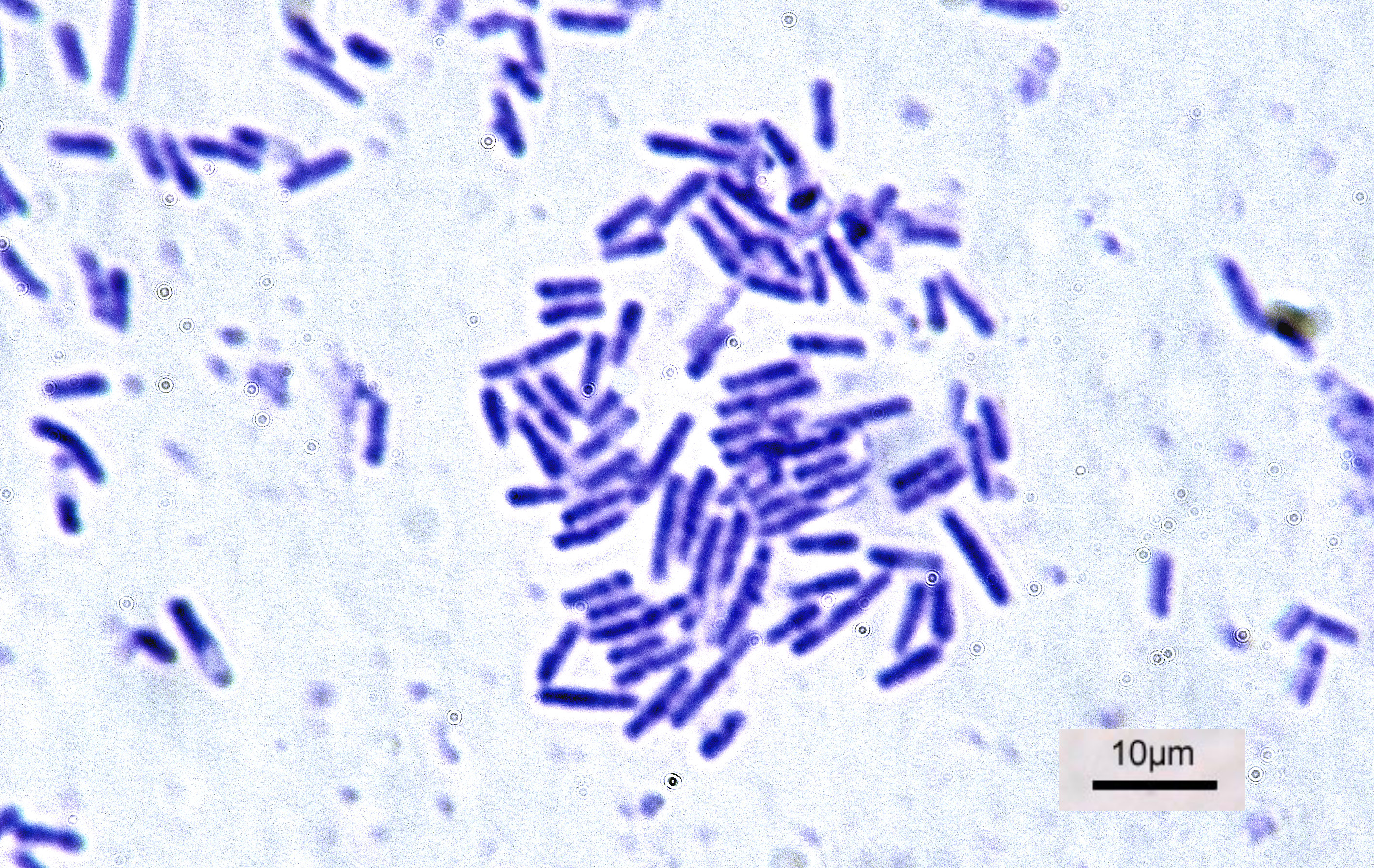
In biology, Kappa organism or Kappa particle refers to inheritable cytoplasmic symbionts, occurring in some strains of the ciliate ''Paramecium''. ''Paramecium'' strains possessing the particles are known as "killer paramecia". They liberate a substance also known as paramecin into the culture medium that is lethal to ''Paramecium'' that do not contain kappa particles. Kappa particles are found in genotypes of ''Paramecium aurelia'' syngen 2 that carry the dominant gene K. Kappa particles are Feulgen-positive and stain with Giemsa after acid hydrolysis. The length of the particles is 0.2–0.5μ. While there was initial confusion over the status of kappa particles as viruses, bacteria, organelles, or mere nucleoprotein, the particles are intracellular bacterial symbionts called '' Caedibacter taeniospiralis''. ''Caedibacter taeniospiralis'' contains cytoplasmic protein inclusions called R bodies which act as a toxin delivery system. References External links * * See also ... [...More Info...] [...Related Items...] OR: [Wikipedia] [Google] [Baidu] |
Cytoplasmic
In cell biology, the cytoplasm is all of the material within a eukaryotic cell, enclosed by the cell membrane, except for the cell nucleus. The material inside the nucleus and contained within the nuclear membrane is termed the nucleoplasm. The main components of the cytoplasm are cytosol (a gel-like substance), the organelles (the cell's internal sub-structures), and various cytoplasmic inclusions. The cytoplasm is about 80% water and is usually colorless. The submicroscopic ground cell substance or cytoplasmic matrix which remains after exclusion of the cell organelles and particles is groundplasm. It is the hyaloplasm of light microscopy, a highly complex, polyphasic system in which all resolvable cytoplasmic elements are suspended, including the larger organelles such as the ribosomes, mitochondria, the plant plastids, lipid droplets, and vacuoles. Most cellular activities take place within the cytoplasm, such as many metabolic pathways including glycolysis, and p ... [...More Info...] [...Related Items...] OR: [Wikipedia] [Google] [Baidu] |
Bacteria
Bacteria (; singular: bacterium) are ubiquitous, mostly free-living organisms often consisting of one biological cell. They constitute a large domain of prokaryotic microorganisms. Typically a few micrometres in length, bacteria were among the first life forms to appear on Earth, and are present in most of its habitats. Bacteria inhabit soil, water, acidic hot springs, radioactive waste, and the deep biosphere of Earth's crust. Bacteria are vital in many stages of the nutrient cycle by recycling nutrients such as the fixation of nitrogen from the atmosphere. The nutrient cycle includes the decomposition of dead bodies; bacteria are responsible for the putrefaction stage in this process. In the biological communities surrounding hydrothermal vents and cold seeps, extremophile bacteria provide the nutrients needed to sustain life by converting dissolved compounds, such as hydrogen sulphide and methane, to energy. Bacteria also live in symbiotic and parasitic re ... [...More Info...] [...Related Items...] OR: [Wikipedia] [Google] [Baidu] |
Oligohymenophorea
The Oligohymenophorea are a large class of ciliates. There is typically a ventral groove containing the mouth and distinct oral cilia, separate from those of the body. These include a paroral membrane to the right of the mouth and membranelles, usually three in number, to its left. The cytopharynx is inconspicuous and never forms the complex cyrtos found in similar classes. Body cilia generally arise from monokinetids, with dikinetids occurring in limited distribution over part of the body. In most groups the body cilia are uniform and often dense, while the oral cilia are inconspicuous and sometimes reduced, but among the peritrichs almost the opposite is the case. Members are widely distributed, and include many free-living (typically fresh-water, but many marine) and symbiotic forms. Most are microphagous, grazing on smaller organisms swept into the mouth by the cilia, but various other feeding habits occur. In one group, the astomes, the mouth and associated struct ... [...More Info...] [...Related Items...] OR: [Wikipedia] [Google] [Baidu] |
Cell Anatomy
Cell most often refers to: * Cell (biology), the functional basic unit of life Cell may also refer to: Locations * Monastic cell, a small room, hut, or cave in which a religious recluse lives, alternatively the small precursor of a monastery with only a few monks or nuns * Prison cell, a room used to hold people in prisons Groups of people * Cell, a group of people in a cell group, a form of Christian church organization * Cell, a unit of a clandestine cell system, a penetration-resistant form of a secret or outlawed organization * Cellular organizational structure, such as in business management Science, mathematics, and technology Computing and telecommunications * Cell (EDA), a term used in an electronic circuit design schematics * Cell (microprocessor), a microprocessor architecture developed by Sony, Toshiba, and IBM * Memory cell (computing), the basic unit of (volatile or non-volatile) computer memory * Cell, a unit in a database table or spreadsheet, formed by the i ... [...More Info...] [...Related Items...] OR: [Wikipedia] [Google] [Baidu] |
R Bodies
R bodies (from ''refractile'' bodies, also R-bodies) are polymeric protein inclusions formed inside the cytoplasm of bacteria. Initially discovered in kappa particles, bacterial endosymbionts of the ciliate ''Paramecium'', R bodies (and genes encoding them) have since been discovered in a variety of taxa. Morphology, assembly, and extension At neutral pH, type 51 R bodies resemble a coil of ribbon approximately 500 nm in diameter and approximately 400 nm deep. Encoded by a single operon containing four open reading frames, R bodies are formed from two small structural proteins, RebA and RebB. A third protein, RebC, is required for the covalent assembly of these two structural proteins into higher-molecular weight products, visualized as a ladder on an SDS-PAGE gel. At low pH, Type 51 R bodies undergo a dramatic structural rearrangement. Much like a paper yo-yo Paper is a thin sheet material produced by mechanically or chemically processing cellulose fibres d ... [...More Info...] [...Related Items...] OR: [Wikipedia] [Google] [Baidu] |
Nucleoprotein
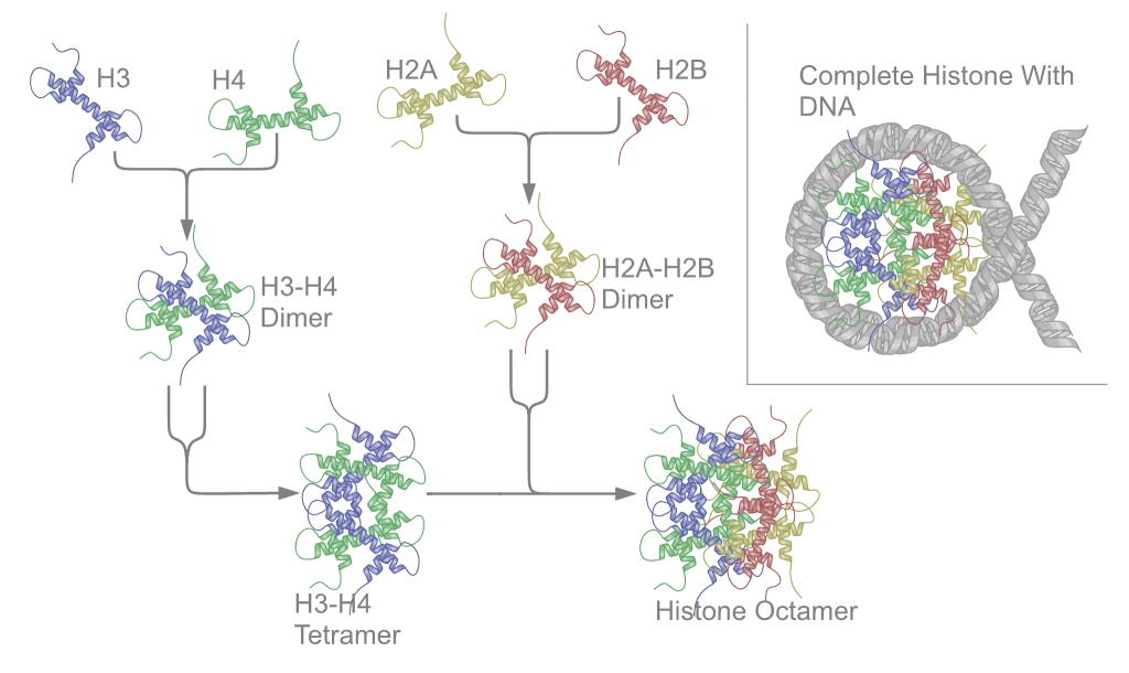
Nucleoproteins are proteins conjugated with nucleic acids (either DNA or RNA). Typical nucleoproteins include ribosomes, nucleosomes and viral nucleocapsid proteins. Structures Nucleoproteins tend to be positively charged, facilitating interaction with the negatively charged nucleic acid chains. The tertiary structures and biological functions of many nucleoproteins are understood.Graeme K. Hunter G. K. (2000): Vital Forces. The discovery of the molecular basis of life. Academic Press, London 2000, . Important techniques for determining the structures of nucleoproteins include X-ray diffraction, nuclear magnetic resonance and cryo-electron microscopy. Viruses Virus genomes (either DNA or RNA) are extremely tightly packed into the viral capsid. Many viruses are therefore little more than an organised collection of nucleoproteins with their binding sites pointing inwards. Structurally characterised viral nucleoproteins include influenza, rabies, Ebola, Bunyamwera, Schmall ... [...More Info...] [...Related Items...] OR: [Wikipedia] [Google] [Baidu] |
Organelles
In cell biology, an organelle is a specialized subunit, usually within a cell, that has a specific function. The name ''organelle'' comes from the idea that these structures are parts of cells, as organs are to the body, hence ''organelle,'' the suffix ''-elle'' being a diminutive. Organelles are either separately enclosed within their own lipid bilayers (also called membrane-bound organelles) or are spatially distinct functional units without a surrounding lipid bilayer (non-membrane bound organelles). Although most organelles are functional units within cells, some function units that extend outside of cells are often termed organelles, such as cilia, the flagellum and archaellum, and the trichocyst. Organelles are identified by microscopy, and can also be purified by cell fractionation. There are many types of organelles, particularly in eukaryotic cells. They include structures that make up the endomembrane system (such as the nuclear envelope, endoplasmic reticulum, a ... [...More Info...] [...Related Items...] OR: [Wikipedia] [Google] [Baidu] |
Viruses
A virus is a submicroscopic infectious agent that replicates only inside the living cells Cell most often refers to: * Cell (biology), the functional basic unit of life Cell may also refer to: Locations * Monastic cell, a small room, hut, or cave in which a religious recluse lives, alternatively the small precursor of a monastery w ... of an organism. Viruses infect all life forms, from animals and plants to microorganisms, including bacteria and archaea. Since Dmitri Ivanovsky's 1892 article describing a non-bacterial pathogen infecting tobacco plants and the discovery of the tobacco mosaic virus by Martinus Beijerinck in 1898,Dimmock p. 4 more than 9,000 virus species have been described in detail of the millions of types of viruses in the environment. Viruses are found in almost every ecosystem on Earth and are the most numerous type of biological entity. The study of viruses is known as virology, a subspeciality of microbiology. When infected, a host cell is ofte ... [...More Info...] [...Related Items...] OR: [Wikipedia] [Google] [Baidu] |
Symbionts
Symbiosis (from Greek , , "living together", from , , "together", and , bíōsis, "living") is any type of a close and long-term biological interaction between two different biological organisms, be it mutualistic, commensalistic, or parasitic. The organisms, each termed a symbiont, must be of different species. In 1879, Heinrich Anton de Bary defined it as "the living together of unlike organisms". The term was subject to a century-long debate about whether it should specifically denote mutualism, as in lichens. Biologists have now abandoned that restriction. Symbiosis can be obligatory, which means that one or more of the symbionts depend on each other for survival, or facultative (optional), when they can generally live independently. Symbiosis is also classified by physical attachment. When symbionts form a single body it is called conjunctive symbiosis, while all other arrangements are called disjunctive symbiosis."symbiosis." Dorland's Illustrated Medical Dictionary. ... [...More Info...] [...Related Items...] OR: [Wikipedia] [Google] [Baidu] |
Giemsa
Giemsa stain (), named after German chemist and bacteriologist Gustav Giemsa, is a nucleic acid stain used in cytogenetics and for the histopathological diagnosis of malaria and other parasites. Uses It is specific for the phosphate groups of DNA and attaches itself to regions of DNA where there are high amounts of adenine-thymine bonding. Giemsa stain is used in Giemsa banding, commonly called G-banding, to stain chromosomes and often used to create a karyogram (chromosome map). It can identify chromosomal aberrations such as translocations and rearrangements. It stains the trophozoite ''Trichomonas vaginalis'', which presents with greenish discharge and motile cells on wet prep. Giemsa stain is also a differential stain, such as when it is combined with Wright stain to form Wright-Giemsa stain. It can be used to study the adherence of pathogenic bacteria to human cells. It differentially stains human and bacterial cells purple and pink respectively. It can be used for ... [...More Info...] [...Related Items...] OR: [Wikipedia] [Google] [Baidu] |


.jpg)