|
Juvenile Myelomonocytic Leukemia
Juvenile myelomonocytic leukemia (JMML) is a serious chronic leukemia (cancer of the blood) that affects children mostly aged 4 and younger. The name JMML now encompasses all diagnoses formerly referred to as juvenile chronic myeloid leukemia (JCML), chronic myelomonocytic leukemia of infancy, and infantile monosomy 7 syndrome. The average age of patients at diagnosis is 2 years old. The World Health Organization has included JMML in the category of myelodysplastic and myeloproliferative disorders. Signs and symptoms The following symptoms are typical ones which lead to testing for JMML, though children with JMML may exhibit any combination of them: pallor, fever, infection, bleeding, cough, poor weight gain, a maculopapular rash (discolored but not raised, or small and raised but not containing pus), lymphadenopathy (enlarged lymph nodes), moderate hepatomegaly (enlarged liver), marked splenomegaly (enlarged spleen), leukocytosis (high white blood cell count in blood), ab ... [...More Info...] [...Related Items...] OR: [Wikipedia] [Google] [Baidu] |
Leukemia
Leukemia ( also spelled leukaemia and pronounced ) is a group of blood cancers that usually begin in the bone marrow and result in high numbers of abnormal blood cells. These blood cells are not fully developed and are called ''blasts'' or ''leukemia cells''. Symptoms may include bleeding and bruising, bone pain, fatigue, fever, and an increased risk of infections. These symptoms occur due to a lack of normal blood cells. Diagnosis is typically made by blood tests or bone marrow biopsy. The exact cause of leukemia is unknown. A combination of genetic factors and environmental (non-inherited) factors are believed to play a role. Risk factors include smoking, ionizing radiation, petrochemicals (such as benzene), prior chemotherapy, and Down syndrome. People with a family history of leukemia are also at higher risk. There are four main types of leukemia— acute lymphoblastic leukemia (ALL), acute myeloid leukemia (AML), chronic lymphocytic leukemia (CLL) and chr ... [...More Info...] [...Related Items...] OR: [Wikipedia] [Google] [Baidu] |
Mutation
In biology, a mutation is an alteration in the nucleic acid sequence of the genome of an organism, virus, or extrachromosomal DNA. Viral genomes contain either DNA or RNA. Mutations result from errors during DNA or viral replication, mitosis, or meiosis or other types of damage to DNA (such as pyrimidine dimers caused by exposure to ultraviolet radiation), which then may undergo error-prone repair (especially microhomology-mediated end joining), cause an error during other forms of repair, or cause an error during replication ( translesion synthesis). Mutations may also result from insertion or deletion of segments of DNA due to mobile genetic elements. Mutations may or may not produce detectable changes in the observable characteristics (phenotype) of an organism. Mutations play a part in both normal and abnormal biological processes including: evolution, cancer, and the development of the immune system, including junctional diversity. Mutation is the ultimate s ... [...More Info...] [...Related Items...] OR: [Wikipedia] [Google] [Baidu] |
White Blood Cell
White blood cells, also called leukocytes or leucocytes, are the cells of the immune system that are involved in protecting the body against both infectious disease and foreign invaders. All white blood cells are produced and derived from multipotent cells in the bone marrow known as hematopoietic stem cells. Leukocytes are found throughout the body, including the blood and lymphatic system. All white blood cells have nuclei, which distinguishes them from the other blood cells, the anucleated red blood cells (RBCs) and platelets. The different white blood cells are usually classified by cell lineage (myeloid cells or lymphoid cells). White blood cells are part of the body's immune system. They help the body fight infection and other diseases. Types of white blood cells are granulocytes (neutrophils, eosinophils, and basophils), and agranulocytes (monocytes, and lymphocytes (T cells and B cells)). Myeloid cells ( myelocytes) include neutrophils, eosinophils, mast cells, ... [...More Info...] [...Related Items...] OR: [Wikipedia] [Google] [Baidu] |
Granulocyte
Granulocytes are cells in the innate immune system characterized by the presence of specific granules in their cytoplasm. Such granules distinguish them from the various agranulocytes. All myeloblastic granulocytes are polymorphonuclear. They have varying shapes (morphology) of the nucleus (segmented, irregular; often lobed into three segments); and are referred to as polymorphonuclear leukocytes (PMN, PML, or PMNL). In common terms, ''polymorphonuclear granulocyte'' refers specifically to "neutrophil granulocytes", the most abundant of the granulocytes; the other types ( eosinophils, basophils, and mast cells) have varying morphology. Granulocytes are produced via granulopoiesis in the bone marrow. Types There are four types of granulocytes (full name polymorphonuclear granulocytes): * Basophils * Eosinophils * Neutrophils * Mast cells Except for the mast cells, their names are derived from their staining characteristics; for example, the most abundant granulocyte is the ... [...More Info...] [...Related Items...] OR: [Wikipedia] [Google] [Baidu] |
Fetal Hemoglobin
Fetal hemoglobin, or foetal haemoglobin (also hemoglobin F, HbF, or α2γ2) is the main oxygen carrier protein in the human fetus. Hemoglobin F is found in fetal red blood cells, and is involved in transporting oxygen from the mother's bloodstream to organs and tissues in the fetus. It is produced at around 6 weeks of pregnancy and the levels remain high after birth until the baby is roughly 2–4 months old. Hemoglobin F has a different composition from the adult forms of hemoglobin, which allows it to bind (or attach to) oxygen more strongly. This way, the developing fetus is able to retrieve oxygen from the mother's bloodstream, which occurs through the placenta found in the mother's uterus. In the newborn, levels of hemoglobin F gradually decrease and reach adult levels (less than 1% of total hemoglobin) usually within the first year, as adult forms of hemoglobin begin to be produced. Diseases such as beta thalassemias, which affect components of the adult hemoglobin, ca ... [...More Info...] [...Related Items...] OR: [Wikipedia] [Google] [Baidu] |
Ras Superfamily
The Ras superfamily, derived from "Rat sarcoma virus", is a protein superfamily of small GTPases. Members of the superfamily are divided into families and subfamilies based on their structure, sequence and function. The five main families are Ras, Rho, Ran, Rab and Arf GTPases. The Ras family itself is further divided into 6 subfamilies: Ras, Ral, Rap, Rheb, Rad and Rit. ''Miro'' is a recent contributor to the superfamily. Each subfamily shares the common core G domain, which provides essential GTPase and nucleotide exchange activity. The surrounding sequence helps determine the functional specificity of the small GTPase, for example the 'Insert Loop', common to the Rho subfamily, specifically contributes to binding to effector proteins such as WASP. In general, the Ras family is responsible for cell proliferation: Rho for cell morphology, Ran for nuclear transport, and Rab and Arf for vesicle transport. Subfamilies and members The following is a list of human proteins be ... [...More Info...] [...Related Items...] OR: [Wikipedia] [Google] [Baidu] |
Splenomegaly
Splenomegaly is an enlargement of the spleen. The spleen usually lies in the left upper quadrant (LUQ) of the human abdomen. Splenomegaly is one of the four cardinal signs of ''hypersplenism'' which include: some reduction in number of circulating blood cells affecting granulocytes, erythrocytes or platelets in any combination; a compensatory proliferative response in the bone marrow; and the potential for correction of these abnormalities by splenectomy. Splenomegaly is usually associated with increased workload (such as in hemolytic anemias), which suggests that it is a response to hyperfunction. It is therefore not surprising that splenomegaly is associated with any disease process that involves abnormal red blood cells being destroyed in the spleen. Other common causes include congestion due to portal hypertension and infiltration by leukemias and lymphomas. Thus, the finding of an enlarged spleen, along with caput medusae, is an important sign of portal hypertension. D ... [...More Info...] [...Related Items...] OR: [Wikipedia] [Google] [Baidu] |
Bone Marrow
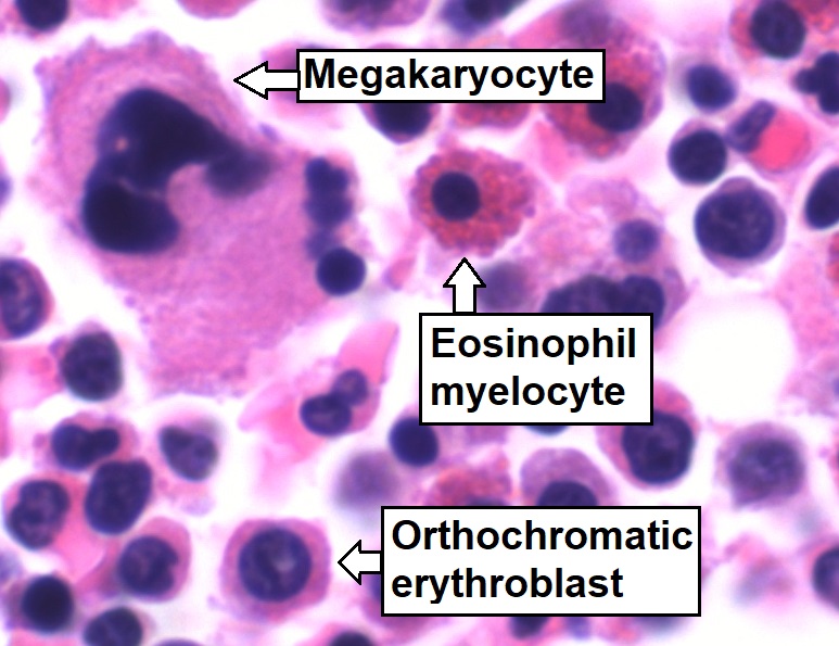
Bone marrow is a semi-solid biological tissue, tissue found within the Spongy bone, spongy (also known as cancellous) portions of bones. In birds and mammals, bone marrow is the primary site of new blood cell production (or haematopoiesis). It is composed of Blood cell, hematopoietic cells, marrow adipose tissue, and supportive stromal cells. In adult humans, bone marrow is primarily located in the Rib cage, ribs, vertebrae, sternum, and Pelvis, bones of the pelvis. Bone marrow comprises approximately 5% of total body mass in healthy adult humans, such that a man weighing 73 kg (161 lbs) will have around 3.7 kg (8 lbs) of bone marrow. Human marrow produces approximately 500 billion blood cells per day, which join the Circulatory system, systemic circulation via permeable vasculature sinusoids within the medullary cavity. All types of hematopoietic cells, including both Myeloid tissue, myeloid and Lymphocyte, lymphoid lineages, are created in bone marrow; howev ... [...More Info...] [...Related Items...] OR: [Wikipedia] [Google] [Baidu] |
Promonocyte
A promonocyte (or premonocyte) is a cell arising from a monoblast Monoblasts are the committed progenitor cells that differentiated from a committed macrophage or dendritic cell precursor (MDP) in the process of hematopoiesis. They are the first developmental stage in the monocyte series leading to a macrophage ... and developing into a monocyte. See also * Pluripotential hemopoietic stem cell Additional images Image:Hematopoiesis (human) diagram en.svg, Hematopoiesis Image:Promonocyte.png, Schematic image of a promonocyte External links "Monocyte Development" at tulane.edu* - "Bone marrow smear" Blood cells Immune system {{cell-biology-stub ... [...More Info...] [...Related Items...] OR: [Wikipedia] [Google] [Baidu] |
Monocytosis
Monocytosis is an increase in the number of monocytes circulating in the blood. Monocytes are white blood cells that give rise to macrophages and dendritic cells in the immune system. In humans, monocytosis occurs when there is a sustained rise in monocyte counts greater than 800/mm3 to 1000/mm3. Monocytosis has sometimes been called mononucleosis, but that name is usually reserved specifically for infectious mononucleosis. Causes Monocytosis often occurs during chronic inflammation. Diseases that produce such a chronic inflammatory state: * Infections: tuberculosis, brucellosis, listeriosis, subacute bacterial endocarditis, syphilis, and other viral infections and many protozoal and rickettsial infections (e.g. kala azar, malaria, Rocky Mountain spotted fever). * Blood and immune causes: chronic neutropenia and myeloproliferative disorders. * Autoimmune diseases and vasculitis: systemic lupus erythematosus, rheumatoid arthritis and inflammatory bowel disease. * Malignancies ... [...More Info...] [...Related Items...] OR: [Wikipedia] [Google] [Baidu] |
Fusion Gene
A fusion gene is a hybrid gene formed from two previously independent genes. It can occur as a result of translocation, interstitial deletion, or chromosomal inversion. Fusion genes have been found to be prevalent in all main types of human neoplasia. The identification of these fusion genes play a prominent role in being a diagnostic and prognostic marker. History The first fusion gene was described in cancer cells in the early 1980s. The finding was based on the discovery in 1960 by Peter Nowell and David Hungerford in Philadelphia of a small abnormal marker chromosome in patients with chronic myeloid leukemia—the first consistent chromosome abnormality detected in a human malignancy, later designated the Philadelphia chromosome. In 1973, Janet Rowley in Chicago showed that the Philadelphia chromosome had originated through a translocation between chromosomes 9 and 22, and not through a simple deletion of chromosome 22 as was previously thought. Several investigators in ... [...More Info...] [...Related Items...] OR: [Wikipedia] [Google] [Baidu] |
Philadelphia Chromosome
The Philadelphia chromosome or Philadelphia translocation (Ph) is a specific genetic abnormality in chromosome 22 of leukemia cancer cells (particularly chronic myeloid leukemia (CML) cells). This chromosome is defective and unusually short because of reciprocal translocation, t(9;22)(q34;q11), of genetic material between chromosome 9 and chromosome 22, and contains a fusion gene called ''BCR-ABL1''. This gene is the ''ABL1'' gene of chromosome 9 juxtaposed onto the breakpoint cluster region '' BCR'' gene of chromosome 22, coding for a hybrid protein: a tyrosine kinase signaling protein that is "always on", causing the cell to divide uncontrollably by interrupting the stability of the genome and impairing various signaling pathways governing the cell cycle. The presence of this translocation is required for diagnosis of CML; in other words, all cases of CML are positive for ''BCR-ABL1''. (Some cases are confounded by either a cryptic translocation that is invisible on G-banded c ... [...More Info...] [...Related Items...] OR: [Wikipedia] [Google] [Baidu] |


