|
Intrafusal Muscle Fibers
Intrafusal muscle fibers are skeletal muscle fibers that serve as specialized sensory organs (proprioceptors). They detect the amount and rate of change in length of a muscle.Casagrand, Janet (2008) ''Action and Movement: Spinal Control of Motor Units and Spinal Reflexes.'' University of Colorado, Boulder. They constitute the muscle spindle, and are innervated by both sensory (afferent) and motor (efferent) fibers. Intrafusal muscle fibers are not to be confused with extrafusal muscle fibers, which contract, generating skeletal movement and are innervated by alpha motor neurons. Structure Types There are two types of intrafusal muscle fibers: nuclear bag fibers and nuclear chain fibers. They bear two types of sensory ending, known as annulospiral and flower-spray endings. Both ends of these fibers contract, but the central region only stretches and does not contract. Intrafusal muscle fibers are walled off from the rest of the muscle by an outer connective tissue she ... [...More Info...] [...Related Items...] OR: [Wikipedia] [Google] [Baidu] |
Proprioception
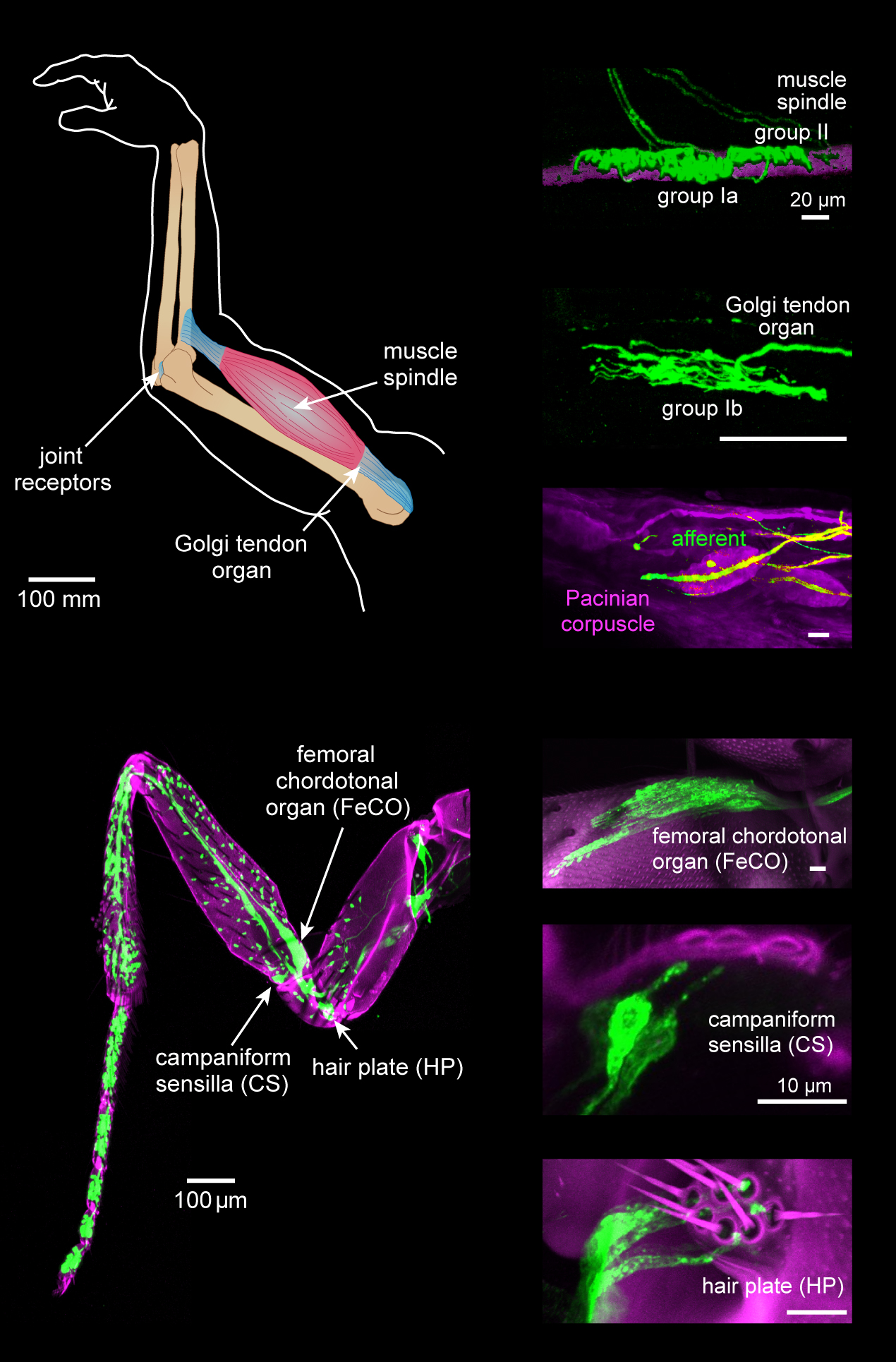
Proprioception ( ), also referred to as kinaesthesia (or kinesthesia), is the sense of self-movement, force, and body position. It is sometimes described as the "sixth sense". Proprioception is mediated by proprioceptors, mechanosensory neurons located within muscles, tendons, and joints. Most animals possess multiple subtypes of proprioceptors, which detect distinct kinematic parameters, such as joint position, movement, and load. Although all mobile animals possess proprioceptors, the structure of the sensory organs can vary across species. Proprioceptive signals are transmitted to the central nervous system, where they are integrated with information from other sensory systems, such as the visual system and the vestibular system, to create an overall representation of body position, movement, and acceleration. In many animals, sensory feedback from proprioceptors is essential for stabilizing body posture and coordinating body movement. System overview In vertebrates, limb ve ... [...More Info...] [...Related Items...] OR: [Wikipedia] [Google] [Baidu] |
Collagen
Collagen () is the main structural protein in the extracellular matrix found in the body's various connective tissues. As the main component of connective tissue, it is the most abundant protein in mammals, making up from 25% to 35% of the whole-body protein content. Collagen consists of amino acids bound together to form a triple helix of elongated fibril known as a collagen helix. It is mostly found in connective tissue such as cartilage, bones, tendons, ligaments, and skin. Depending upon the degree of mineralization, collagen tissues may be rigid (bone) or compliant (tendon) or have a gradient from rigid to compliant (cartilage). Collagen is also abundant in corneas, blood vessels, the gut, intervertebral discs, and the dentin in teeth. In muscle tissue, it serves as a major component of the endomysium. Collagen constitutes one to two percent of muscle tissue and accounts for 6% of the weight of the skeletal muscle tissue. The fibroblast is the most common cell that crea ... [...More Info...] [...Related Items...] OR: [Wikipedia] [Google] [Baidu] |
Type Ia Sensory Fiber
A type Ia sensory fiber, or a primary afferent fiber is a type of afferent nerve fiber. It is the sensory fiber of a stretch receptor called the muscle spindle found in muscles, which constantly monitors the rate at which a muscle stretch changes. The information carried by type Ia fibers contributes to the sense of proprioception. Function of muscle spindles For the body to keep moving properly and with finesse, the nervous system has to have a constant input of sensory data coming from areas such as the muscles and joints. In order to receive a continuous stream of sensory data, the body has developed special sensory receptors called proprioceptors. Muscle spindles are a type of proprioceptor, and they are found inside the muscle itself. They lie parallel with the contractile fibers. This gives them the ability to monitor muscle length with precision. Types of sensory fibers This change in length of the spindle is transduced (transformed into electric membrane potentials ... [...More Info...] [...Related Items...] OR: [Wikipedia] [Google] [Baidu] |
Gamma Motor Neuron
A gamma motor neuron (γ motor neuron), also called gamma motoneuron, or fusimotor neuron, is a type of lower motor neuron that takes part in the process of muscle contraction, and represents about 30% of ( Aγ) fibers going to the muscle. Like alpha motor neurons, their cell bodies are located in the anterior grey column of the spinal cord. They receive input from the reticular formation of the pons in the brainstem. Their axons are smaller than those of the alpha motor neurons, with a diameter of only 5 μm. Unlike the alpha motor neurons, gamma motor neurons do not directly adjust the lengthening or shortening of muscles. However, their role is important in keeping muscle spindles taut, thereby allowing the continued firing of alpha neurons, leading to muscle contraction. These neurons also play a role in adjusting the sensitivity of muscle spindles. The presence of myelination in gamma motor neurons allows a conduction velocity of 4 to 24 meters per second, significantl ... [...More Info...] [...Related Items...] OR: [Wikipedia] [Google] [Baidu] |
Extrafusal Muscle Fiber
Extrafusal muscle fibers are the standard skeletal muscle fibers that are innervated by alpha motor neurons and generate tension by contracting, thereby allowing for skeletal movement. They make up the large mass of skeletal striated muscle tissue and are attached to bone by fibrous tissue extensions (tendons). Each alpha motor neuron and the extrafusal muscle fibers innervated by it make up a motor unit. The connection between the alpha motor neuron and the extrafusal muscle fiber is a neuromuscular junction, where the neuron's signal, the action potential, is transduced to the muscle fiber by the neurotransmitter acetylcholine. Extrafusal muscle fibers are not to be confused with intrafusal muscle fibers, which are innervated by sensory nerve endings in central noncontractile parts and by gamma motor neurons in contractile ends and thus serve as a sensory proprioceptor. Extrafusal muscle fibers can be generated in vitro (in a dish) from pluripotent stem cells through dire ... [...More Info...] [...Related Items...] OR: [Wikipedia] [Google] [Baidu] |
Beta Motor Neuron
Beta motor neurons (β motor neurons), also called beta motoneurons, are a kind of lower motor neuron, along with alpha motor neurons and gamma motor neurons. Beta motor neurons innervate intrafusal fibers of muscle spindles with collaterals to extrafusal fibers - a type of slow twitch fiber. Also, axons of alpha, beta, and gamma motor neurons become myelinated. Moreover, these efferent neurons originate from the anterior grey column of the spinal cord and travel to skeletal muscles. However, the larger diameter alpha motor fibers require higher conduction velocity than beta and gamma. Types There are two kinds of beta motor neuron (as gamma motor neuron) that include: *Static beta motor neurons. These motor neurons innervate nuclear chain fibers of muscle spindles, with collaterals to extrafusal muscle fibers. *Dynamic beta motor neurons. The dynamic type innervates nuclear bag fibers of muscle spindles, with collaterals to extrafusal muscle fibers. Gamma motor neurons innervate ... [...More Info...] [...Related Items...] OR: [Wikipedia] [Google] [Baidu] |
Alpha Motor Neuron
Alpha (α) motor neurons (also called alpha motoneurons), are large, multipolar lower motor neurons of the brainstem and spinal cord. They innervate extrafusal muscle fibers of skeletal muscle and are directly responsible for initiating their contraction. Alpha motor neurons are distinct from gamma motor neurons, which innervate intrafusal muscle fibers of muscle spindles. While their cell bodies are found in the central nervous system (CNS), α motor neurons are also considered part of the somatic nervous system—a branch of the peripheral nervous system (PNS)—because their axons extend into the periphery to innervate skeletal muscles. An alpha motor neuron and the muscle fibers it innervates is a motor unit. A motor neuron pool contains the cell bodies of all the alpha motor neurons involved in contracting a single muscle. Location Alpha motor neurons (α-MNs) innervating the head and neck are found in the brainstem; the remaining α-MNs innervate the rest of the body and ... [...More Info...] [...Related Items...] OR: [Wikipedia] [Google] [Baidu] |
Muscle Spindle
Muscle spindles are stretch receptors within the body of a skeletal muscle that primarily detect changes in the length of the muscle. They convey length information to the central nervous system via afferent nerve fibers. This information can be processed by the brain as proprioception. The responses of muscle spindles to changes in length also play an important role in regulating the contraction of muscles, for example, by activating motor neurons via the stretch reflex to resist muscle stretch. The muscle spindle has both sensory and motor components. * Sensory information conveyed by primary type Ia sensory fibers which spiral around muscle fibres within the spindle, and secondary type II sensory fibers * Activation of muscle fibres within the spindle by up to a dozen gamma motor neurons and to a lesser extent by one or two beta motor neurons Structure Muscle spindles are found within the belly of a skeletal muscle. Muscle spindles are fusiform (spindle-shaped), and the spec ... [...More Info...] [...Related Items...] OR: [Wikipedia] [Google] [Baidu] |
Multipolar Neuron
A multipolar neuron is a type of neuron that possesses a single axon and many dendrites (and dendritic branches), allowing for the integration of a great deal of information from other neurons. These processes are projections from the neuron cell body. Multipolar neurons constitute the majority of neurons in the central nervous system. They include motor neurons and interneurons/relaying neurons are most commonly found in the cortex of the brain and the spinal cord. Peripherally, multipolar neurons are found in autonomic ganglia. See also * Dogiel cells * Ganglion cell * Purkinje cell Purkinje cells, or Purkinje neurons, are a class of GABAergic inhibitory neurons located in the cerebellum. They are named after their discoverer, Czech people, Czech anatomist Jan Evangelista Purkyně, who characterized the cells in 1839. Stru ... * Pyramidal cell Additional images File:Blausen 0672 NeuralTissue.png, Neural tissue References External links DiagramDiagramImage {{neuro ... [...More Info...] [...Related Items...] OR: [Wikipedia] [Google] [Baidu] |
Beta Motor Neuron
Beta motor neurons (β motor neurons), also called beta motoneurons, are a kind of lower motor neuron, along with alpha motor neurons and gamma motor neurons. Beta motor neurons innervate intrafusal fibers of muscle spindles with collaterals to extrafusal fibers - a type of slow twitch fiber. Also, axons of alpha, beta, and gamma motor neurons become myelinated. Moreover, these efferent neurons originate from the anterior grey column of the spinal cord and travel to skeletal muscles. However, the larger diameter alpha motor fibers require higher conduction velocity than beta and gamma. Types There are two kinds of beta motor neuron (as gamma motor neuron) that include: *Static beta motor neurons. These motor neurons innervate nuclear chain fibers of muscle spindles, with collaterals to extrafusal muscle fibers. *Dynamic beta motor neurons. The dynamic type innervates nuclear bag fibers of muscle spindles, with collaterals to extrafusal muscle fibers. Gamma motor neurons innervate ... [...More Info...] [...Related Items...] OR: [Wikipedia] [Google] [Baidu] |
Gamma Motor Neuron
A gamma motor neuron (γ motor neuron), also called gamma motoneuron, or fusimotor neuron, is a type of lower motor neuron that takes part in the process of muscle contraction, and represents about 30% of ( Aγ) fibers going to the muscle. Like alpha motor neurons, their cell bodies are located in the anterior grey column of the spinal cord. They receive input from the reticular formation of the pons in the brainstem. Their axons are smaller than those of the alpha motor neurons, with a diameter of only 5 μm. Unlike the alpha motor neurons, gamma motor neurons do not directly adjust the lengthening or shortening of muscles. However, their role is important in keeping muscle spindles taut, thereby allowing the continued firing of alpha neurons, leading to muscle contraction. These neurons also play a role in adjusting the sensitivity of muscle spindles. The presence of myelination in gamma motor neurons allows a conduction velocity of 4 to 24 meters per second, significantl ... [...More Info...] [...Related Items...] OR: [Wikipedia] [Google] [Baidu] |
Fibroblasts
A fibroblast is a type of biological cell that synthesizes the extracellular matrix and collagen, produces the structural framework ( stroma) for animal tissues, and plays a critical role in wound healing. Fibroblasts are the most common cells of connective tissue in animals. Structure Fibroblasts have a branched cytoplasm surrounding an elliptical, speckled nucleus having two or more nucleoli. Active fibroblasts can be recognized by their abundant rough endoplasmic reticulum. Inactive fibroblasts (called fibrocytes) are smaller, spindle-shaped, and have a reduced amount of rough endoplasmic reticulum. Although disjointed and scattered when they have to cover a large space, fibroblasts, when crowded, often locally align in parallel clusters. Unlike the epithelial cells lining the body structures, fibroblasts do not form flat monolayers and are not restricted by a polarizing attachment to a basal lamina on one side, although they may contribute to basal lamina components in s ... [...More Info...] [...Related Items...] OR: [Wikipedia] [Google] [Baidu] |


.jpg)