|
Intercostal Muscle
Intercostal muscles are many different groups of muscles that run between the ribs, and help form and move the chest wall. The intercostal muscles are mainly involved in the mechanical aspect of breathing by helping expand and shrink the size of the chest cavity. Structure There are three principal layers; # External intercostal muscles also known as intercostalis externus aid in quiet and forced inhalation. They originate on ribs 1–11 and have their insertion on ribs 2–12. The external intercostals are responsible for the elevation of the ribs and bending them more open, thus expanding the transverse dimensions of the thoracic cavity. The muscle fibers are directed downwards, forwards and medially in the anterior part. #Internal intercostal muscles also known as intercostalis internus aid in forced expiration (quiet expiration is a passive process). They originate on ribs 2–12 and have their insertions on ribs 1–11.Their fibers pass anterior and superi ... [...More Info...] [...Related Items...] OR: [Wikipedia] [Google] [Baidu] |
Intercostal Arteries
The intercostal arteries are a group of arteries that supply the area between the ribs ("costae"), called the intercostal space. The highest intercostal artery (supreme intercostal artery or superior intercostal artery) is an artery in the human body that usually gives rise to the first and second posterior intercostal arteries, which supply blood to their corresponding intercostal space. It usually arises from the costocervical trunk, which is a branch of the subclavian artery. Some anatomists may contend that there is no supreme intercostal artery, only a supreme intercostal vein. The anterior intercostal branches of internal thoracic artery supply the upper five or six intercostal spaces. The internal thoracic artery (previously called as internal mammary artery) then divides into the superior epigastric artery and musculophrenic artery. The latter gives out the remaining anterior intercostal branches. Two in number in each space, these small vessels pass lateralward, ... [...More Info...] [...Related Items...] OR: [Wikipedia] [Google] [Baidu] |
Exhalation
Exhalation (or expiration) is the flow of the breath out of an organism. In animals, it is the movement of air from the lungs out of the airways, to the external environment during breathing. This happens due to elastic properties of the lungs, as well as the internal intercostal muscles which lower the rib cage and decrease thoracic volume. As the thoracic diaphragm relaxes during exhalation it causes the tissue it has depressed to rise superiorly and put pressure on the lungs to expel the air. During forced exhalation, as when blowing out a candle, expiratory muscles including the abdominal muscles and internal intercostal muscles generate abdominal and thoracic pressure, which forces air out of the lungs. Exhaled air is 4% carbon dioxide, a waste product of cellular respiration during the production of energy, which is stored as ATP. Exhalation has a complementary relationship to inhalation which together make up the respiratory cycle of a breath. Exhalation and gas excha ... [...More Info...] [...Related Items...] OR: [Wikipedia] [Google] [Baidu] |
Inhalation
Inhalation (or Inspiration) happens when air or other gases enter the lungs. Inhalation of air Inhalation of air, as part of the cycle of breathing, is a vital process for all human life. The process is autonomic (though there are exceptions in some disease states) and does not need conscious control or effort. However, breathing can be consciously controlled or interrupted (within limits). Breathing allows oxygen (which humans and a lot of other species need for survival) to enter the lungs, from where it can be absorbed into the bloodstream. Other substances – accidental Examples of accidental inhalation includes inhalation of water (e.g. in drowning), smoke, food, vomitus and less common foreign substances (e.g. tooth fragments, coins, batteries, small toy parts, needles). Other substances – deliberate Recreational use Legal – helium, nitrous oxide (" laughing gas") Illegal – various gaseous, vaporised or aerosolized recreational drugs Medical use D ... [...More Info...] [...Related Items...] OR: [Wikipedia] [Google] [Baidu] |
Thoracic Diaphragm
The thoracic diaphragm, or simply the diaphragm ( grc, διάφραγμα, diáphragma, partition), is a sheet of internal skeletal muscle in humans and other mammals that extends across the bottom of the thoracic cavity. The diaphragm is the most important muscle of respiration, and separates the thoracic cavity, containing the heart and lungs, from the abdominal cavity: as the diaphragm contracts, the volume of the thoracic cavity increases, creating a negative pressure there, which draws air into the lungs. Its high oxygen consumption is noted by the many mitochondria and capillaries present; more than in any other skeletal muscle. The term ''diaphragm'' in anatomy, created by Gerard of Cremona, can refer to other flat structures such as the urogenital diaphragm or pelvic diaphragm, but "the diaphragm" generally refers to the thoracic diaphragm. In humans, the diaphragm is slightly asymmetric—its right half is higher up (superior) to the left half, since the large ... [...More Info...] [...Related Items...] OR: [Wikipedia] [Google] [Baidu] |
Scalene Muscles
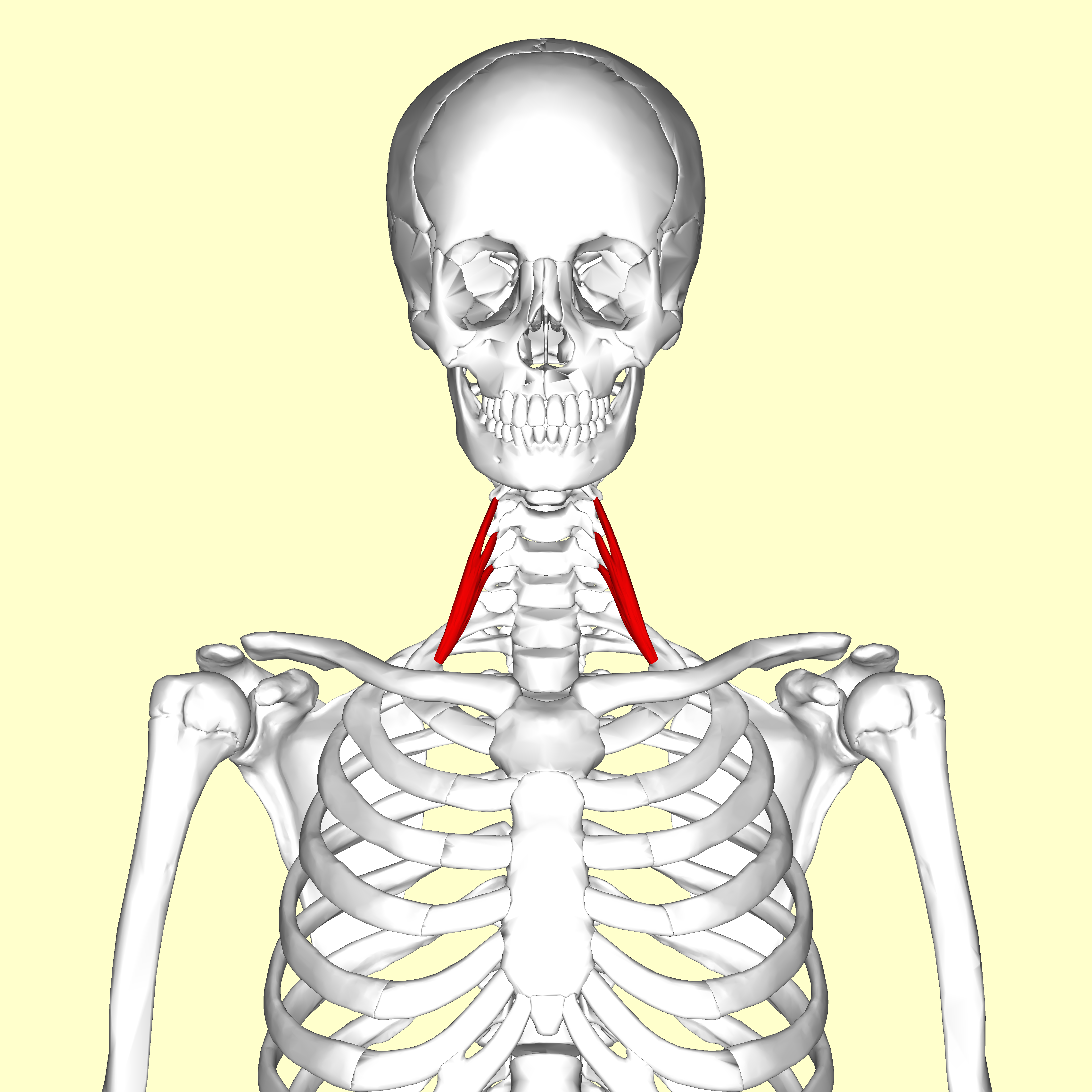
The scalene muscles are a group of three pairs of muscles in the lateral neck, namely the anterior scalene, middle scalene, and posterior scalene. They are innervated by the third to the eight cervical spinal nerves (C3-C8). The anterior and middle scalene muscles lift the first rib and bend the neck to the same side; the posterior scalene lifts the second rib and tilts the neck to the same side. The muscles are named . Structure The scalene muscles originate from the transverse processes from the cervical vertebrae of C2 to C7 and insert onto the first and second ribs. Anterior scalene The anterior scalene muscle ( la, scalenus anterior), lies deeply at the side of the neck, behind the sternocleidomastoid muscle. It arises from the anterior tubercles of the transverse processes of the third, fourth, fifth, and sixth cervical vertebrae, and descending, almost vertically, is inserted by a narrow, flat tendon into the scalene tubercle on the inner border of the first rib, ... [...More Info...] [...Related Items...] OR: [Wikipedia] [Google] [Baidu] |
Intercostal Veins
The intercostal veins are a group of veins which drain the area between the ribs ("costae"), called the intercostal space. They can be divided as follows: * Anterior intercostal veins * Posterior intercostal veins ** Posterior intercost vein that drain into the Supreme intercostal vein - 1st intercostal space ** Posterior intercost veins that drain into the Superior intercostal vein - 2nd, 3rd, and 4th intercostal spaces. The superior intercostal vein then drains into the Azygous vein. ** Posterior intercost veins that drain directly into the Azygous vein - in spaces 5-11. ** Subcostal vein -- below bottom (12th) rib and also drains into the Azygous vein. See also * Intercostal nerves The intercostal nerves are part of the somatic nervous system, and arise from the anterior rami of the thoracic spinal nerves from T1 to T11. The intercostal nerves are distributed chiefly to the thoracic pleura and abdominal peritoneum, and dif ... External links * * http://www.instantanatomy. ... [...More Info...] [...Related Items...] OR: [Wikipedia] [Google] [Baidu] |
Thoracic Spinal Nerves
A spinal nerve is a mixed nerve, which carries motor, sensory, and autonomic signals between the spinal cord and the body. In the human body there are 31 pairs of spinal nerves, one on each side of the vertebral column. These are grouped into the corresponding cervical, thoracic, lumbar, sacral and coccygeal regions of the spine. There are eight pairs of cervical nerves, twelve pairs of thoracic nerves, five pairs of lumbar nerves, five pairs of sacral nerves, and one pair of coccygeal nerves. The spinal nerves are part of the peripheral nervous system. Structure Each spinal nerve is a mixed nerve, formed from the combination of nerve fibers from its dorsal and ventral roots. The dorsal root is the afferent sensory root and carries sensory information to the brain. The ventral root is the efferent motor root and carries motor information from the brain. The spinal nerve emerges from the spinal column through an opening (intervertebral foramen) between adjacent vertebra ... [...More Info...] [...Related Items...] OR: [Wikipedia] [Google] [Baidu] |
Ventral Rami
The ventral ramus (pl. ''rami'') (Latin for ''branch'') is the anterior division of a spinal nerve. The ventral rami supply the antero-lateral parts of the trunk and the limbs. They are mainly larger than the dorsal rami. Shortly after a spinal nerve exits the intervertebral foramen, it branches into the dorsal ramus, the ventral ramus, and the ramus communicans. Each of these three structures carries both sensory Sensory may refer to: Biology * Sensory ecology, how organisms obtain information about their environment * Sensory neuron, nerve cell responsible for transmitting information about external stimuli * Sensory perception, the process of acquiri ... and motor information. Each spinal nerve carries both sensory and motor information, via efferent and afferent nerve fibers - ultimately via the motor cortex in the frontal lobe and to somatosensory cortex in the parietal lobe - but also through the phenomenon of reflex. Spinal nerves are referred to as "mixed ner ... [...More Info...] [...Related Items...] OR: [Wikipedia] [Google] [Baidu] |
Neurovascular Bundle
A neurovascular bundle is a structure that binds nerves and veins (and in some cases arteries and lymphatics) with connective tissue so that they travel in tandem through the body. Structure There are two types of neurovascular bundles: superficial bundles and deep bundles. As arteries do not travel within the superficial fascia (loose connective tissue under the skin), superficial neurovascular bundles differ from deep neurovascular bundles in both composition and function. Superficial bundles Superficial neurovascular bundles do not include arteries, and consist primarily of capillaries and nerves. Because capillaries function as the sites for substance exchange between interstitial fluid and blood, they tend to have large surface area and short diffusion path. Normally, capillaries consist of a central lumen lined with an endothelium, a single layer of smooth epithelial cells. Deep bundles Deep neurovascular bundles, which often include arteries, have a more complicated ... [...More Info...] [...Related Items...] OR: [Wikipedia] [Google] [Baidu] |
Innermost Intercostal Muscle
The innermost intercostal muscle is a layer of intercostal muscles. It may also be called the intima of the internal intercostal muscles. It is the deepest muscular layer of the thorax, with muscle fibres running vertically (in parallel with the internal intercostal muscles). It is present only in the middle of each intercostal space, and often not present higher up the rib cage. It lies deep to the plane that contains the intercostal nerves The intercostal nerves are part of the somatic nervous system, and arise from the anterior rami of the thoracic spinal nerves from T1 to T11. The intercostal nerves are distributed chiefly to the thoracic pleura and abdominal peritoneum, and dif ... and intercostal vessels, and the internal intercostal muscles. The diaphragm is continuous with the innermost intercostal muscle. Additional images File:Innermost intercostal muscles animation.gif, Innermost intercostal muscle (shown in red). Animation. File:1114 Thorax zoom.png, A cutout o ... [...More Info...] [...Related Items...] OR: [Wikipedia] [Google] [Baidu] |
Internal Intercostal Muscles
The internal intercostal muscles (intercostales interni) are a group of skeletal muscles located between the ribs. They are eleven in number on either side. They commence anteriorly at the sternum, in the intercostal spaces between the cartilages of the true ribs, and at the anterior extremities of the cartilages of the false ribs, and extend backward as far as the angles of the ribs, hence they are continued to the vertebral column by thin aponeuroses, the posterior intercostal membranes. They pull the sternum and ribs upward and inward. Structure Their fibers are also directed obliquely, but pass in a direction opposite to those of the external intercostal muscles. The internal intercostal muscles originate from the costal groove of the rib and insert into the superior aspect of the rib below in a direction perpendicular to the external intercostal muscles. It is this arrangement that allows these muscles to facilitate exhalation. For the most part, they are muscles of exha ... [...More Info...] [...Related Items...] OR: [Wikipedia] [Google] [Baidu] |
Intercostal Nerves
The intercostal nerves are part of the somatic nervous system, and arise from the anterior rami of the thoracic spinal nerves from T1 to T11. The intercostal nerves are distributed chiefly to the thoracic pleura and abdominal peritoneum, and differ from the anterior rami of the other spinal nerves in that each pursues an independent course without plexus formation. The first two nerves supply fibers to the upper limb and thorax; the next four distribute to the walls of the thorax; the lower five supply the walls of the thorax and abdomen. The 7th intercostal nerve end at the xyphoid process of the sternum. The 10th intercostal nerve terminates at the navel. The 12th ( subcostal) thoracic is distributed to the walls of the abdomen and groin. Each of these fibers contains around 1300 axons. Unlike the nerves from the autonomic nervous system that innervate the visceral pleura of the thoracic cavity, the intercostal nerves arise from the somatic nervous system. This enables them ... [...More Info...] [...Related Items...] OR: [Wikipedia] [Google] [Baidu] |


