|
In Vitro Muscle Testing
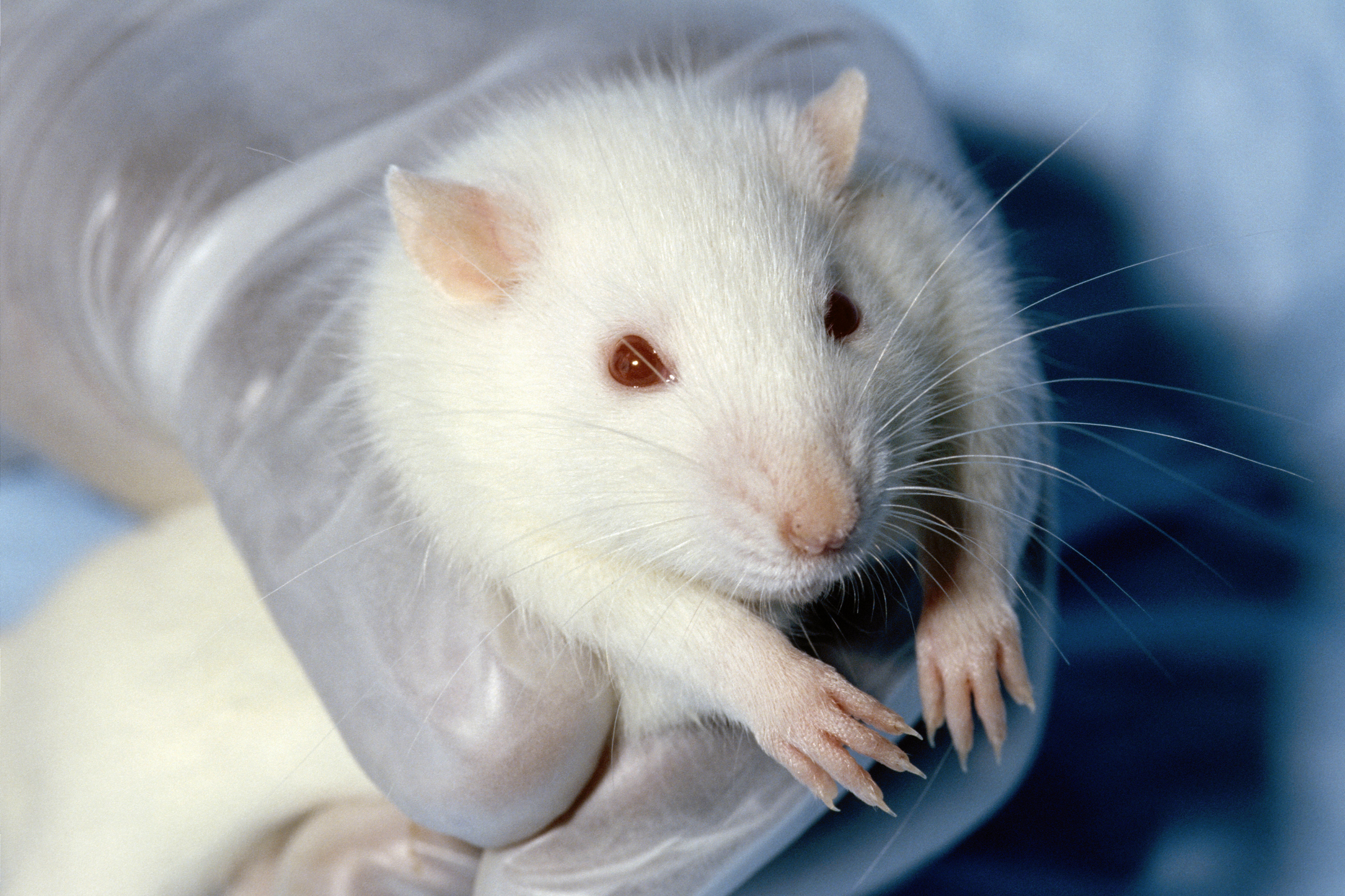
''In vitro'' muscle testing is a method used to characterize properties of living muscle tissue after removing it from an organism, which allows more extensive and precise quantification of its properties than ''in vivo'' testing. ''In vitro'' muscle testing has provided the bulk of scientific knowledge of muscle structure and physiology, and how both relate to organismal performance. Isolation of tissue Once an appropriate animal has been selected—whether for a specific locomotor function (i.e. frogs for jumping); or a specific animal strain, to answer a research question—a specific muscle is identified based on its ''in vivo'' function and fibre type distribution. Following ethical approval, and if necessary, government approval, the animal is humanely euthanised. Humane methods differ by country, with the most appropriate based on ethical approval and researcher skill level. A number of further criteria should be followed to ensure the animal is completely dead without the ... [...More Info...] [...Related Items...] OR: [Wikipedia] [Google] [Baidu] |
Muscle
Skeletal muscles (commonly referred to as muscles) are Organ (biology), organs of the vertebrate muscular system and typically are attached by tendons to bones of a skeleton. The muscle cells of skeletal muscles are much longer than in the other types of muscle tissue, and are often known as Skeletal muscle#Skeletal muscle cells, muscle fibers. The muscle tissue of a skeletal muscle is striated muscle tissue, striated – having a striped appearance due to the arrangement of the sarcomeres. Skeletal muscles are voluntary muscles under the control of the somatic nervous system. The other types of muscle are cardiac muscle which is also striated and smooth muscle which is non-striated; both of these types of muscle tissue are classified as involuntary, or, under the control of the autonomic nervous system. A skeletal muscle contains multiple muscle fascicle, fascicles – bundles of muscle fibers. Each individual fiber, and each muscle is surrounded by a type of connective tissue ... [...More Info...] [...Related Items...] OR: [Wikipedia] [Google] [Baidu] |
Physiology
Physiology (; ) is the scientific study of functions and mechanisms in a living system. As a sub-discipline of biology, physiology focuses on how organisms, organ systems, individual organs, cells, and biomolecules carry out the chemical and physical functions in a living system. According to the classes of organisms, the field can be divided into medical physiology, animal physiology, plant physiology, cell physiology, and comparative physiology. Central to physiological functioning are biophysical and biochemical processes, homeostatic control mechanisms, and communication between cells. ''Physiological state'' is the condition of normal function. In contrast, '' pathological state'' refers to abnormal conditions, including human diseases. The Nobel Prize in Physiology or Medicine is awarded by the Royal Swedish Academy of Sciences for exceptional scientific achievements in physiology related to the field of medicine. Foundations Cells Although there are ... [...More Info...] [...Related Items...] OR: [Wikipedia] [Google] [Baidu] |
Animal Testing
Animal testing, also known as animal experimentation, animal research, and ''in vivo'' testing, is the use of non-human animals in experiments that seek to control the variables that affect the behavior or biological system under study. This approach can be contrasted with field studies in which animals are observed in their natural environments or habitats. Experimental research with animals is usually conducted in universities, medical schools, pharmaceutical companies, defense establishments, and commercial facilities that provide animal-testing services to the industry. The focus of animal testing varies on a continuum from pure research, focusing on developing fundamental knowledge of an organism, to applied research, which may focus on answering some questions of great practical importance, such as finding a cure for a disease. Examples of applied research include testing disease treatments, breeding, defense research, and toxicology, including cosmetics testing. In ed ... [...More Info...] [...Related Items...] OR: [Wikipedia] [Google] [Baidu] |
Cardiac Muscle Cell
Cardiac muscle (also called heart muscle, myocardium, cardiomyocytes and cardiac myocytes) is one of three types of vertebrate muscle tissues, with the other two being skeletal muscle and smooth muscle. It is an involuntary, striated muscle that constitutes the main tissue of the wall of the heart. The cardiac muscle (myocardium) forms a thick middle layer between the outer layer of the heart wall (the pericardium) and the inner layer (the endocardium), with blood supplied via the coronary circulation. It is composed of individual cardiac muscle cells joined by intercalated discs, and encased by collagen fibers and other substances that form the extracellular matrix. Cardiac muscle contracts in a similar manner to skeletal muscle, although with some important differences. Electrical stimulation in the form of a cardiac action potential triggers the release of calcium from the cell's internal calcium store, the sarcoplasmic reticulum. The rise in calcium causes the ... [...More Info...] [...Related Items...] OR: [Wikipedia] [Google] [Baidu] |
Sarcomere
A sarcomere (Greek σάρξ ''sarx'' "flesh", μέρος ''meros'' "part") is the smallest functional unit of striated muscle tissue. It is the repeating unit between two Z-lines. Skeletal muscles are composed of tubular muscle cells (called muscle fibers or myofibers) which are formed during embryonic myogenesis. Muscle fibers contain numerous tubular myofibrils. Myofibrils are composed of repeating sections of sarcomeres, which appear under the microscope as alternating dark and light bands. Sarcomeres are composed of long, fibrous proteins as filaments that slide past each other when a muscle contracts or relaxes. The costamere is a different component that connects the sarcomere to the sarcolemma. Two of the important proteins are myosin, which forms the thick filament, and actin, which forms the thin filament. Myosin has a long, fibrous tail and a globular head, which binds to actin. The myosin head also binds to ATP, which is the source of energy for muscle movement. Myo ... [...More Info...] [...Related Items...] OR: [Wikipedia] [Google] [Baidu] |
Muscle Fiber
A muscle cell is also known as a myocyte when referring to either a cardiac muscle cell (cardiomyocyte), or a smooth muscle cell as these are both small cells. A skeletal muscle cell is long and threadlike with many nuclei and is called a muscle fiber. Muscle cells (including myocytes and muscle fibers) develop from embryonic precursor cells called myoblasts. Myoblasts fuse to form multinucleated skeletal muscle cells known as syncytia in a process known as myogenesis. Skeletal muscle cells and cardiac muscle cells both contain myofibrils and sarcomeres and form a striated muscle tissue. Cardiac muscle cells form the cardiac muscle in the walls of the heart chambers, and have a single central nucleus. Cardiac muscle cells are joined to neighboring cells by intercalated discs, and when joined in a visible unit they are described as a ''cardiac muscle fiber''. Smooth muscle cells control involuntary movements such as the peristalsis contractions in the esophagus and stom ... [...More Info...] [...Related Items...] OR: [Wikipedia] [Google] [Baidu] |
Sonomicrometry
Sonomicrometry is a technique of measuring the distance between piezoelectric crystals based on the speed of acoustic signals through the medium they are embedded in. Typically, the crystals will be coated with an epoxy 'lens' and placed into the material facing each other. An electrical signal sent to either crystal will be transformed into sound, which passes through the medium, eventually reaching the other crystal, which converts the sound into electricity, detected by a receiver. From the time taken for sound to move between the crystals and the speed of sound in the medium, the distance between the crystals can be calculated. History Sonomicrometry was originally applied in the study of cardiac function in research animals by Dean Franklin in 1956, and was quickly adopted by biologists working in biomechanics as well as other physiological organ systems and structures (gastro-intestinal, uro-genital and musculo-skeletal). Medical device companies also use sonomicrometry to ... [...More Info...] [...Related Items...] OR: [Wikipedia] [Google] [Baidu] |
Pennate Muscles
A pennate or pinnate muscle (also called a penniform muscle) is a type of skeletal muscle with fascicles that attach obliquely (in a slanting position) to its tendon. This type of muscle generally allows higher force production but a smaller range of motion When a muscle contracts and shortens, the pennation angle increases. Etymology From the Latin ''pinnātus'' “feathered, winged,” from ''pinna'' “feather, wing.” Types of pennate muscle In skeletal muscle tissue, 10-100 endomysium-sheathed muscle fibers are organized into perimysium-wrapped bundles known as fascicles. Each muscle is composed of a number of fascicles grouped together by a sleeve of connective tissue, known as an epimysium. In a pennate muscle, aponeuroses run along each side of the muscle and attach to the tendon. The fascicles attach to the aponeuroses and form an angle (the pennation angle) to the load axis of the muscle. If all the fascicles are on the same side of the tendon, the pennate muscle ... [...More Info...] [...Related Items...] OR: [Wikipedia] [Google] [Baidu] |
Motor Unit
A motor unit is made up of a motor neuron and all of the skeletal muscle fibers innervated by the neuron's axon terminals, including the neuromuscular junctions between the neuron and the fibres. Groups of motor units often work together as a motor pool to coordinate the contractions of a single muscle. The concept was proposed by Charles Scott Sherrington. All muscle fibers in a motor unit are of the same fiber type. When a motor unit is activated, all of its fibers contract. In vertebrates, the force of a muscle contraction is controlled by the number of activated motor units. The number of muscle fibers within each unit can vary within a particular muscle and even more from muscle to muscle; the muscles that act on the largest body masses have motor units that contain more muscle fibers, whereas smaller muscles contain fewer muscle fibers in each motor unit. For instance, thigh muscles can have a thousand fibers in each unit, while extraocular muscles might have ten. Muscles ... [...More Info...] [...Related Items...] OR: [Wikipedia] [Google] [Baidu] |
In Vivo
Studies that are ''in vivo'' (Latin for "within the living"; often not italicized in English) are those in which the effects of various biological entities are tested on whole, living organisms or cells, usually animals, including humans, and plants, as opposed to a tissue extract or dead organism. This is not to be confused with experiments done ''in vitro'' ("within the glass"), i.e., in a laboratory environment using test tubes, Petri dishes, etc. Examples of investigations ''in vivo'' include: the pathogenesis of disease by comparing the effects of bacterial infection with the effects of purified bacterial toxins; the development of non-antibiotics, antiviral drugs, and new drugs generally; and new surgical procedures. Consequently, animal testing and clinical trials are major elements of ''in vivo'' research. ''In vivo'' testing is often employed over ''in vitro'' because it is better suited for observing the overall effects of an experiment on a living subject. In dr ... [...More Info...] [...Related Items...] OR: [Wikipedia] [Google] [Baidu] |
Muscle Contraction
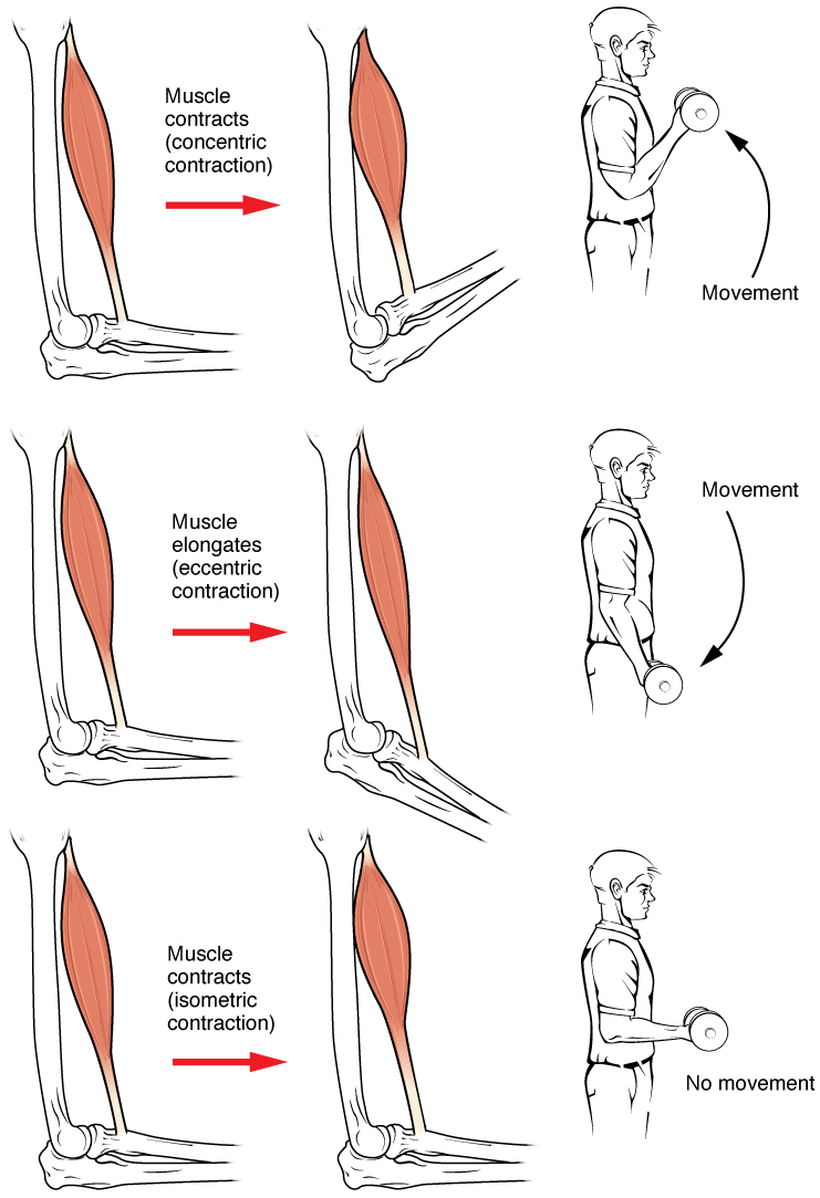
Muscle contraction is the activation of tension-generating sites within muscle cells. In physiology, muscle contraction does not necessarily mean muscle shortening because muscle tension can be produced without changes in muscle length, such as when holding something heavy in the same position. The termination of muscle contraction is followed by muscle relaxation, which is a return of the muscle fibers to their low tension-generating state. For the contractions to happen, the muscle cells must rely on the interaction of two types of filaments which are the thin and thick filaments. Thin filaments are two strands of actin coiled around each, and thick filaments consist of mostly elongated proteins called myosin. Together, these two filaments form myofibrils which are important organelles in the skeletal muscle system. Muscle contraction can also be described based on two variables: length and tension. A muscle contraction is described as isometric if the muscle tension changes ... [...More Info...] [...Related Items...] OR: [Wikipedia] [Google] [Baidu] |


