|
ILO Classification
The ILO International Classification of Radiographs of Pneumoconioses is a system of classifying chest radiographs (X-rays) for persons with a (or, rarely, more than one) form of pneumoconiosis. The intent is to provide a standardized, uniform method of interpreting and describing abnormalities in chest x-rays that are thought to be caused by prolonged dust inhalation. In use, it provides a system for both epidemiological comparisons of many individuals exposed to dust and evaluation of an individual's potential disease relative to established standards. History Since 1946, the International Labour Organization has been a specialized agency of the United Nations, with objectives including establishing and overseeing international labor standards and labor rights. The International Labour Office ("ILO") is the Organization’s research body and publishing house. Since 1950, the ILO has periodically published guidelines on how to classify chest X-rays for pneumoconiosis. The purpose of ... [...More Info...] [...Related Items...] OR: [Wikipedia] [Google] [Baidu] |
Chest X-rays
A chest radiograph, called a chest X-ray (CXR), or chest film, is a Projectional radiography, projection radiograph of the chest used to diagnose conditions affecting the chest, its contents, and nearby structures. Chest radiographs are the most common film taken in medicine. Like all methods of radiography, chest radiography employs ionizing radiation in the form of X-rays to generate images of the chest. The mean radiation dose to an adult from a chest radiograph is around 0.02 Sievert, mSv (2 Roentgen equivalent man, mrem) for a front view (PA, or posteroanterior) and 0.08 mSv (8 mrem) for a side view (LL, or latero-lateral). Together, this corresponds to a background radiation equivalent time of about 10 days. Medical uses Conditions commonly identified by chest radiography * Pneumonia * Pneumothorax * Interstitial lung disease * Heart failure * Fracture (bone), Bone fracture * Hiatal hernia Chest radiographs are used to diagnose many conditions involving the chest wall, i ... [...More Info...] [...Related Items...] OR: [Wikipedia] [Google] [Baidu] |
Emphysema
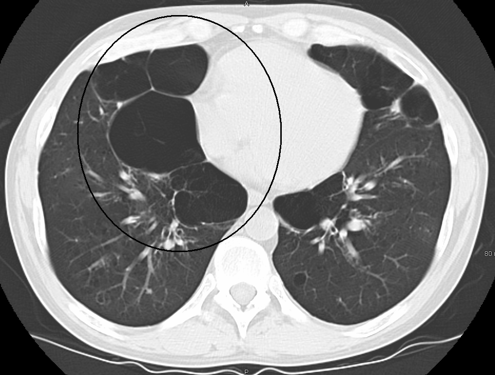
Emphysema, or pulmonary emphysema, is a lower respiratory tract disease, characterised by air-filled spaces ( pneumatoses) in the lungs, that can vary in size and may be very large. The spaces are caused by the breakdown of the walls of the alveoli and they replace the spongy lung parenchyma. This reduces the total alveolar surface available for gas exchange leading to a reduction in oxygen supply for the blood. Emphysema usually affects the middle aged or older population because it takes time to develop with the effects of tobacco smoking, and other risk factors. Alpha-1 antitrypsin deficiency is a genetic risk factor that may lead to the condition presenting earlier. When associated with significant airflow limitation, emphysema is a major subtype of chronic obstructive pulmonary disease (COPD), a progressive lung disease characterized by long-term breathing problems and poor airflow. Without COPD, the finding of emphysema on a CT lung scan still confers a higher mortality r ... [...More Info...] [...Related Items...] OR: [Wikipedia] [Google] [Baidu] |
Tuberculosis
Tuberculosis (TB) is an infectious disease usually caused by '' Mycobacterium tuberculosis'' (MTB) bacteria. Tuberculosis generally affects the lungs, but it can also affect other parts of the body. Most infections show no symptoms, in which case it is known as latent tuberculosis. Around 10% of latent infections progress to active disease which, if left untreated, kill about half of those affected. Typical symptoms of active TB are chronic cough with blood-containing mucus, fever, night sweats, and weight loss. It was historically referred to as consumption due to the weight loss associated with the disease. Infection of other organs can cause a wide range of symptoms. Tuberculosis is spread from one person to the next through the air when people who have active TB in their lungs cough, spit, speak, or sneeze. People with Latent TB do not spread the disease. Active infection occurs more often in people with HIV/AIDS and in those who smoke. Diagnosis of active TB is ... [...More Info...] [...Related Items...] OR: [Wikipedia] [Google] [Baidu] |
Pneumothorax
A pneumothorax is an abnormal collection of air in the pleural space between the lung and the chest wall. Symptoms typically include sudden onset of sharp, one-sided chest pain and shortness of breath. In a minority of cases, a one-way valve is formed by an area of damaged tissue, and the amount of air in the space between chest wall and lungs increases; this is called a tension pneumothorax. This can cause a steadily worsening oxygen shortage and low blood pressure. This leads to a type of shock called obstructive shock, which can be fatal unless reversed. Very rarely, both lungs may be affected by a pneumothorax. It is often called a "collapsed lung", although that term may also refer to atelectasis. A primary spontaneous pneumothorax is one that occurs without an apparent cause and in the absence of significant lung disease. A secondary spontaneous pneumothorax occurs in the presence of existing lung disease. Smoking increases the risk of primary spontaneous pneumothora ... [...More Info...] [...Related Items...] OR: [Wikipedia] [Google] [Baidu] |
Atelectasis
Atelectasis is the collapse or closure of a lung resulting in reduced or absent gas exchange. It is usually unilateral, affecting part or all of one lung. It is a condition where the alveoli are deflated down to little or no volume, as distinct from pulmonary consolidation, in which they are filled with liquid. It is often called a ''collapsed lung'', although that term may also refer to pneumothorax. It is a very common finding in chest X-rays and other radiological studies, and may be caused by normal exhalation or by various medical conditions. Although frequently described as a ''collapse of lung tissue'', atelectasis is not synonymous with a pneumothorax, which is a more specific condition that can cause atelectasis. Acute atelectasis may occur as a post-operative complication or as a result of surfactant deficiency. In premature babies, this leads to infant respiratory distress syndrome. The term uses combining forms of ''atel-'' + ''ectasis'', from el, ἀτελής, ... [...More Info...] [...Related Items...] OR: [Wikipedia] [Google] [Baidu] |
Pleural
The pleural cavity, pleural space, or interpleural space is the potential space between the pleurae of the pleural sac that surrounds each lung. A small amount of serous pleural fluid is maintained in the pleural cavity to enable lubrication between the membranes, and also to create a pressure gradient. The serous membrane that covers the surface of the lung is the visceral pleura and is separated from the outer membrane the parietal pleura by just the film of pleural fluid in the pleural cavity. The visceral pleura follows the fissures of the lung and the root of the lung structures. The parietal pleura is attached to the mediastinum, the upper surface of the diaphragm, and to the inside of the ribcage. Structure In humans, the left and right lungs are completely separated by the mediastinum, and there is no communication between their pleural cavities. Therefore, in cases of a unilateral pneumothorax, the contralateral lung will remain functioning normally unless there is a ... [...More Info...] [...Related Items...] OR: [Wikipedia] [Google] [Baidu] |
Kerley Lines
Kerley lines are a sign seen on chest radiographs with interstitial pulmonary edema. They are thin linear pulmonary opacities caused by fluid or cellular infiltration into the interstitium of the lungs. They are named after Irish neurologist and radiologist Peter Kerley. Associated conditions They are suggestive for the diagnosis of congestive heart failure, but are also seen in various non-cardiac conditions such as pulmonary fibrosis, interstitial deposition of heavy metal particles or carcinomatosis of the lung. Chronic Kerley B lines may be caused by fibrosis or hemosiderin deposition caused by recurrent pulmonary edema. Types Kerley A lines These are longer (at least 2cm and up to 6cm) unbranching lines coursing diagonally from the hila out to the periphery of the lungs. They are caused by distension of anastomotic channels between peripheral and central lymphatics of the lungs. Kerley A lines are less commonly seen than Kerley B lines. Kerley A lines are never seen with ... [...More Info...] [...Related Items...] OR: [Wikipedia] [Google] [Baidu] |
Rib Fracture
A rib fracture is a break in a rib bone. This typically results in chest pain that is worse with inspiration. Bruising may occur at the site of the break. When several ribs are broken in several places a flail chest results. Potential complications include a pneumothorax, pulmonary contusion, and pneumonia. Rib fractures usually occur from a direct blow to the chest such as during a motor vehicle collision or from a crush injury. Coughing or metastatic cancer may also result in a broken rib. The middle ribs are most commonly fractured. Fractures of the first or second ribs are more likely to be associated with complications. Diagnosis can be made based on symptoms and supported by medical imaging. Pain control is an important part of treatment. This may include the use of paracetamol (acetaminophen), NSAIDs, or opioids. A nerve block may be another option. While fractured ribs can be wrapped, this may increase complications. In those with a flail chest, surgery may improve ou ... [...More Info...] [...Related Items...] OR: [Wikipedia] [Google] [Baidu] |
Emphysema
Emphysema, or pulmonary emphysema, is a lower respiratory tract disease, characterised by air-filled spaces ( pneumatoses) in the lungs, that can vary in size and may be very large. The spaces are caused by the breakdown of the walls of the alveoli and they replace the spongy lung parenchyma. This reduces the total alveolar surface available for gas exchange leading to a reduction in oxygen supply for the blood. Emphysema usually affects the middle aged or older population because it takes time to develop with the effects of tobacco smoking, and other risk factors. Alpha-1 antitrypsin deficiency is a genetic risk factor that may lead to the condition presenting earlier. When associated with significant airflow limitation, emphysema is a major subtype of chronic obstructive pulmonary disease (COPD), a progressive lung disease characterized by long-term breathing problems and poor airflow. Without COPD, the finding of emphysema on a CT lung scan still confers a higher mortality r ... [...More Info...] [...Related Items...] OR: [Wikipedia] [Google] [Baidu] |
Pleural Effusion
A pleural effusion is accumulation of excessive fluid in the pleural space, the potential space that surrounds each lung. Under normal conditions, pleural fluid is secreted by the parietal pleural capillaries at a rate of 0.6 millilitre per kilogram weight per hour, and is cleared by lymphatic absorption leaving behind only 5–15 millilitres of fluid, which helps to maintain a functional vacuum between the parietal and visceral pleurae. Excess fluid within the pleural space can impair inspiration by upsetting the functional vacuum and hydrostatically increasing the resistance against lung expansion, resulting in a fully or partially collapsed lung. Various kinds of fluid can accumulate in the pleural space, such as serous fluid (hydrothorax), blood (hemothorax), pus (pyothorax, more commonly known as pleural empyema), chyle ( chylothorax), or very rarely urine (urinothorax). When unspecified, the term "pleural effusion" normally refers to hydrothorax. A pleural effusion can a ... [...More Info...] [...Related Items...] OR: [Wikipedia] [Google] [Baidu] |
Cor Pulmonale
Pulmonary heart disease, also known as cor pulmonale, is the enlargement and failure of the right ventricle of the heart as a response to increased vascular resistance (such as from pulmonic stenosis) or high blood pressure in the lungs. Chronic pulmonary heart disease usually results in right ventricular hypertrophy (RVH), whereas acute pulmonary heart disease usually results in dilatation. Hypertrophy is an adaptive response to a long-term increase in pressure. Individual muscle cells grow larger (in thickness) and change to drive the increased contractile force required to move the blood against greater resistance. Dilatation is a stretching (in length) of the ventricle in response to acute increased pressure. To be classified as pulmonary heart disease, the cause must originate in the pulmonary circulation system; RVH due to a systemic defect is not classified as pulmonary heart disease. Two causes are vascular changes as a result of tissue damage (e.g. disease, hypoxic in ... [...More Info...] [...Related Items...] OR: [Wikipedia] [Google] [Baidu] |
Granuloma
A granuloma is an aggregation of macrophages that forms in response to chronic inflammation. This occurs when the immune system attempts to isolate foreign substances that it is otherwise unable to eliminate. Such substances include infectious organisms including bacteria and fungi, as well as other materials such as foreign objects, keratin, and suture fragments. Definition In pathology, a granuloma is an organized collection of macrophages. In medical practice, doctors occasionally use the term ''granuloma'' in its more literal meaning: "a small nodule". Since a small nodule can represent any tissue from a harmless nevus to a malignant tumor, this use of the term is not very specific. Examples of this use of the term ''granuloma'' are the lesions known as vocal cord granuloma (known as contact granuloma), pyogenic granuloma, and intubation granuloma, all of which are examples of granulation tissue, not granulomas. "Pulmonary hyalinizing granuloma" is a lesion characterized ... [...More Info...] [...Related Items...] OR: [Wikipedia] [Google] [Baidu] |


