|
Intravoxel Incoherent Motion
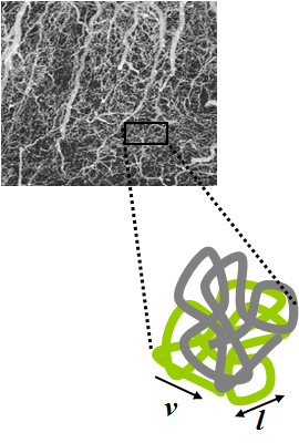
Intravoxel incoherent motion (IVIM) imaging is a concept and a method initially introduced and developed by Le Bihan et al. to quantitatively assess all the microscopic translational motions that could contribute to the signal acquired with diffusion MRI. In this model, biological tissue contains two distinct environments: molecular diffusion of water in the tissue (sometimes referred to as 'true diffusion'), and microcirculation of blood in the capillary network (perfusion). The concept introduced by D. Le Bihan is that water flowing in capillaries (at the voxel level) mimics a random walk (“pseudo-diffusion” ) (Fig.1), as long as the assumption that all directions are represented in the capillaries (i.e. there is no net coherent flow in any direction) is satisfied. It is responsible for a signal attenuation in diffusion MRI, which depends on the velocity of the flowing blood and the vascular architecture. Similarly to molecular diffusion, the effect of pseudodiffusion on the ... [...More Info...] [...Related Items...] OR: [Wikipedia] [Google] [Baidu] |
Diffusion MRI
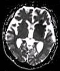
Diffusion-weighted magnetic resonance imaging (DWI or DW-MRI) is the use of specific MRI sequences as well as software that generates images from the resulting data that uses the diffusion of water molecules to generate contrast in MR images. It allows the mapping of the diffusion process of molecules, mainly water, in biological tissues, in vivo and non-invasively. Molecular diffusion in tissues is not random, but reflects interactions with many obstacles, such as macromolecules, fibers, and membranes. Water molecule diffusion patterns can therefore reveal microscopic details about tissue architecture, either normal or in a diseased state. A special kind of DWI, diffusion tensor imaging (DTI), has been used extensively to map white matter tractography in the brain. Introduction In diffusion weighted imaging (DWI), the intensity of each image element (voxel) reflects the best estimate of the rate of water diffusion at that location. Because the mobility of water is driven by th ... [...More Info...] [...Related Items...] OR: [Wikipedia] [Google] [Baidu] |
Diffusion-weighted MRI
Diffusion-weighted magnetic resonance imaging (DWI or DW-MRI) is the use of specific MRI sequences as well as software that generates images from the resulting data that uses the diffusion of water molecules to generate contrast in MR images. It allows the mapping of the diffusion process of molecules, mainly water, in biological tissues, in vivo and non-invasively. Molecular diffusion in tissues is not random, but reflects interactions with many obstacles, such as macromolecules, fibers, and membranes. Water molecule diffusion patterns can therefore reveal microscopic details about tissue architecture, either normal or in a diseased state. A special kind of DWI, diffusion tensor imaging (DTI), has been used extensively to map white matter tractography in the brain. Introduction In diffusion weighted imaging (DWI), the intensity of each image element (voxel) reflects the best estimate of the rate of water diffusion at that location. Because the mobility of water is driven by the ... [...More Info...] [...Related Items...] OR: [Wikipedia] [Google] [Baidu] |
IVIM2
Intravoxel incoherent motion (IVIM) imaging is a concept and a method initially introduced and developed by Le Bihan et al. to quantitatively assess all the microscopic translational motions that could contribute to the signal acquired with diffusion MRI. In this model, biological tissue contains two distinct environments: molecular diffusion of water in the tissue (sometimes referred to as 'true diffusion'), and microcirculation of blood in the capillary network (perfusion). The concept introduced by D. Le Bihan is that water flowing in capillaries (at the voxel level) mimics a random walk (“pseudo-diffusion” ) (Fig.1), as long as the assumption that all directions are represented in the capillaries (i.e. there is no net coherent flow in any direction) is satisfied. It is responsible for a signal attenuation in diffusion MRI, which depends on the velocity of the flowing blood and the vascular architecture. Similarly to molecular diffusion, the effect of pseudodiffusion on the ... [...More Info...] [...Related Items...] OR: [Wikipedia] [Google] [Baidu] |
NMR In Biomedicine
''NMR in Biomedicine'' is a monthly peer-reviewed medical journal published since 1988 by John Wiley & Sons. It publishes original full-length papers, rapid communications, and review articles in which magnetic resonance spectroscopy or imaging methods are used to investigate physiological, biochemical, biophysical, or medical problems. The current editor-in-chief is John R. Griffiths (Cancer Research UK). Highest cited articles The following articles have been cited most frequently: # "The basis of anisotropic water diffusion in the nervous system - a technical review", 15 (7-8) Nov-Dec 2002: 435–455, Beaulieu C. # "A review of chemical issues in H-1-NMR spectroscopy - n-acetyl-l-aspartate, creatine and choline", 4 (2) Apr 1991: 47–52, Miller BL. # "Fiber tracking: principles and strategies - a technical review", 15 (7-8) Nov-Dec 2002: 468–480, DMori S, van Zijl PCM. # "Inferring microstructural features and the physiological state of tissues from diffusion-weighted images ... [...More Info...] [...Related Items...] OR: [Wikipedia] [Google] [Baidu] |
FMRI
Functional magnetic resonance imaging or functional MRI (fMRI) measures brain activity by detecting changes associated with blood flow. This technique relies on the fact that cerebral blood flow and neuronal activation are coupled. When an area of the brain is in use, blood flow to that region also increases. The primary form of fMRI uses the blood-oxygen-level dependent (BOLD) contrast, discovered by Seiji Ogawa in 1990. This is a type of specialized brain and body scan used to map neural activity in the brain or spinal cord of humans or other animals by imaging the change in blood flow (hemodynamic response) related to energy use by brain cells. Since the early 1990s, fMRI has come to dominate brain mapping research because it does not involve the use of injections, surgery, the ingestion of substances, or exposure to ionizing radiation. This measure is frequently corrupted by noise from various sources; hence, statistical procedures are used to extract the underlying signal. T ... [...More Info...] [...Related Items...] OR: [Wikipedia] [Google] [Baidu] |
Arterial Spin Labeling
Arterial spin labeling (ASL), also known as arterial spin tagging, is a magnetic resonance imaging technique used to quantify cerebral blood perfusion by labelling blood water as it flows throughout the brain. ASL specifically refers to magnetic labeling of arterial blood below or in the imaging slab, without the need of gadolinium contrast. A number of ASL schemes are possible, the simplest being flow alternating inversion recovery (FAIR) which requires two acquisitions of identical parameters with the exception of the out-of-slice saturation; the difference in the two images is theoretically only from inflowing spins, and may be considered a 'perfusion map'. The ASL technique was developed by Alan P. Koretsky, Donald S. Williams, John A. Detre and John S. Leigh, Jr in 1992. Physics Arterial spin labeling utilizes the water molecules circulating with the brain, and using a radiofrequency pulse, tracks the blood water as it circulates throughout the brain. After a period of time in m ... [...More Info...] [...Related Items...] OR: [Wikipedia] [Google] [Baidu] |
Echo Time
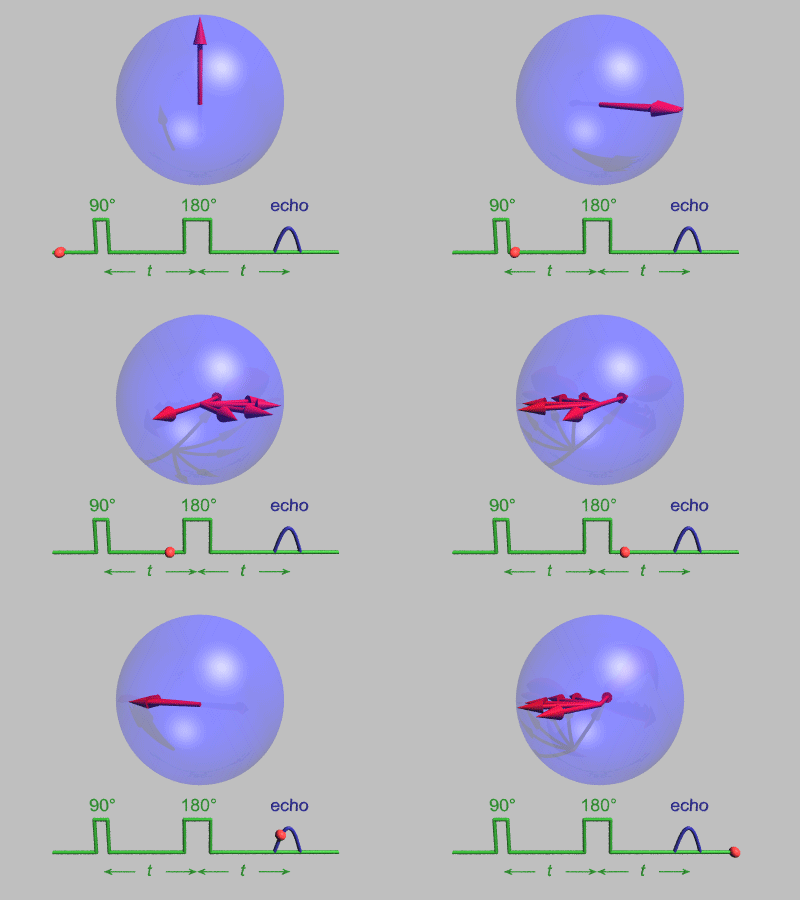
In magnetic resonance, a spin echo or Hahn echo is the refocusing of spin magnetisation by a pulse of resonant electromagnetic radiation. Modern nuclear magnetic resonance (NMR) and magnetic resonance imaging (MRI) make use of this effect. The NMR signal observed following an initial excitation pulse decays with time due to both spin relaxation and any ''inhomogeneous'' effects which cause spins in the sample to precess at different rates. The first of these, relaxation, leads to an irreversible loss of magnetisation. But the inhomogeneous dephasing can be removed by applying a 180° ''inversion'' pulse that inverts the magnetisation vectors. Examples of inhomogeneous effects include a magnetic field gradient and a distribution of chemical shifts. If the inversion pulse is applied after a period ''t'' of dephasing, the inhomogeneous evolution will rephase to form an echo at time 2''t''. In simple cases, the intensity of the echo relative to the initial signal is given by ''e� ... [...More Info...] [...Related Items...] OR: [Wikipedia] [Google] [Baidu] |
Nephrogenic Systemic Fibrosis
Nephrogenic systemic fibrosis is a rare syndrome that involves fibrosis of skin, joints, eyes, and internal organs. NSF is caused by exposure to gadolinium in gadolinium-based MRI contrast agents (GBCAs) in patients with impaired kidney function. Epidemiological studies suggest that the incidence of NSF is unrelated to gender or ethnicity and it is not thought to have a genetic basis. After GBCAs were identified as a cause of the disorder in 2006, and screening and prevention measures put in place, it is now considered rare. Signs and symptoms Clinical features of NSF develop within days to months and, in some cases, years following exposure to some GBCAs. The main symptoms are the thickening and hardening of the skin associated with brawny hyperpigmentation, typically presenting in a symmetric fashion. The skin gradually becomes fibrotic and adheres to the underlying fascia. The symptoms initiate distally in the limbs and progress proximally, sometimes involving the trunk. Join ... [...More Info...] [...Related Items...] OR: [Wikipedia] [Google] [Baidu] |


