|
Interstitial Cells
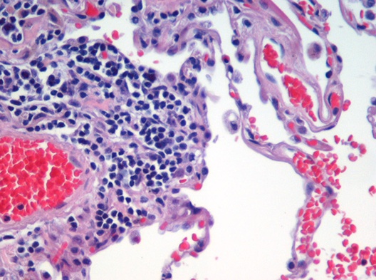
Interstitial cell refers to any cell that lies in the spaces between the functional cells of a tissue. Examples include: * Interstitial cell of Cajal (ICC) * Leydig cells, cells present in the male testes responsible for the production of androgen (male sex hormone) * A portion of the stroma of ovary * Certain cells in the pineal gland * Renal interstitial cells * neuroglial cells See also *List of human cell types derived from the germ layers This is a list of cells in humans derived from the three embryonic germ layers – ectoderm, mesoderm, and endoderm. Cells derived from ectoderm Surface ectoderm Skin * Trichocyte * Keratinocyte Anterior pituitary * Gonadotrope * Corticotro ... References *Sybil B Parker (ed). "Interstitial cell". McGraw Hill Dictionary of Scientific and Technical Terms. Fifth Edition. International Edition. 1994. Page 1041. {{DEFAULTSORT:Interstitial cells Cell biology Biology-related lists ... [...More Info...] [...Related Items...] OR: [Wikipedia] [Google] [Baidu] |
Cell (biology)
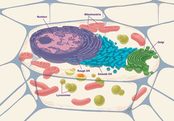
The cell is the basic structural and functional unit of life forms. Every cell consists of a cytoplasm enclosed within a membrane, and contains many biomolecules such as proteins, DNA and RNA, as well as many small molecules of nutrients and metabolites.Cell Movements and the Shaping of the Vertebrate Body in Chapter 21 of Molecular Biology of the Cell '' fourth edition, edited by Bruce Alberts (2002) published by Garland Science. The Alberts text discusses how the "cellular building blocks" move to shape developing embryos. It is also common to describe small molecules such as ... [...More Info...] [...Related Items...] OR: [Wikipedia] [Google] [Baidu] |
Tissue (anatomy)
In biology, tissue is a biological organizational level between cells and a complete organ. A tissue is an ensemble of similar cells and their extracellular matrix from the same origin that together carry out a specific function. Organs are then formed by the functional grouping together of multiple tissues. The English word "tissue" derives from the French word "tissu", the past participle of the verb tisser, "to weave". The study of tissues is known as histology or, in connection with disease, as histopathology. Xavier Bichat is considered as the "Father of Histology". Plant histology is studied in both plant anatomy and physiology. The classical tools for studying tissues are the paraffin block in which tissue is embedded and then sectioned, the histological stain, and the optical microscope. Developments in electron microscopy, immunofluorescence, and the use of frozen tissue-sections have enhanced the detail that can be observed in tissues. With these tools, the c ... [...More Info...] [...Related Items...] OR: [Wikipedia] [Google] [Baidu] |
Interstitial Cell Of Cajal
Interstitial cells of Cajal (ICC) are interstitial cells found in the gastrointestinal tract. There are different types of ICC with different functions. ICC and another type of interstitial cell, known as platelet-derived growth factor receptor alpha (PDGFRα) cells, are electrically coupled to smooth muscle cells via gap junctions, that work together as an SIP functional syncytium. Myenteric interstitial cells of Cajal (ICC-MY) serve as pacemaker cells that generate the bioelectrical events known as slow waves. Slow waves conduct to smooth muscle cells and cause phasic contractions. The picture to the right shows an isolated Interstitial cell of Cajal from the Myenteric plexus of the mouse small intestine grown in a primary cell culture. This cell type can be characterized morphologically as having a small cell body often triangular or stellate-shaped with several long processes branching out into secondary and tertiary extensions - these processes often contact smooth muscl ... [...More Info...] [...Related Items...] OR: [Wikipedia] [Google] [Baidu] |
Leydig Cells
Leydig cells, also known as interstitial cells of the testes and interstitial cells of Leydig, are found adjacent to the seminiferous tubules in the testicle and produce testosterone in the presence of luteinizing hormone (LH). They are polyhedral in shape and have a large, prominent nucleus, an eosinophilic cytoplasm, and numerous lipid-filled vesicles. Structure The mammalian Leydig cell is a polyhedral epithelioid cell with a single eccentrically located ovoid nucleus. The nucleus contains one to three prominent nucleoli and large amounts of dark-staining peripheral heterochromatin. The acidophilic cytoplasm usually contains numerous membrane-bound lipid droplets and large amounts of smooth endoplasmic reticulum (SER). Besides the abundance of SER with scattered patches of rough endoplasmic reticulum, several mitochondria are also prominent within the cytoplasm. Reinke crystals have lipofuscin pigment and rod-shaped crystal-like structures 3 to 20 micrometres in diameter. Adult ... [...More Info...] [...Related Items...] OR: [Wikipedia] [Google] [Baidu] |
Stroma Of Ovary
The stroma of the ovary is a unique type of connective tissue abundantly supplied with blood vessels, consisting for the most part of spindle-shaped stroma cells. These appear similar to fibroblasts. The stroma also contains ordinary connective tissue such as reticular fibers and collagen. Ovarian stroma differs from typical connective tissue in that it contains a high number of cells. The stoma cells are distributed in such a way that the tissue appears to be whorled. Stromal cells associated with maturing follicles may acquire endocrine function and secrete estrogens. The entire ovarian stroma is highly vascular. On the surface of the organ this tissue is much condensed, and forms a layer ( tunica albuginea) composed of short connective-tissue fibers, with fusiform cells between them. The stroma of the ovary may contain interstitial cells resembling those of the testis. See also * stroma (other) * Stromal cell Stromal cells, or mesenchymal stromal cells, are dif ... [...More Info...] [...Related Items...] OR: [Wikipedia] [Google] [Baidu] |
Pineal Gland
The pineal gland, conarium, or epiphysis cerebri, is a small endocrine gland in the brain of most vertebrates. The pineal gland produces melatonin, a serotonin-derived hormone which modulates sleep, sleep patterns in both circadian rhythm, circadian and Season, seasonal cycles. The shape of the gland resembles a pine cone, which gives it its name. The pineal gland is located in the epithalamus, near the center of the brain, between the two cerebral hemisphere, hemispheres, tucked in a groove where the two halves of the thalamus join. The pineal gland is one of the neuroendocrinology, neuroendocrine Circumventricular organs, secretory circumventricular organs in which capillaries are mostly Vascular permeability, permeable to solutes in the blood. Nearly all vertebrate species possess a pineal gland. The most important exception is a primitive vertebrate, the hagfish. Even in the hagfish, however, there may be a "pineal equivalent" structure in the dorsal diencephalon. The lanc ... [...More Info...] [...Related Items...] OR: [Wikipedia] [Google] [Baidu] |
Neuroglial Cells
Glia, also called glial cells (gliocytes) or neuroglia, are non-neuronal cells in the central nervous system (brain and spinal cord) and the peripheral nervous system that do not produce electrical impulses. They maintain homeostasis, form myelin in the peripheral nervous system, and provide support and protection for neurons. In the central nervous system, glial cells include oligodendrocytes, astrocytes, ependymal cells, and microglia, and in the peripheral nervous system they include Schwann cells and satellite cells. Function They have four main functions: *to surround neurons and hold them in place *to supply nutrients and oxygen to neurons *to insulate one neuron from another *to destroy pathogens and remove dead neurons. They also play a role in neurotransmission and synaptic connections, and in physiological processes such as breathing. While glia were thought to outnumber neurons by a ratio of 10:1, recent studies using newer methods and reappraisal of historical quanti ... [...More Info...] [...Related Items...] OR: [Wikipedia] [Google] [Baidu] |
List Of Human Cell Types Derived From The Germ Layers
This is a list of cells in humans derived from the three embryonic germ layers – ectoderm, mesoderm, and endoderm. Cells derived from ectoderm Surface ectoderm Skin * Trichocyte * Keratinocyte Anterior pituitary * Gonadotrope * Corticotrope * Thyrotrope * Somatotrope * Lactotroph Tooth enamel * Ameloblast Neural crest Peripheral nervous system * Neuron * Glia ** Schwann cell ** Satellite glial cell Neuroendocrine system * Chromaffin cell * Glomus cell Skin * Melanocyte ** Nevus cell * Merkel cell Teeth * Odontoblast * Cementoblast Eyes * Corneal keratocyte Neural tube Central nervous system * Neuron * Glia ** Astrocyte ** Ependymocytes ** Muller glia (retina) ** Oligodendrocyte ** Oligodendrocyte progenitor cell ** Pituicyte (posterior pituitary) Pineal gland * Pinealocyte Cells derived from mesoderm Paraxial mesoderm Mesenchymal stem cell =Osteochondroprogenitor cell= * Bone ( Osteoblast → Osteocyte) * Cartilage (Chondroblast → Chondrocyte) =Myofibroblast ... [...More Info...] [...Related Items...] OR: [Wikipedia] [Google] [Baidu] |
Cell Biology
Cell biology (also cellular biology or cytology) is a branch of biology that studies the structure, function, and behavior of cells. All living organisms are made of cells. A cell is the basic unit of life that is responsible for the living and functioning of organisms. Cell biology is the study of structural and functional units of cells. Cell biology encompasses both prokaryotic and eukaryotic cells and has many subtopics which may include the study of cell metabolism, cell communication, cell cycle, biochemistry, and cell composition. The study of cells is performed using several microscopy techniques, cell culture, and cell fractionation. These have allowed for and are currently being used for discoveries and research pertaining to how cells function, ultimately giving insight into understanding larger organisms. Knowing the components of cells and how cells work is fundamental to all biological sciences while also being essential for research in biomedical fields such as ... [...More Info...] [...Related Items...] OR: [Wikipedia] [Google] [Baidu] |


