|
Interphalangeal Joints Of Hand
The interphalangeal joints of the hand are the hinge joints between the phalanges of the fingers that provide flexion towards the palm of the hand. There are two sets in each finger (except in the thumb, which has only one joint): * "proximal interphalangeal joints" (PIJ or PIP), those between the first (also called proximal) and second (intermediate) phalanges * "distal interphalangeal joints" (DIJ or DIP), those between the second (intermediate) and third (distal) phalanges Anatomically, the proximal and distal interphalangeal joints are very similar. There are some minor differences in how the palmar plates are attached proximally and in the segmentation of the flexor tendon sheath, but the major differences are the smaller dimension and reduced mobility of the distal joint. Joint structure The PIP joint exhibits great lateral stability. Its transverse diameter is greater than its antero-posterior diameter and its thick collateral ligaments are tight in all positions during ... [...More Info...] [...Related Items...] OR: [Wikipedia] [Google] [Baidu] |
Metacarpophalangeal Joint
The metacarpophalangeal joints (MCP) are situated between the metacarpal bones and the proximal phalanges of the fingers. These joints are of the condyloid kind, formed by the reception of the rounded heads of the metacarpal bones into shallow cavities on the proximal ends of the proximal phalanges. Being condyloid, they allow the movements of flexion, extension, abduction, adduction and circumduction at the joint. Structure Ligaments Each joint has: * palmar ligaments of metacarpophalangeal articulations * collateral ligaments of metacarpophalangeal articulations Dorsal surfaces The dorsal surfaces of these joints are covered by the expansions of the Extensor tendons, together with some loose areolar tissue which connects the deep surfaces of the tendons to the bones. Function The movements which occur in these joints are flexion, extension, adduction, abduction, and circumduction; the movements of abduction and adduction are very limited, and cannot be performed while th ... [...More Info...] [...Related Items...] OR: [Wikipedia] [Google] [Baidu] |
Flexor Digitorum Profundus
The flexor digitorum profundus is a muscle in the forearm of humans that flexes the fingers (also known as digits). It is considered an extrinsic hand muscle because it acts on the hand while its muscle belly is located in the forearm. Together the flexor pollicis longus, pronator quadratus, and flexor digitorum profundus form the deep layer of ventral forearm muscles.Platzer 2004, p 162 The muscle is named . Structure Flexor digitorum profundus originates in the upper 3/4 of the anterior and medial surfaces of the ulna, interosseous membrane and deep fascia of the forearm. The muscle fans out into four tendons (one to each of the second to fifth fingers) to the palmar base of the distal phalanx. Along with the flexor digitorum superficialis, it has long tendons that run down the arm and through the carpal tunnel and attach to the palmar side of the phalanges of the fingers. Flexor digitorum profundus lies deep to the superficialis, but it attaches more distally. Therefore, ... [...More Info...] [...Related Items...] OR: [Wikipedia] [Google] [Baidu] |
Metacarpophalangeal Joints
The metacarpophalangeal joints (MCP) are situated between the metacarpal bones and the proximal phalanges of the fingers. These joints are of the condyloid kind, formed by the reception of the rounded heads of the metacarpal bones into shallow cavities on the proximal ends of the proximal phalanges. Being condyloid, they allow the movements of flexion, extension, abduction, adduction and circumduction at the joint. Structure Ligaments Each joint has: * palmar ligaments of metacarpophalangeal articulations * collateral ligaments of metacarpophalangeal articulations Dorsal surfaces The dorsal surfaces of these joints are covered by the expansions of the Extensor tendons, together with some loose areolar tissue which connects the deep surfaces of the tendons to the bones. Function The movements which occur in these joints are flexion, extension, adduction, abduction, and circumduction; the movements of abduction and adduction are very limited, and cannot be performed while the ... [...More Info...] [...Related Items...] OR: [Wikipedia] [Google] [Baidu] |
Interphalangeal Joints Of Foot
The interphalangeal joints of the foot are between the phalanx bones of the toes in the foot, feet. Since the Toe#Hallux, great toe only has two phalanx bones (proximal and distal phalanges), it only has one interphalangeal joint, which is often abbreviated as the "IP joint". The rest of the toes each have three phalanx bones (proximal, middle, and distal phalanges), so they have two interphalangeal joints: the proximal interphalangeal joint between the proximal and middle phalanges (abbreviated "PIP joint") and the distal interphalangeal joint between the middle and distal phalanges (abbreviated "DIP joint"). All interphalangeal joints are ginglymoid (hinge) joints, and each has a plantar (underside) and two Collateral ligaments of interphalangeal articulations of foot, collateral ligaments. In the arrangement of these ligaments, Extension (kinesiology), extensor tendons supply the places of Anatomical terms of location#Dorsal and ventral, dorsal ligaments, which is similar to t ... [...More Info...] [...Related Items...] OR: [Wikipedia] [Google] [Baidu] |
UpToDate
UpToDate, Inc. is a company in the Wolters Kluwer Health division of Wolters Kluwer whose main product is UpToDate, a software system that is a point of care, point-of-care medical resource. The UpToDate system is an Evidence-based medicine, evidence-based clinical resource. It includes a collection of medical and patient information, access to Lexi-comp drug monographs and Drug interaction, drug-to-drug interactions, and a number of medical calculators. UpToDate is written by over 7,100 physician authors, editors, and peer reviewers. It is available both via the Internet and offline on personal computers or mobile devices. It requires a subscription for full access. The company was launched in 1992 by Dr. Burton Rose along with Dr. Joseph Rush out of Rose's home. They started with nephrology and have since added over twenty other specialties, with more in development. Controversies UpToDate's articles are Scholarly peer review#Anonymous, anonymously peer-reviewed and it mandates ... [...More Info...] [...Related Items...] OR: [Wikipedia] [Google] [Baidu] |
Psoriatic Arthritis
Psoriatic arthritis is a long-term inflammatory arthritis that occurs in people affected by the autoimmune disease psoriasis. The classic feature of psoriatic arthritis is swelling of entire fingers and toes with a sausage-like appearance. This often happens in association with changes to the nails such as small depressions in the nail (pitting), thickening of the nails, and detachment of the nail from the nailbed. Skin changes consistent with psoriasis (e.g., red, scaly, and itchy plaques) frequently occur before the onset of psoriatic arthritis but psoriatic arthritis can precede the rash in 15% of affected individuals. It is classified as a type of seronegative spondyloarthropathy. Genetics are thought to be strongly involved in the development of psoriatic arthritis. Obesity and certain forms of psoriasis are thought to increase the risk. Psoriatic arthritis affects up to 30% of people with psoriasis and occurs in both children and adults. Approximately 40–50% ... [...More Info...] [...Related Items...] OR: [Wikipedia] [Google] [Baidu] |
Osteoarthritis
Osteoarthritis (OA) is a type of degenerative joint disease that results from breakdown of joint cartilage and underlying bone which affects 1 in 7 adults in the United States. It is believed to be the fourth leading cause of disability in the world. The most common symptoms are joint pain and stiffness. Usually the symptoms progress slowly over years. Initially they may occur only after exercise but can become constant over time. Other symptoms may include joint swelling, decreased range of motion, and, when the back is affected, weakness or numbness of the arms and legs. The most commonly involved joints are the two near the ends of the fingers and the joint at the base of the thumbs; the knee and hip joints; and the joints of the neck and lower back. Joints on one side of the body are often more affected than those on the other. The symptoms can interfere with work and normal daily activities. Unlike some other types of arthritis, only the joints, not internal organs, are af ... [...More Info...] [...Related Items...] OR: [Wikipedia] [Google] [Baidu] |
Johns Hopkins Hospital
The Johns Hopkins Hospital (JHH) is the teaching hospital and biomedical research facility of the Johns Hopkins School of Medicine, located in Baltimore, Maryland, U.S. It was founded in 1889 using money from a bequest of over $7 million (1873 money, worth 163.9 million dollars in 2021) by city merchant, banker/financier, civic leader and philanthropist Johns Hopkins (1795–1873). Johns Hopkins Hospital and its School of Medicine are considered to be the founding institutions of modern American medicine and the birthplace of numerous famous medical traditions including rounds, residents and house staff. Many medical specialties were formed at the hospital including neurosurgery, by Harvey Cushing and Walter Dandy; cardiac surgery by Alfred Blalock; and child psychiatry, by Leo Kanner. Attached to the hospital is the Johns Hopkins Children’s Center which serves infants, children, teens, and young adults aged 0–21. Johns Hopkins Hospital is widely regarded as one of the world' ... [...More Info...] [...Related Items...] OR: [Wikipedia] [Google] [Baidu] |
Rheumatoid Arthritis
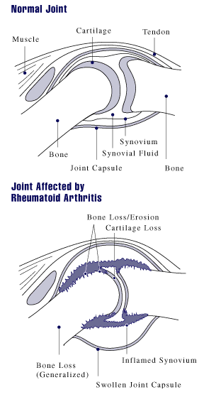
Rheumatoid arthritis (RA) is a long-term autoimmune disorder that primarily affects joints. It typically results in warm, swollen, and painful joints. Pain and stiffness often worsen following rest. Most commonly, the wrist and hands are involved, with the same joints typically involved on both sides of the body. The disease may also affect other parts of the body, including skin, eyes, lungs, heart, nerves and blood. This may result in a low red blood cell count, inflammation around the lungs, and inflammation around the heart. Fever and low energy may also be present. Often, symptoms come on gradually over weeks to months. While the cause of rheumatoid arthritis is not clear, it is believed to involve a combination of genetic and environmental factors. The underlying mechanism involves the body's immune system attacking the joints. This results in inflammation and thickening of the joint capsule. It also affects the underlying bone and cartilage. The diagnosis is made mos ... [...More Info...] [...Related Items...] OR: [Wikipedia] [Google] [Baidu] |
Extensor Pollicis Longus
In human anatomy, the extensor pollicis longus muscle (EPL) is a skeletal muscle located dorsally on the forearm. It is much larger than the extensor pollicis brevis, the origin of which it partly covers and acts to stretch the thumb together with this muscle. Structure The extensor pollicis longus arises from the dorsal surface of the ulna and from the interosseous membrane, next to the origins of abductor pollicis longus and extensor pollicis brevis. Passing through the third tendon compartment, lying in a narrow, oblique groove on the back of the lower end of the radius,''Gray's Anatomy'' 1918, see infobox it crosses the wrist close to the dorsal midline before turning towards the thumb using Lister's tubercle on the distal end of the radius as a pulley. It obliquely crosses the tendons of the extensores carpi radialis longus and brevis, and is separated from the extensor pollicis brevis by a triangular interval, the anatomical snuff box in which the radial artery is found. ... [...More Info...] [...Related Items...] OR: [Wikipedia] [Google] [Baidu] |
Flexor Pollicis Longus
The flexor pollicis longus (; FPL, Latin ''flexor'', bender; ''pollicis'', of the thumb; ''longus'', long) is a muscle in the forearm and hand that flexes the thumb. It lies in the same plane as the flexor digitorum profundus. This muscle is unique to humans, being either rudimentary or absent in other primates. A meta-analysis indicated accessory flexor pollicis longus is present in around 48% of the population. Human anatomy Origin and insertion It arises from the grooved anterior (side of palm) surface of the body of the radius, extending from immediately below the radial tuberosity and oblique line to within a short distance of the pronator quadratus muscle.Gray 1918, ''Flexor Pollicis Longus'', paras 20, 25 An occasionally present accessory long head of the flexor pollicis longus muscle is called 'Gantzer's muscle'. It may cause compression of the anterior interosseous nerve. It arises also from the adjacent part of the interosseous membrane of the forearm, and generally ... [...More Info...] [...Related Items...] OR: [Wikipedia] [Google] [Baidu] |
Interossei
{{short description, Muscles between certain bones Interossei refer to muscles between certain bones. There are many interossei in a human body. Specific interossei include: On the hands * Dorsal interossei muscles of the hand * Palmar interossei muscles File:Gray428.png, Dorsal interossei muscles of the hand File:Gray429.png, Palmar interossei muscles On the feet * Dorsal interossei muscles of the foot * Plantar interossei muscles In human anatomy, plantar interossei muscles are three muscles located between the metatarsal bones in the foot. Structure The three plantar interosseous muscles are unipennate, as opposed to the bipennate structure of dorsal interosseous muscles ... File:Gray446.png, Dorsal interossei muscles of the foot File:Gray447.png, Plantar interossei muscles Muscular system ... [...More Info...] [...Related Items...] OR: [Wikipedia] [Google] [Baidu] |


