|
Instruments Used In Gastroenterology
This is a list of instruments used specially in Gastroenterology Gastroenterology (from the Greek gastߪŚr- ÔÇťbellyÔÇŁ, -├ęnteron ÔÇťintestineÔÇŁ, and -log├şa "study of") is the branch of medicine focused on the digestive system and its disorders. The digestive system consists of the gastrointestinal tract .... __TOC__ Instrument list Image gallery Image:Gastroscope.jpg, Video gastroscope Image:Colonoscope.jpg, Video colonoscope Image:Endoscopy snare.jpg, Endoscopy snare used to perform polypectomy Image:PEG tube kit.jpg, PEG tube References Medical equipment Gastroenterology {{Medical-equipment-stub ... [...More Info...] [...Related Items...] OR: [Wikipedia] [Google] [Baidu] |
Gastroenterology
Gastroenterology (from the Greek gastߪŚr- ÔÇťbellyÔÇŁ, -├ęnteron ÔÇťintestineÔÇŁ, and -log├şa "study of") is the branch of medicine focused on the digestive system and its disorders. The digestive system consists of the gastrointestinal tract, sometimes referred to as the ''GI tract,'' which includes the esophagus, stomach, small intestine and large intestine as well as the accessory organs of digestion which includes the pancreas, gallbladder, and liver. The digestive system functions to move material through the GI tract via peristalsis, break down that material via digestion, absorb nutrients for use throughout the body, and remove waste from the body via defecation. Physicians who specialize in the medical specialty of gastroenterology are called gastroenterologists or sometimes ''GI doctors''. Some of the most common conditions managed by gastroenterologists include gastroesophageal reflux disease, gastrointestinal bleeding, irritable bowel syndrome, irritable bowel dise ... [...More Info...] [...Related Items...] OR: [Wikipedia] [Google] [Baidu] |
Banding (medical)
Banding is a medical procedure which uses elastic bands for constriction. Banding may be used to tie off blood vessels in order to stop bleeding, as in the treatment of bleeding esophageal varices. The band restricts blood flow to the ligated tissue, so that it eventually dies and sloughs away from the supporting tissue. This same principle underlies banding as treatment for hemorrhoids. Banding may also be used to restrict the function of an organ without killing it. In gastric banding to treat obesity, the size of the stomach is reduced so that digestion is slowed and the patient feels full more quickly. Banding as a medical procedure is commonly used in livestock for male castration of sheep and cattle. Banding is also commonly done in tail docking of lambs to prevent flystrike, and less commonly, used to dock tails of dairy cattle and draft horses. The bands are applied at the base of the scrotum or desired tail site, restricting blood flow to the scrotum or tail tissue, w ... [...More Info...] [...Related Items...] OR: [Wikipedia] [Google] [Baidu] |
Liver Biopsy
Liver biopsy is the biopsy (removal of a small sample of tissue) from the liver. It is a medical test that is done to aid diagnosis of liver disease, to assess the severity of known liver disease, and to monitor the progress of treatment. Medical uses Liver biopsy is often required for the diagnosis of a liver problem (jaundice, abnormal blood tests) where blood tests, such as hepatitis A serology, have not been able to identify a cause. It is also required if hepatitis is possibly the result of medication, but the exact nature of the reaction is unclear. Alcoholic liver disease and tuberculosis of the liver may be diagnosed through biopsy. Direct biopsy of tumors of the liver may aid the diagnosis, although this may be avoided if the source is clear (e.g. spread from previously known colorectal cancer). Liver biopsy will likely remain particularly important in the diagnosis of unexplained liver disease. Non-invasive tests for liver fibrosis in alcoholic, nonalcoholic and viral li ... [...More Info...] [...Related Items...] OR: [Wikipedia] [Google] [Baidu] |
Percutaneous Endoscopic Gastrostomy
Percutaneous endoscopic gastrostomy (PEG) is an endoscopic medical procedure in which a tube (PEG tube) is passed into a patient's stomach through the abdominal wall, most commonly to provide a means of feeding when oral intake is not adequate (for example, because of dysphagia or sedation). This provides enteral nutrition (making use of the natural digestion process of the gastrointestinal tract) despite bypassing the mouth; enteral nutrition is generally preferable to parenteral nutrition (which is only used when the GI tract must be avoided). The PEG procedure is an alternative to open surgical gastrostomy insertion, and does not require a general anesthetic; mild sedation is typically used. PEG tubes may also be extended into the small intestine by passing a jejunal extension tube (PEG-J tube) through the PEG tube and into the jejunum via the pylorus. PEG administration of enteral feeds is the most commonly used method of nutritional support for patients in the community. Man ... [...More Info...] [...Related Items...] OR: [Wikipedia] [Google] [Baidu] |
Argon Plasma Coagulation
Argon plasma coagulation (APC) is a medical endoscopic procedure used to control bleeding from certain lesions in the gastrointestinal tract. It is administered during esophagogastroduodenoscopy or colonoscopy. Medical use APC involves the use of a jet of ionized argon gas ( plasma) directed through a probe passed through the endoscope. The probe is placed at some distance from the bleeding lesion, and argon gas is emitted, then ionized by a high-voltage discharge (approx 6 kV). High-frequency electric current is then conducted through the jet of gas, resulting in coagulation of the bleeding lesion. As no physical contact is made with the lesion, the procedure is safe if the bowel has been cleaned of colonic gases, and can be used to treat bleeding in parts of the gastrointestinal tract with thin walls, such as the cecum. The depth of coagulation is usually only a few millimetres. APC is used to treat the following conditions: * angiodysplasias, anywhere in the GI tract * gastri ... [...More Info...] [...Related Items...] OR: [Wikipedia] [Google] [Baidu] |
Esophageal Dilatation
Esophageal dilatation is a therapeutic endoscopy, endoscopic procedure that enlarges the lumen (anatomy), lumen of the esophagus. Indications It can be used to treat a number of medical conditions that result in narrowing of the esophageal lumen, or decrease motility in the distal esophagus. These include the following: * Esophageal stricture, Peptic stricture * Eosinophilic esophagitis * Schatzki rings * Achalasia * Scleroderma esophagus * Rarely esophageal cancer Types of dilators There are three major classes of dilators: * Mercury-weighted bougies are blindly inserted Bougie (medical instrument), bougies placed into the esophagus by the treating physician. They are passed in sequentially increasing sizes to dilate the obstructed area. They must be used with precaution in patients with narrow strictures, as they may curl proximal to the obstruction. * Bougie over guidewire dilators are used at the time of esophagogastroduodenoscopy, gastroscopy or fluoroscopy. An endoscopy ... [...More Info...] [...Related Items...] OR: [Wikipedia] [Google] [Baidu] |
Esophageal Varices
Esophageal varices are extremely dilated sub-mucosal veins in the lower third of the esophagus. They are most often a consequence of portal hypertension, commonly due to cirrhosis. People with esophageal varices have a strong tendency to develop severe bleeding which left untreated can be fatal. Esophageal varices are typically diagnosed through an esophagogastroduodenoscopy. Pathogenesis The upper two thirds of the esophagus are drained via the esophageal veins, which carry deoxygenated blood from the esophagus to the azygos vein, which in turn drains directly into the superior vena cava. These veins have no part in the development of esophageal varices. The lower one third of the esophagus is drained into the superficial veins lining the esophageal mucosa, which drain into the left gastric vein, which in turn drains directly into the portal vein. These superficial veins (normally only approximately 1 mm in diameter) become distended up to 1ÔÇô2 cm in diameter in as ... [...More Info...] [...Related Items...] OR: [Wikipedia] [Google] [Baidu] |
SengstakenÔÇôBlakemore Tube
A SengstakenÔÇôBlakemore tube is a medical device inserted through the nose or mouth and used occasionally in the management of upper gastrointestinal hemorrhage due to esophageal varices (distended and fragile veins in the esophageal wall, usually a result of cirrhosis). The use of the tube was originally described in 1950, although similar approaches to bleeding varices were described by Westphal in 1930. With the advent of modern endoscopic techniques which can rapidly and definitively control variceal bleeding, SengstakenÔÇôBlakemore tubes are rarely used at present. __TOC__ Device The device consists of a flexible plastic tube containing several internal channels and two inflatable balloons. Apart from the balloons, the tube has an opening at the bottom (gastric tip) of the device. More modern models also have an opening near the upper esophagus; such devices are properly termed Minnesota tubes. The tube is passed down into the esophagus and the gastric balloon is inflated in ... [...More Info...] [...Related Items...] OR: [Wikipedia] [Google] [Baidu] |
Endoscopic Foreign Body Retrieval
Endoscopic foreign body retrieval refers to the removal of ingested objects from the esophagus, stomach and duodenum by endoscopic techniques. It does not involve surgery, but rather encompasses a variety of techniques employed through the gastroscope for grasping foreign bodies, manipulating them, and removing them while protecting the esophagus and trachea. It is of particular importance with children, people with mental illness, and prison inmates as these groups have a high rate of foreign body ingestion. Commonly swallowed objects include coins, buttons, batteries, and small bones (such as fish bones), but can include more complex objects, such as eyeglasses,Grover SC, Kim YI, Kortan PP, Marcon NE. Endoscopic removal of eight gastric foreign bodies ingested sequentially in twelve days: a case of creative endoscopy. Abstract presented at ''World Congress of Gastroenterology'', Montreal, Canada, September 2005. spoons, and toothbrushes (see image). Indications and contra ... [...More Info...] [...Related Items...] OR: [Wikipedia] [Google] [Baidu] |
Esophagogastroduodenoscopy
Esophagogastroduodenoscopy (EGD) or oesophagogastroduodenoscopy (OGD), also called by various other names, is a diagnostic endoscopic procedure that visualizes the upper part of the gastrointestinal tract down to the duodenum. It is considered a minimally invasive procedure since it does not require an incision into one of the major body cavities and does not require any significant recovery after the procedure (unless sedation or anesthesia has been used). However, a sore throat is common. Alternative names The words ''esophagogastroduodenoscopy'' (EGD; American English) and ''oesophagogastroduodenoscopy'' (OGD; British English; see spelling differences) are both pronounced . It is also called ''panendoscopy'' (PES) and ''upper GI endoscopy''. It is also often called just ''upper endoscopy'', ''upper GI'', or even just ''endoscopy''; because EGD is the most commonly performed type of endoscopy, the ambiguous term ''endoscopy'' is sometimes informally used to refer to EGD b ... [...More Info...] [...Related Items...] OR: [Wikipedia] [Google] [Baidu] |
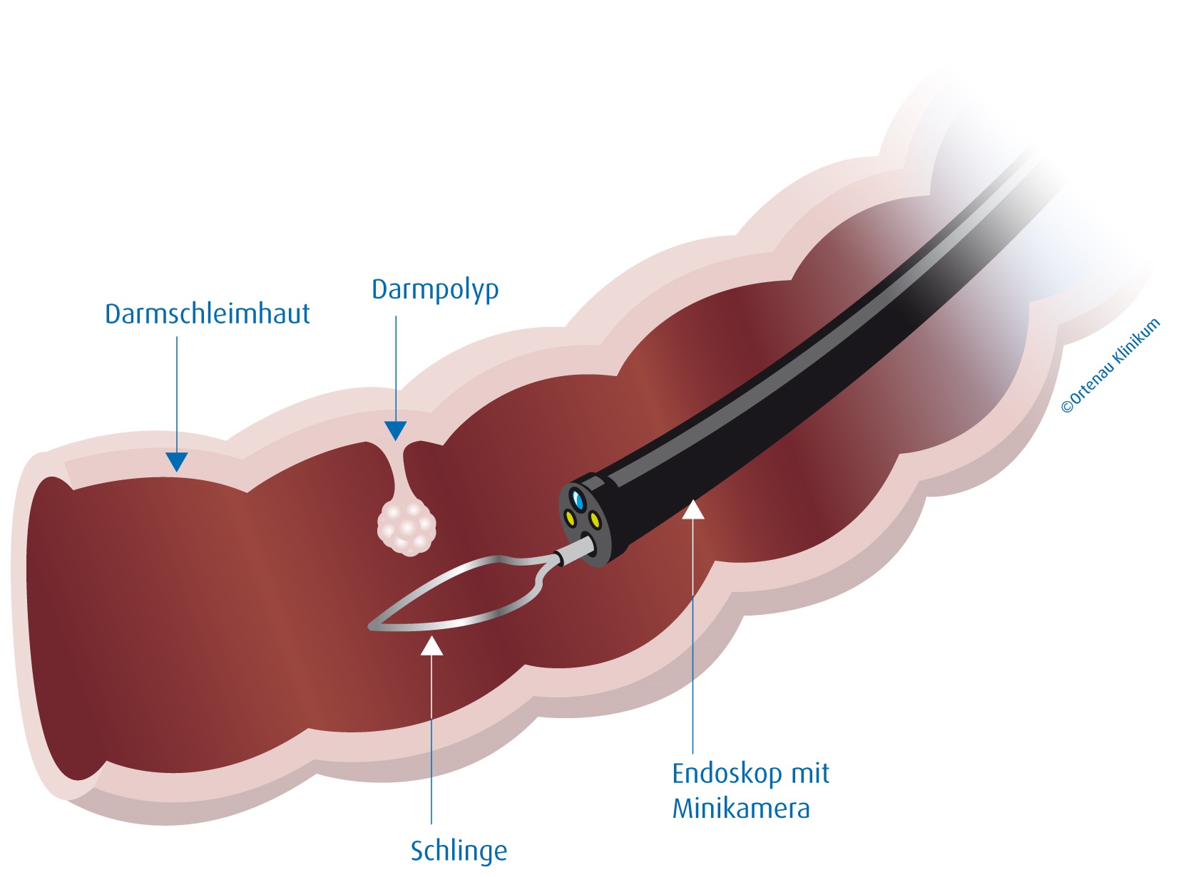
Polypectomy
In medicine, a polypectomy is the removal of an abnormal growth of tissue called a polyp. Polypectomy can be performed by excision if the polyp is external (on the skin). See also * Colonic polypectomy * Non-lifting sign The non-lifting sign is a finding on endoscopic examination that provides information on the suitability of large flat or sessile colorectal polyps for polypectomy by endoscopic mucosal resection (EMR). When fluid is injected under a polyp in prepa ... References {{surgery-stub Surgical procedures and techniques ... [...More Info...] [...Related Items...] OR: [Wikipedia] [Google] [Baidu] |
Capsule Endoscopy
Capsule endoscopy is a medical procedure used to record internal images of the gastrointestinal tract for use in disease diagnosis. Newer developments are also able to take biopsies and release medication at specific locations of the entire gastrointestinal tract. Unlike the more widely used endoscope, capsule endoscopy provides the ability to see the middle portion of the small intestine. It can be applied to the detection of various gastrointestinal cancers, digestive diseases, ulcers, unexplained bleedings, and general abdominal pains. After a patient swallows the capsule, it passes along the gastrointestinal tract, taking a number of images per second which are transmitted wirelessly to an array of receivers connected to a portable recording device carried by the patient. General advantages of capsule endoscopy over standard endoscopy include the minimally invasive procedure setup, ability to visualize more of the gastrointestinal tract, and lower cost of the procedure. ... [...More Info...] [...Related Items...] OR: [Wikipedia] [Google] [Baidu] |


