|
Infraorbital Vein
The infraorbital vein is a vein that drains structures of the floor of the orbit. It arises on the face and passes backwards through the orbit alongside infraorbital artery and nerve, exiting the orbit through the inferior orbital fissure to drain into the pterygoid venous plexus. Anatomy Origin The infraorbital vein arises on the face by the union of several tributaries. Course and relations Accompanied by the infraorbital artery and the infraorbital nerve, it passes posteriorly through the infraorbital foramen, infraorbital canal, and infraorbital groove. It exits the orbit through the inferior orbital fissure to drain into the pterygoid venous plexus. Distribution The infraorbital vein drains structures of the floor of the orbit; receives tributaries from structures that lie close to the floor of the orbit. Anastomoses The infraorbital vein communicates with the inferior ophthalmic vein. It may sometimes additionally also communicate with the facial vein The facial ... [...More Info...] [...Related Items...] OR: [Wikipedia] [Google] [Baidu] |
Pterygoid Venous Plexus
The pterygoid plexus (; in Merriam-Webster Online Dictionary '. from ''pteryx'', "wing" and ''eidos'', "shape") is a of considerable size, and is situated between the and |
Infraorbital Artery
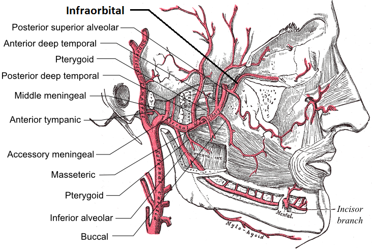
The infraorbital artery is an artery in the head that branches off the maxillary artery, emerging through the infraorbital foramen, just under the orbit of the eye. Course The infraorbital artery appears, from its direction, to be the continuation of the trunk of the maxillary artery, but often arises in conjunction with the posterior superior alveolar artery. It runs along the infraorbital groove and canal with the infraorbital nerve, and emerges on the face through the infraorbital foramen, beneath the infraorbital head of the levator labii superioris muscle. Branches While in the canal, it gives off * (a) orbital branches which assist in supplying the inferior rectus and inferior oblique and the lacrimal sac, and * (b) anterior superior alveolar arteries - branches which descend through the anterior alveolar canals to supply the upper incisor and canine teeth and the mucous membrane of the maxillary sinus. On the face, some branches pass upward to the medial angle of the o ... [...More Info...] [...Related Items...] OR: [Wikipedia] [Google] [Baidu] |
Infraorbital Nerve
The infraorbital nerve is a branch of the maxillary nerve, itself a branch of the trigeminal nerve (CN V). It travels through the orbit and enters the infraorbital canal to exit onto the face through the infraorbital foramen. It provides sensory innervation to the skin and mucous membranes around the middle of the face. Structure The infraorbital nerve is a branch of the maxillary nerve (CN V2), itself a branch of the trigeminal nerve (CN V). It travels with the infraorbital artery and vein. It branches from the maxillary nerve in the pterygopalatine fossa and travels through the inferior orbital fissure to enter the orbit. It runs anteriorly along the floor of the orbit in the infraorbital groove to the infraorbital canal of the maxilla. Within the infraorbital canal it has three branches, the posterior superior alveolar nerve, middle superior alveolar nerve and anterior superior alveolar nerve. After traversing the canal it emerges onto the anterior surface of the maxilla thr ... [...More Info...] [...Related Items...] OR: [Wikipedia] [Google] [Baidu] |
Inferior Orbital Fissure
The inferior orbital fissure is formed by the sphenoid bone and the maxilla. It is located posteriorly along the boundary of the floor and lateral wall of the orbit. It transmits a number of structures, including: * the zygomatic branch of the maxillary nerve * the ascending branches from the pterygopalatine ganglion * the infraorbital vessels, which travel down the infraorbital groove into the infraorbital canal and exit through the infraorbital foramen * the inferior division of the ophthalmic vein Images File:Gray189.png, Left infratemporal fossa. File:Gray191.png, Horizontal section of nasal and orbital cavities. File:Gray787.png, Dissection showing origins of right ocular muscles, and nerves entering by the superior orbital fissure. File:Slide2rome.JPG, Inferior orbital fissure. See also *Foramina of skull *Superior orbital fissure The superior orbital fissure is a foramen or cleft of the skull between the lesser and greater wings of the sphenoid bone. It gives ... [...More Info...] [...Related Items...] OR: [Wikipedia] [Google] [Baidu] |
Pterygoid Venous Plexus
The pterygoid plexus (; in Merriam-Webster Online Dictionary '. from ''pteryx'', "wing" and ''eidos'', "shape") is a of considerable size, and is situated between the and |
Infraorbital Foramen
In human anatomy, the infraorbital foramen is one of two small holes in the skull's upper jawbone (maxillary bone), located below the eye socket and to the left and right of the nose. Both holes are used for blood vessels and nerves. In anatomical terms, it is located below the infraorbital margin of the orbit. It transmits the infraorbital artery and vein, and the infraorbital nerve, a branch of the maxillary nerve. It is typically from the infraorbital margin. Structure Forming the exterior end of the infraorbital canal, the infraorbital foramen communicates with the infraorbital groove, the canal's opening on the interior side. The ramifications of the three principal branches of the trigeminal nerve—at the supraorbital, infraorbital, and mental foramen—are distributed on a vertical line (in anterior view) passing through the middle of the pupil. The infraorbital foramen is used as a pressure point to test the sensitivity of the infraorbital nerve. Palpation of the inf ... [...More Info...] [...Related Items...] OR: [Wikipedia] [Google] [Baidu] |
Infraorbital Canal
The infraorbital canal is a canal found at the base of the orbit that opens on to the maxilla. It is continuous with the infraorbital groove and opens onto the maxilla at the infraorbital foramen. The infraorbital nerve and infraorbital artery travel through the canal. Structure One of the canals of the orbital surface of the maxilla, the infraorbital canal, opens just below the margin of the orbit, the area of the skull containing the eye and related structures. It should not be confused with the infraorbital foramen, with which it is continuous. Function It transmits the infraorbital nerve as well as infraorbital artery, both of which enter this canal at the infraorbital groove and after coursing through the maxillary sinus exit via the infraorbital foramen. Before exiting, the anterior superior alveolar nerve, middle superior alveolar nerve The middle superior alveolar nerve is a nerve that drops from the infraorbital portion of the maxillary nerve to supply the sinus mu ... [...More Info...] [...Related Items...] OR: [Wikipedia] [Google] [Baidu] |
Infraorbital Groove
The infraorbital groove (or sulcus) is located in the middle of the posterior part of the orbital surface of the maxilla. Its function is to act as the passage of the infraorbital artery, the infraorbital vein, and the infraorbital nerve. Structure The infraorbital groove begins at the middle of the posterior border of the maxilla (with which it is continuous). This is near the upper edge of the infratemporal surface of the maxilla. It passes forward, and ends in a canal which subdivides into two branches. The infraorbital groove has an average length of 16.7 mm, with a small amount of variation between people. It is similar in men and women. Function The infraorbital groove creates space that allows for passage of the infraorbital artery, the infraorbital vein, and the infraorbital nerve. Clinical significance The infraorbital groove is an important surgical landmark for local anaesthesia of the infraorbital nerve. See also * Infraorbital foramen In human anatomy ... [...More Info...] [...Related Items...] OR: [Wikipedia] [Google] [Baidu] |
Inferior Ophthalmic Vein
The inferior ophthalmic vein is a vein of the orbit that - together with the superior ophthalmic vein - represents the principal drainage system of the orbit. It begins from a venous network in the front of the orbit, then passes backwards through the lower orbit. It drains several structures of the orbit. It may end by splitting into two branches, one draining into the pterygoid venous plexus and the other ultimately (i.e. directly or indirectly) into the cavernous sinus. Structure The inferior ophthalmic vein - together with the superior ophthalmic vein - represents the principal drainage system of the orbit. Origin The inferior ophthalmic vein originates from a venous network at the anterior part of the floor and anterior part of the medial wall of the orbit. Course The inferior ophthalmic vein passes posterior-ward through the inferior orbit. Distribution The inferior ophthalmic vein drains venous blood from the inferior rectus muscle, inferior oblique muscle, lat ... [...More Info...] [...Related Items...] OR: [Wikipedia] [Google] [Baidu] |
Facial Vein
The facial vein (or anterior facial vein) is a relatively large vein in the human face. It commences at the side of the root of the nose and is a direct continuation of the angular vein where it also receives a small nasal branch. It lies behind the facial artery and follows a less tortuous course. It receives blood from the external palatine vein before it either joins the anterior branch of the retromandibular vein to form the common facial vein, or drains directly into the internal jugular vein. A common misconception states that the facial vein has no valves, but this has been contradicted by recent studies. Its walls are not so flaccid as most superficial veins. Path From its origin it runs obliquely downward and backward, beneath the zygomaticus major muscle and zygomatic head of the quadratus labii superioris, descends along the anterior border and then on the superficial surface of the masseter, crosses over the body of the mandible, and passes obliquely backward, benea ... [...More Info...] [...Related Items...] OR: [Wikipedia] [Google] [Baidu] |
