|
Inferior Vesical Arteries
The inferior vesical artery (or inferior vesicle artery) is an artery of the pelvis which arises from the internal iliac artery and supplies parts of the urinary bladder as well as other structures of the urinary system and structures of the male reproductive system. Some sources consider this vessel to be present only in males, and cite the vaginal artery as the homologous structure in females; others consider it to be present in both sexes, with the vessel taking the form of a small branch of a vaginal artery in females. Structure Origin The inferior vesical artery is a branch of the anterior division of the internal iliac artery. It frequently has a common origin with the middle rectal artery. Course The inferior vesical artery passes medially across the pelvic floor. Distribution The inferior vesical artery is distributed to the trigone and inferior portion of the urinary bladder, the ureter, prostate, vas deferens, and seminal vesicles.vas deferens The branches to th ... [...More Info...] [...Related Items...] OR: [Wikipedia] [Google] [Baidu] |
Internal Iliac Artery
The internal iliac artery (formerly known as the hypogastric artery) is the main artery of the pelvis. Structure The internal iliac artery supplies the walls and viscera of the pelvis, the buttock, the reproductive organs, and the medial compartment of the thigh. The vesicular branches of the internal iliac arteries supply the bladder. It is a short, thick vessel, smaller than the external iliac artery, and about 3 to 4 cm in length. Course The internal iliac artery arises at the bifurcation of the common iliac artery, opposite the lumbosacral articulation, and, passing downward to the upper margin of the greater sciatic foramen, divides into two large trunks, an anterior and a posterior. It is posterior to the ureter, anterior to the internal iliac vein, anterior to the lumbosacral trunk, and anterior to the piriformis muscle. Near its origin, it is medial to the external iliac vein, which lies between it and the psoas major muscle. It is above the obturator nerve. Bra ... [...More Info...] [...Related Items...] OR: [Wikipedia] [Google] [Baidu] |
Vesical Venous Plexus
The vesical plexus envelops the lower part of the bladder and the base of the prostate and communicates with the pudendal and prostatic plexuses. It is drained, by means of several vesical veins The vesical veins are veins in the pelvis that drain blood from the urinary bladder. The vesical veins receive blood from the vesical venous plexus The vesical plexus envelops the lower part of the bladder and the base of the prostate The ..., into the internal iliac veins. References External links Anatomy at umich.edu Veins of the torso {{circulatory-stub ... [...More Info...] [...Related Items...] OR: [Wikipedia] [Google] [Baidu] |
Prostate
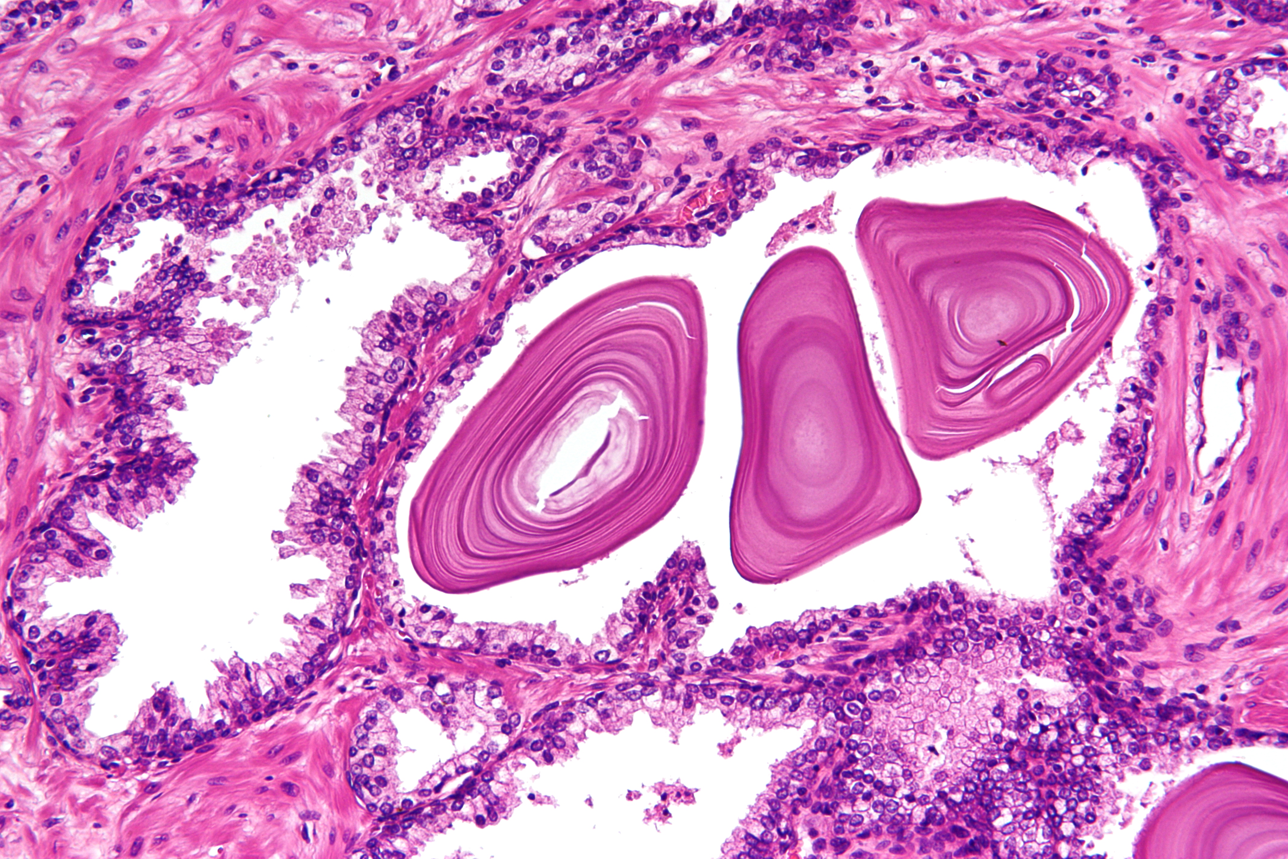
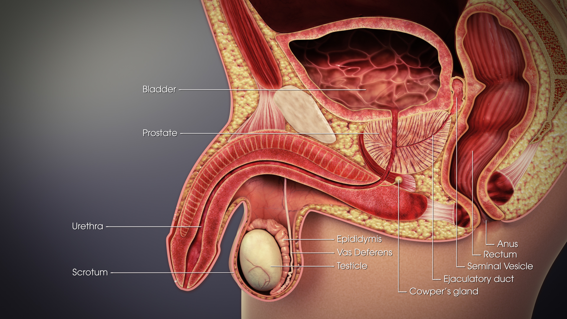
The prostate is both an Male accessory gland, accessory gland of the male reproductive system and a muscle-driven mechanical switch between urination and ejaculation. It is found only in some mammals. It differs between species anatomically, chemically, and physiologically. Anatomically, the prostate is found below the Urinary bladder, bladder, with the urethra passing through it. It is described in gross anatomy as consisting of lobes and in microanatomy by zone. It is surrounded by an elastic, fibromuscular capsule and contains glandular tissue as well as connective tissue. The prostate glands produce and contain fluid that forms part of semen, the substance emitted during ejaculation as part of the male Human sexual response cycle, sexual response. This prostatic fluid is slightly alkaline, milky or white in appearance. The alkalinity of semen helps neutralize the acidity of the vagina, vaginal tract, prolonging the lifespan of sperm. The prostatic fluid is expelled in the ... [...More Info...] [...Related Items...] OR: [Wikipedia] [Google] [Baidu] |
Seminal Vesicle
The seminal vesicles (also called vesicular glands, or seminal glands) are a pair of two convoluted tubular glands that lie behind the urinary bladder of some male mammals. They secrete fluid that partly composes the semen. The vesicles are 5–10 cm in size, 3–5 cm in diameter, and are located between the bladder and the rectum. They have multiple outpouchings which contain secretory glands, which join together with the vas deferens at the ejaculatory duct. They receive blood from the vesiculodeferential artery, and drain into the vesiculodeferential veins. The glands are lined with column-shaped and cuboidal cells. The vesicles are present in many groups of mammals, but not marsupials, monotremes or carnivores. Inflammation of the seminal vesicles is called seminal vesiculitis, most often is due to bacterial infection as a result of a sexually transmitted disease or following a surgical procedure. Seminal vesiculitis can cause pain in the lower abdomen, scrotum, ... [...More Info...] [...Related Items...] OR: [Wikipedia] [Google] [Baidu] |
Urinary Bladder
The urinary bladder, or simply bladder, is a hollow organ in humans and other vertebrates that stores urine from the kidneys before disposal by urination. In humans the bladder is a distensible organ that sits on the pelvic floor. Urine enters the bladder via the ureters and exits via the urethra. The typical adult human bladder will hold between 300 and (10.14 and ) before the urge to empty occurs, but can hold considerably more. The Latin phrase for "urinary bladder" is ''vesica urinaria'', and the term ''vesical'' or prefix ''vesico -'' appear in connection with associated structures such as vesical veins. The modern Latin word for "bladder" – ''cystis'' – appears in associated terms such as cystitis (inflammation of the bladder). Structure In humans, the bladder is a hollow muscular organ situated at the base of the pelvis. In gross anatomy, the bladder can be divided into a broad , a body, an apex, and a neck. The apex (also called the vertex) is directed forward ... [...More Info...] [...Related Items...] OR: [Wikipedia] [Google] [Baidu] |
Vas Deferens
The vas deferens or ductus deferens is part of the male reproductive system of many vertebrates. The ducts transport sperm from the epididymis to the ejaculatory ducts in anticipation of ejaculation. The vas deferens is a partially coiled tube which exits the abdominal cavity through the inguinal canal. Etymology ''Vas deferens'' is Latin, meaning "carrying-away vessel"; the plural version is ''vasa deferentia''. ''Ductus deferens'' is also Latin, meaning "carrying-away duct"; the plural version is ''ducti deferentes''. Structure There are two vasa deferentia, connecting the left and right epididymis with the seminal vesicles to form the ejaculatory duct in order to move sperm. The (human) vas deferens measures 30–35 cm in length, and 2–3 mm in diameter. The vas deferens is continuous proximally with the tail of the epididymis. The vas deferens exhibits a tortuous, convoluted initial/proximal section (which measures 2–3 cm in length). Distally, it forms ... [...More Info...] [...Related Items...] OR: [Wikipedia] [Google] [Baidu] |
Pelvis
The pelvis (plural pelves or pelvises) is the lower part of the trunk, between the abdomen and the thighs (sometimes also called pelvic region), together with its embedded skeleton (sometimes also called bony pelvis, or pelvic skeleton). The pelvic region of the trunk includes the bony pelvis, the pelvic cavity (the space enclosed by the bony pelvis), the pelvic floor, below the pelvic cavity, and the perineum, below the pelvic floor. The pelvic skeleton is formed in the area of the back, by the sacrum and the coccyx and anteriorly and to the left and right sides, by a pair of hip bones. The two hip bones connect the spine with the lower limbs. They are attached to the sacrum posteriorly, connected to each other anteriorly, and joined with the two femurs at the hip joints. The gap enclosed by the bony pelvis, called the pelvic cavity, is the section of the body underneath the abdomen and mainly consists of the reproductive organs (sex organs) and the rectum, while the pelvic f ... [...More Info...] [...Related Items...] OR: [Wikipedia] [Google] [Baidu] |
Urinary System
The urinary system, also known as the urinary tract or renal system, consists of the kidneys, ureters, urinary bladder, bladder, and the urethra. The purpose of the urinary system is to eliminate waste from the body, regulate blood volume and blood pressure, control levels of Electrolyte, electrolytes and Metabolite, metabolites, and regulate Acid–base homeostasis, blood pH. The urinary tract is the body's drainage system for the eventual removal of urine. The kidneys have an extensive blood supply via the Renal artery, renal arteries which leave the kidneys via the renal vein. Each kidney consists of functional units called nephrons. Following filtration of blood and further processing, wastes (in the form of urine) exit the kidney via the ureters, tubes made of smooth muscle fibres that propel urine towards the urinary bladder, where it is stored and subsequently expelled from the body by urination (voiding). The female and male urinary system are very similar, differing only ... [...More Info...] [...Related Items...] OR: [Wikipedia] [Google] [Baidu] |
Male Reproductive System
The male reproductive system consists of a number of sex organs that play a role in the process of human reproduction. These organs are located on the outside of the body and within the pelvis. The main male sex organs are the penis and the testicles which produce semen and sperm, which, as part of sexual intercourse, fertilize an ovum in the female's body; the fertilized ovum (zygote) develops into a fetus, which is later born as an infant. The corresponding system in females is the female reproductive system. External genital organs Penis The penis is the male intromittent organ. It has a long shaft and an enlarged bulbous-shaped tip called the glans penis, which supports and is protected by the foreskin. When the male becomes sexually aroused, the penis becomes erect and ready for sexual activity. Erection occurs because sinuses within the erectile tissue of the penis become filled with blood. The arteries of the penis are dilated while the veins are compressed so ... [...More Info...] [...Related Items...] OR: [Wikipedia] [Google] [Baidu] |
Vaginal Artery
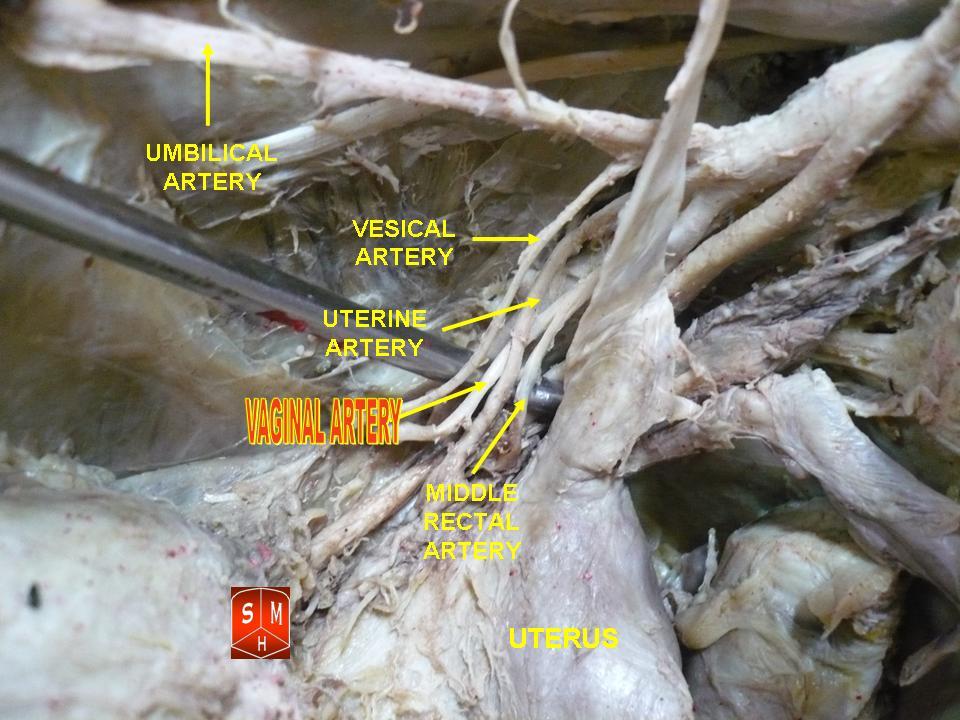
The vaginal artery is an artery in females that supplies blood to the vagina and the base of the bladder. Structure The vaginal artery is usually a branch of the internal iliac artery. Some sources say that the vaginal artery can arise from the uterine artery, but the phrase vaginal branches of uterine artery is the term for blood supply to the vagina coming from the uterine artery. The vaginal artery is frequently represented by two or three branches. These descend to the vagina, supplying its mucous membrane. They anastomose with branches from the uterine artery. It can send branches to the bulb of the vestibule, the fundus of the bladder, and the contiguous part of the rectum. Function The vaginal artery supplies oxygenated blood to the muscular wall of the vagina, along with the uterine artery The uterine artery is an artery that supplies blood to the uterus in females. Structure The uterine artery usually arises from the anterior division of the internal iliac artery. ... [...More Info...] [...Related Items...] OR: [Wikipedia] [Google] [Baidu] |
Middle Rectal Artery
The middle rectal artery is an artery in the pelvis that supplies blood to the rectum. Structure The middle rectal artery usually arises from the internal iliac artery. It is distributed to the rectum above the pectinate line. It anastomoses with the inferior vesical artery, superior rectal artery, and inferior rectal artery. In males, the middle rectal artery may give off branches to the prostate and the seminal vesicles. In females, the middle rectal artery gives off branches to the vagina. Function The middle rectal artery supplies the rectum above the pectinate line. Additional images File:Gray538.png, Sigmoid colon and rectum, showing distribution of branches of inferior mesenteric artery and their anastomoses. File:Middle rectal artery.jpg, Middle rectal artery See also * Superior rectal artery * Inferior rectal artery The inferior rectal artery (inferior hemorrhoidal artery) is an artery that supplies blood to the lower third of the anal canal below the pectin ... [...More Info...] [...Related Items...] OR: [Wikipedia] [Google] [Baidu] |


