|
Inferior Fascia Of The Urogenital Diaphragm
The perineal membrane is an anatomical term for a fibrous membrane in the perineum. The term "inferior fascia of urogenital diaphragm", used in older texts, is considered equivalent to the perineal membrane. It is the superior border of the superficial perineal pouch, and the inferior border of the deep perineal pouch. Structure The perineal membrane is triangular in shape. It attaches to both ischiopubic rami of the pelvis. It also attaches to the perineal body. It is about 4 cm. in depth. Its apex is directed forward, and is separated from the arcuate pubic ligament by an oval opening for the transmission of the deep dorsal vein of the penis or the deep dorsal vein of the clitoris. Its lateral margins are attached on either side to the inferior rami of the pubis and ischium, above the crus penis. Its base is directed toward the rectum, and connected to the central tendinous point of the perineum. The base is fused with both the pelvic fascia and Colle's fascia. ... [...More Info...] [...Related Items...] OR: [Wikipedia] [Google] [Baidu] |
Perineum
The perineum in humans is the space between the anus and scrotum in the male, or between the anus and the vulva in the female. The perineum is the region of the body between the pubic symphysis (pubic arch) and the coccyx (tail bone), including the perineal body and surrounding structures. There is some variability in how the boundaries are defined. The perineal raphe is visible and pronounced to varying degrees. The perineum is an erogenous zone. The word perineum entered English from late Latin via Greek περίναιος ~ περίνεος ''perinaios, perineos'', itself from περίνεος, περίνεοι 'male genitals' and earlier περίς ''perís'' 'penis' through influence from πηρίς ''pērís'' 'scrotum'. The term was originally understood as a purely male body-part with the perineal raphe seen as a continuation of the scrotal septum since masculinization causes the development of a large anogenital distance in men, in comparison to the corresponding lack ... [...More Info...] [...Related Items...] OR: [Wikipedia] [Google] [Baidu] |
Rectum
The rectum is the final straight portion of the large intestine in humans and some other mammals, and the Gastrointestinal tract, gut in others. The adult human rectum is about long, and begins at the rectosigmoid junction (the end of the sigmoid colon) at the level of the third sacral vertebra or the sacral promontory depending upon what definition is used. Its diameter is similar to that of the sigmoid colon at its commencement, but it is dilated near its termination, forming the rectal ampulla. It terminates at the level of the anorectal ring (the level of the puborectalis sling) or the dentate line, again depending upon which definition is used. In humans, the rectum is followed by the anal canal which is about long, before the gastrointestinal tract terminates at the anal verge. The word rectum comes from the Latin ''Wikt:rectum, rectum Wikt:intestinum, intestinum'', meaning ''straight intestine''. Structure The rectum is a part of the lower gastrointestinal tract ... [...More Info...] [...Related Items...] OR: [Wikipedia] [Google] [Baidu] |
Deep Transverse Perineal Muscle
The transverse perineal muscles (transversus perinei) are the superficial and the deep transverse perineal muscles. Superficial transverse perineal The superficial transverse perineal muscle (transversus superficialis perinei or Lloyd-Beanie muscle) is a narrow muscular slip, which passes more or less transversely across the perineal space in front of the anus. It arises by tendinous fibers from the inner and forepart of the ischial tuberosity and, running medially, is inserted into the central tendinous point of the perineum (perineal body), joining in this situation with the muscle of the opposite side, with the external anal sphincter muscle behind, and with the bulbospongiosus muscle in front. In some cases, the fibers of the deeper layer of the external anal sphincter cross over in front of the anus and are continued into this mus ... [...More Info...] [...Related Items...] OR: [Wikipedia] [Google] [Baidu] |
Membranous Portion Of The Urethra
The membranous urethra or intermediate part of male urethra is the shortest, least dilatable, and, with the exception of the urinary meatus, the narrowest part of the urethra. It extends downward and forward, with a slight anterior concavity, between the apex of the prostate and the bulb of the urethra, perforating the urogenital diaphragm about 2.5 cm below and behind the pubic symphysis. The hinder part of the urethral bulb lies in apposition with the inferior fascia of the urogenital diaphragm, but its upper portion diverges somewhat from this fascia: the anterior wall of the membranous urethra is thus prolonged for a short distance in front of the urogenital diaphragm; it measures about 2 cm in length, while the posterior wall which is between the two fasciæ of the diaphragm is only 1.25 cm long. The anatomical variation in membranous urethral length measurements in men have been reported to range from 0.5 cm to 3.4 cm. The membranous portion of the ... [...More Info...] [...Related Items...] OR: [Wikipedia] [Google] [Baidu] |
Perineal Vessels
The perineal artery (superficial perineal artery) arises from the internal pudendal artery, and turns upward, crossing either over or under the superficial transverse perineal muscle, and runs forward, parallel to the pubic arch, in the interspace between the bulbospongiosus and ischiocavernosus muscles, both of which it supplies, and finally divides into several posterior scrotal branches which are distributed to the skin and dartos tunic of the scrotum. As it crosses the superficial transverse perineal muscle it gives off the ''transverse perineal artery'' which runs transversely on the cutaneous surface of the muscle, and anastomoses with the corresponding vessel of the opposite side and with the perineal and inferior hemorrhoidal arteries. It supplies the Transversus perinæi superficialis and the structures between the anus and the urethral bulb Just before each crus of the penis meets its fellow, it presents a slight enlargement, which Georg Ludwig Kobelt named the bu ... [...More Info...] [...Related Items...] OR: [Wikipedia] [Google] [Baidu] |
Pubic Arch
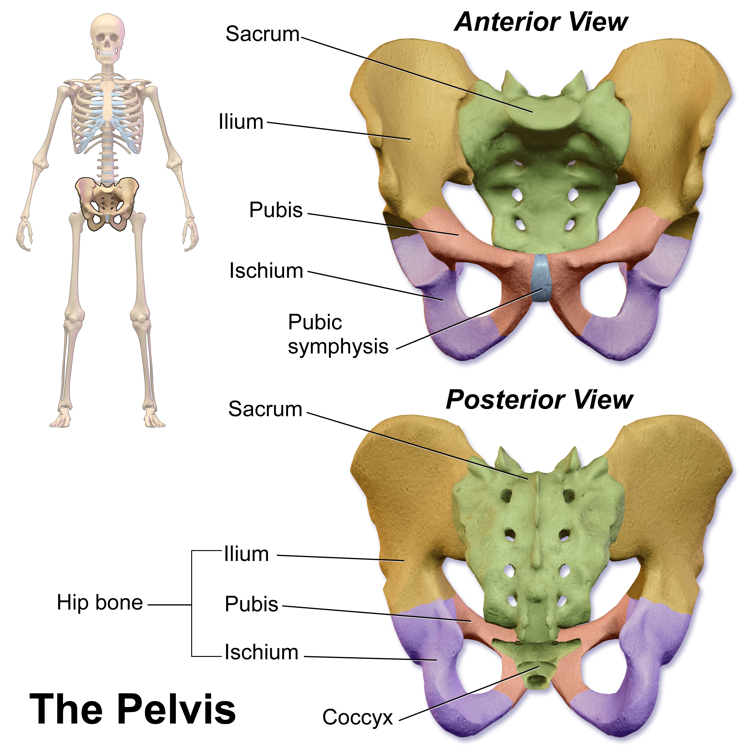
The pubic arch, also referred to as the ''ischiopubic arch'', is part of the pelvis. It is formed by the convergence of the inferior rami of the ischium and pubis on either side, below the pubic symphysis. The angle at which they converge is known as the subpubic angle. Function The pubic arch is one of three notches (the one in front) that separate the eminences of the lower circumference of the true pelvis. Clinical significance Subpubic angle The subpubic angle (or pubic angle) is the angle in the human body as the apex of the pubic arch, formed by the convergence of the inferior rami of the ischium and pubis on either side. The subpubic angle is important in forensic anthropology, in determining the sex of someone from skeletal remains. A subpubic angle of 50-82 degrees indicates a male; an angle of 90 degrees indicates a female.Anthony J. Bertino. Forensic Science - Fundamentals and Investigations. South-Western Cengage Learning, 2000. . Page 368 Other sources operate w ... [...More Info...] [...Related Items...] OR: [Wikipedia] [Google] [Baidu] |
Deep Arteries Of The Penis
The deep artery of the penis (artery to the corpus cavernosum), one of the terminal branches of the internal pudendal, arises from that vessel while it is situated between the two fasciæ of the urogenital diaphragm (deep perineal pouch). It pierces the inferior fascia, and, entering the crus penis For their anterior three-fourths the corpora cavernosa penis lie in intimate apposition with one another, but behind they diverge in the form of two tapering processes, known as the crura, which are firmly connected to the ischial rami. Traced ... obliquely, runs forward in the center of the corpus cavernosum penis, to which its branches are distributed. Additional images File:Penvein.png, Arteries and veins of the penis (Spanish) File:Gray588.png, The penis in transverse section, showing the blood vessels. File:Penis cross section.svg, The penis in transverse section, showing the blood vessels, including the deep artery References External links * * Arteries of t ... [...More Info...] [...Related Items...] OR: [Wikipedia] [Google] [Baidu] |
Internal Urethral Orifice
The internal urethral orifice is the opening of the urinary bladder into the urethra. It is placed at the apex of the trigonum vesicae, in the most dependent part of the bladder, and is usually somewhat crescent-shaped; the mucous membrane immediately behind it presents a slight elevation in males, the uvula vesicae, caused by the middle lobe of the prostate. See also * Internal sphincter muscle of urethra The internal urethral sphincter is a urethral sphincter muscle which constricts the internal urethral orifice. It is located at the junction of the urethra with the urinary bladder and is continuous with the detrusor muscle, but anatomically and ... References External links * - "The Male Pelvis: The Urethra" Urinary system Urethra {{genitourinary-stub ... [...More Info...] [...Related Items...] OR: [Wikipedia] [Google] [Baidu] |
Bulbourethral Glands
The bulbourethral glands or Cowper's glands (named for English anatomist William Cowper) are two small exocrine glands in the reproductive system of many male mammals (of all domesticated animals, they are absent only in dogs). They are homologous to Bartholin's glands in females. The bulbouretheral glands are responsible for producing a pre-ejaculate fluid called Cowper's fluid (known colloquially as ''pre-ejaculate'' or ''pre-cum''), which is secreted during sexual arousal, neutralizing the acidity of the urethra in preparation for the passage of sperm cells. Location Bulbourethral glands are located posterior and lateral to the membranous portion of the urethra at the base of the penis, between the two layers of the fascia of the urogenital diaphragm, in the deep perineal pouch. They are enclosed by transverse fibers of the sphincter urethrae membranaceae muscle. Structure The bulbourethral glands are compound tubulo-alveolar glands, each approximately the size of a pea i ... [...More Info...] [...Related Items...] OR: [Wikipedia] [Google] [Baidu] |
Urethra
The urethra (from Greek οὐρήθρα – ''ourḗthrā'') is a tube that connects the urinary bladder to the urinary meatus for the removal of urine from the body of both females and males. In human females and other primates, the urethra connects to the urinary meatus above the vagina, whereas in marsupials, the female's urethra empties into the urogenital sinus. Females use their urethra only for urinating, but males use their urethra for both urination and ejaculation. The external urethral sphincter is a striated muscle that allows voluntary control over urination. The internal sphincter, formed by the involuntary smooth muscles lining the bladder neck and urethra, receives its nerve supply by the sympathetic division of the autonomic nervous system. The internal sphincter is present both in males and females. Structure The urethra is a fibrous and muscular tube which connects the urinary bladder to the external urethral meatus. Its length differs between the sexes, ... [...More Info...] [...Related Items...] OR: [Wikipedia] [Google] [Baidu] |
Pubic Symphysis
The pubic symphysis is a secondary cartilaginous joint between the left and right superior rami of the pubis of the hip bones. It is in front of and below the urinary bladder. In males, the suspensory ligament of the penis attaches to the pubic symphysis. In females, the pubic symphysis is close to the clitoris. In most adults it can be moved roughly 2 mm and with 1 degree rotation. This increases for women at the time of childbirth. The name comes from the Greek word ''symphysis'', meaning 'growing together'. Structure The pubic symphysis is a nonsynovial amphiarthrodial joint. The width of the pubic symphysis at the front is 3–5 mm greater than its width at the back. This joint is connected by fibrocartilage and may contain a fluid-filled cavity; the center is avascular, possibly due to the nature of the compressive forces passing through this joint, which may lead to harmful vascular disease. The ends of both pubic bones are covered by a thin layer of hyaline ... [...More Info...] [...Related Items...] OR: [Wikipedia] [Google] [Baidu] |
Inferior Fascia Of Pelvic Diaphragm
The pelvic fasciae are the fascia of the pelvis and can be divided into: * (a) the fascial sheaths of ** the Obturator internus muscle ( Fascia of the Obturator internus) ** the Piriformis muscle ( Fascia of the Piriformis) ** the pelvic floor * (b) fascia associated with the organs of the pelvis. Structure Fascia of pelvic organs Pelvic fascia extends to cover the organs within the pelvis. It is attached to the fascia that runs along the pelvic floor along the tendinous arch. The fascia which covers pelvic organs can be divided according to the organs that are covered: * The front is known as the "vesical layer". It forms the anterior and lateral ligaments of the bladder. * In males, its middle lamina crosses the floor of the pelvis between the rectum and vesiculæ seminales as the ''rectovesical septum''; in the female this is perforated by the cervix and is named the transverse cervical ligament. * At the back, the fascia passes to the side of the rectum; it forms a loose s ... [...More Info...] [...Related Items...] OR: [Wikipedia] [Google] [Baidu] |


