|
Incisive Canal
The incisive canals (also: "''nasopalatine canals''") are two bony canals of the anterior hard palate connecting the nasal cavity and the oral cavity. An incisive canal courses through each maxilla. Below, the two incisive canals typically converge medially. Each incisive canal transmits a nasopalatine nerve, and an anastomosis of the greater palatine artery and a posterior septal branch of the sphenopalatine artery. Anatomy An incisive canal has an average length of 10 mm, and an average width of up to 6 mm at the incisive fossa (the dimensions of the canal change with age, trauma, and loss of teeth). Course and openings The two incisive canals usually (in 60% of individuals) have a characteristic "Y"-shaped or "V"-shaped morphology: above, each incisive canal opens into the nasal cavity on either side of the nasal septum as the nasal foramina; below, the two incisive canals converge medially to open into the oral cavity at midline at the incisive fossa as several incisive ... [...More Info...] [...Related Items...] OR: [Wikipedia] [Google] [Baidu] |
Bony Palate
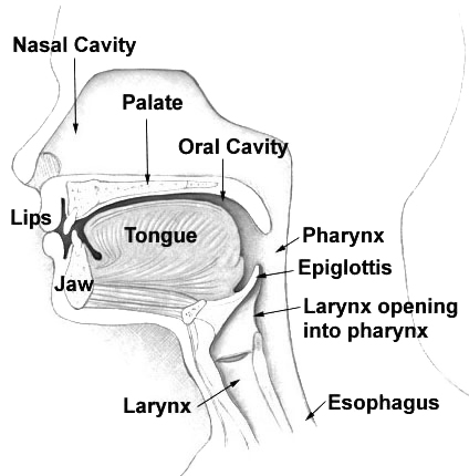
The hard palate is a thin horizontal bony plate made up of two bones of the facial skeleton, located in the roof of the mouth. The bones are the palatine process of the maxilla and the horizontal plate of palatine bone. The hard palate spans the alveolar arch formed by the alveolar process that holds the upper teeth (when these are developed). Structure The hard palate is formed by the palatine process of the maxilla and horizontal plate of palatine bone. It forms a partition between the nasal passages and the mouth. On the anterior portion of the hard palate are the plicae, irregular ridges in the mucous membrane that help facilitate the movement of food backward towards the larynx. This partition is continued deeper into the mouth by a fleshy extension called the soft palate. On the ventral surface of hard palate, some projections or transverse ridges are present which are called as palatine rugae. Function The hard palate is important for feeding and speech. Mammals with a ... [...More Info...] [...Related Items...] OR: [Wikipedia] [Google] [Baidu] |
Alveolar Arch
The alveolar process () or alveolar bone is the thickened ridge of bone that contains the tooth sockets on the jaw bones (in humans, the maxilla and the mandible). The structures are covered by gums as part of the oral cavity. The synonymous terms ''alveolar ridge'' and ''alveolar margin'' are also sometimes used more specifically to refer to the ridges on the inside of the mouth which can be felt with the tongue, either on roof of the mouth between the upper teeth and the hard palate or on the bottom of the mouth behind the lower teeth. Terminology The term ''alveolar'' () ('hollow') refers to the cavities of the tooth sockets, known as dental alveoli. The alveolar process is also called the ''alveolar bone'' or ''alveolar ridge''. The curved portion is referred to as the alveolar arch. The alveolar bone proper, also called bundle bone, directly surrounds the teeth. The term alveolar crest describes the extreme rim of the bone nearest to the crowns of the teeth. The ... [...More Info...] [...Related Items...] OR: [Wikipedia] [Google] [Baidu] |
Hard Palate
The hard palate is a thin horizontal bony plate made up of two bones of the facial skeleton, located in the roof of the mouth. The bones are the palatine process of the maxilla and the horizontal plate of palatine bone. The hard palate spans the alveolar arch formed by the alveolar process that holds the upper teeth (when these are developed). Structure The hard palate is formed by the palatine process of the maxilla and horizontal plate of palatine bone. It forms a partition between the nasal passages and the mouth. On the anterior portion of the hard palate are the plicae, irregular ridges in the mucous membrane that help facilitate the movement of food backward towards the larynx. This partition is continued deeper into the mouth by a fleshy extension called the soft palate. On the ventral surface of hard palate, some projections or transverse ridges are present which are called as palatine rugae. Function The hard palate is important for feeding and speech. Mammals with a ... [...More Info...] [...Related Items...] OR: [Wikipedia] [Google] [Baidu] |
Nasal Cavity
The nasal cavity is a large, air-filled space above and behind the nose in the middle of the face. The nasal septum divides the cavity into two cavities, also known as fossae. Each cavity is the continuation of one of the two nostrils. The nasal cavity is the uppermost part of the respiratory system and provides the nasal passage for inhaled air from the nostrils to the nasopharynx and rest of the respiratory tract. The paranasal sinuses surround and drain into the nasal cavity. Structure The term "nasal cavity" can refer to each of the two cavities of the nose, or to the two sides combined. The lateral wall of each nasal cavity mainly consists of the maxilla. However, there is a deficiency that is compensated for by the perpendicular plate of the palatine bone, the medial pterygoid plate, the labyrinth of ethmoid and the inferior concha. The paranasal sinuses are connected to the nasal cavity through small orifices called ostia. Most of these ostia communicate with ... [...More Info...] [...Related Items...] OR: [Wikipedia] [Google] [Baidu] |
Oral Cavity
In animal anatomy, the mouth, also known as the oral cavity, or in Latin cavum oris, is the opening through which many animals take in food and issue vocal sounds. It is also the cavity lying at the upper end of the alimentary canal, bounded on the outside by the lips and inside by the pharynx. In tetrapods, it contains the tongue and, except for some like birds, teeth. This cavity is also known as the buccal cavity, from the Latin ''bucca'' ("cheek"). Some animal phyla, including arthropods, molluscs and chordates, have a complete digestive system, with a mouth at one end and an anus at the other. Which end forms first in ontogeny is a criterion used to classify bilaterian animals into protostomes and deuterostomes. Development In the first multicellular animals, there was probably no mouth or gut and food particles were engulfed by the cells on the exterior surface by a process known as endocytosis. The particles became enclosed in vacuoles into which enzymes were secret ... [...More Info...] [...Related Items...] OR: [Wikipedia] [Google] [Baidu] |
Maxilla
The maxilla (plural: ''maxillae'' ) in vertebrates is the upper fixed (not fixed in Neopterygii) bone of the jaw formed from the fusion of two maxillary bones. In humans, the upper jaw includes the hard palate in the front of the mouth. The two maxillary bones are fused at the intermaxillary suture, forming the anterior nasal spine. This is similar to the mandible (lower jaw), which is also a fusion of two mandibular bones at the mandibular symphysis. The mandible is the movable part of the jaw. Structure In humans, the maxilla consists of: * The body of the maxilla * Four processes ** the zygomatic process ** the frontal process of maxilla ** the alveolar process ** the palatine process * three surfaces – anterior, posterior, medial * the Infraorbital foramen * the maxillary sinus * the incisive foramen Articulations Each maxilla articulates with nine bones: * two of the cranium: the frontal and ethmoid * seven of the face: the nasal, zygomatic, lacrimal, ... [...More Info...] [...Related Items...] OR: [Wikipedia] [Google] [Baidu] |
Nasopalatine Nerve
The nasopalatine nerve (long sphenopalatine nerve) is a nerve of the head. It is a branch of the pterygopalatine ganglion, a continuation from the maxillary nerve (V2). It supplies parts of the palate and nasal septum. Structure The nasopalatine nerve communicates with the corresponding nerve of the opposite side and with the greater palatine nerve. The medial superior posterior nasal branches of the maxillary nerve usually branch from the nasopalatine nerve. Origin The nasopalatine nerve is a branch of the pterygopalatine ganglion, a continuation from the maxillary nerve (V2), itself a branch of the trigeminal nerve. It enters the nasal cavity through the sphenopalatine foramen. Course It passes across the roof of the nasal cavity below the orifice of the sphenoidal sinus to reach the nasal septum. It then runs obliquely downward and forward between the periosteum and mucous membrane of the lower part of the nasal septum. It descends to the roof of the mouth ... [...More Info...] [...Related Items...] OR: [Wikipedia] [Google] [Baidu] |
Anastomosis
An anastomosis (, plural anastomoses) is a connection or opening between two things (especially cavities or passages) that are normally diverging or branching, such as between blood vessels, leaf veins, or streams. Such a connection may be normal (such as the foramen ovale in a fetus's heart) or abnormal (such as the patent foramen ovale in an adult's heart); it may be acquired (such as an arteriovenous fistula) or innate (such as the arteriovenous shunt of a metarteriole); and it may be natural (such as the aforementioned examples) or artificial (such as a surgical anastomosis). The reestablishment of an anastomosis that had become blocked is called a reanastomosis. Anastomoses that are abnormal, whether congenital or acquired, are often called fistulas. The term is used in medicine, biology, mycology, geology, and geography. Etymology Anastomosis: medical or Modern Latin, from Greek ἀναστόμωσις, anastomosis, "outlet, opening", Gr ana- "up, on, upon", stoma "mouth", ... [...More Info...] [...Related Items...] OR: [Wikipedia] [Google] [Baidu] |
Greater Palatine Artery
The greater palatine artery is a branch of the descending palatine artery (a terminal branch of the maxillary artery) and contributes to the blood supply of the hard palate and nasal septum. Course The descending palatine artery branches off of the maxillary artery in the pterygopalatine fossa and descends through the greater palatine canal along with the greater palatine nerve (from the pterygopalatine ganglion). Once emerging from the greater palatine foramen, it changes names to the greater palatine artery and begins to supply the hard palate. As it terminates it travels through the incisive canal to anastomose with the sphenopalatine artery to supply the nasal septum The nasal septum () separates the left and right airways of the nasal cavity, dividing the two nostrils. It is depressed by the depressor septi nasi muscle. Structure The fleshy external end of the nasal septum is called the columella or col .... See also * Greater palatine nerve References E ... [...More Info...] [...Related Items...] OR: [Wikipedia] [Google] [Baidu] |
Posterior Septal Branches Of Sphenopalatine Artery
Crossing the under surface of the sphenoid the sphenopalatine artery ends on the nasal septum as the posterior septal branches; these anastomose with the ethmoidal arteries and the septal branch of the superior labial; one branch descends in a groove on the vomer The vomer (; lat, vomer, lit=ploughshare) is one of the unpaired facial bones of the skull. It is located in the midsagittal line, and articulates with the sphenoid, the ethmoid, the left and right palatine bones, and the left and right maxil ... to the incisive canal and anastomoses with the descending palatine artery. References Arteries of the head and neck {{circulatory-stub ... [...More Info...] [...Related Items...] OR: [Wikipedia] [Google] [Baidu] |
Sphenopalatine Artery
The sphenopalatine artery (nasopalatine artery) is an artery of the head, commonly known as the artery of epistaxis. Course The sphenopalatine artery is a branch of the maxillary artery which passes through the sphenopalatine foramen into the cavity of the nose, at the back part of the superior meatus. Here it gives off its posterior lateral nasal branches. Crossing the under surface of the sphenoid, the sphenopalatine artery ends on the nasal septum as the posterior septal branches. Here it will anastomose with the branches of the greater palatine artery The greater palatine artery is a branch of the descending palatine artery (a terminal branch of the maxillary artery) and contributes to the blood supply of the hard palate and nasal septum. Course The descending palatine artery branches off of .... Clinical significance The sphenopalatine artery is the artery responsible for the most serious, posterior nosebleeds (also known as epistaxis). It can be ligated surgically ... [...More Info...] [...Related Items...] OR: [Wikipedia] [Google] [Baidu] |
Incisive Foramen
In the human mouth, the incisive foramen (also known as: "''anterior palatine foramen''", or "''nasopalatine foramen''") is the opening of the incisive canals on the hard palate immediately behind the incisor teeth. It gives passage to blood vessels and nerves. The incisive foramen is situated within the incisive fossa of the maxilla. The incisive foramen is used as an anatomical landmark for defining the severity of cleft lip and cleft palate. The incisive foramen exists in a variety of species. Structure The incisive foramen is a funnel-shaped opening of the in the bone of the oral hard palate representing the inferior termination of the incisive canal. An oral prominence - the incisive papilla - overlies the incisive fossa. The incisive foramen is situated immediately behind the incisor teeth, and in between the two premaxillae. Contents The incisive foramen allows for blood vessels and nerves to pass. These include: * the pterygopalatine nerves to the hard palate. ... [...More Info...] [...Related Items...] OR: [Wikipedia] [Google] [Baidu] |
.jpg)


