|
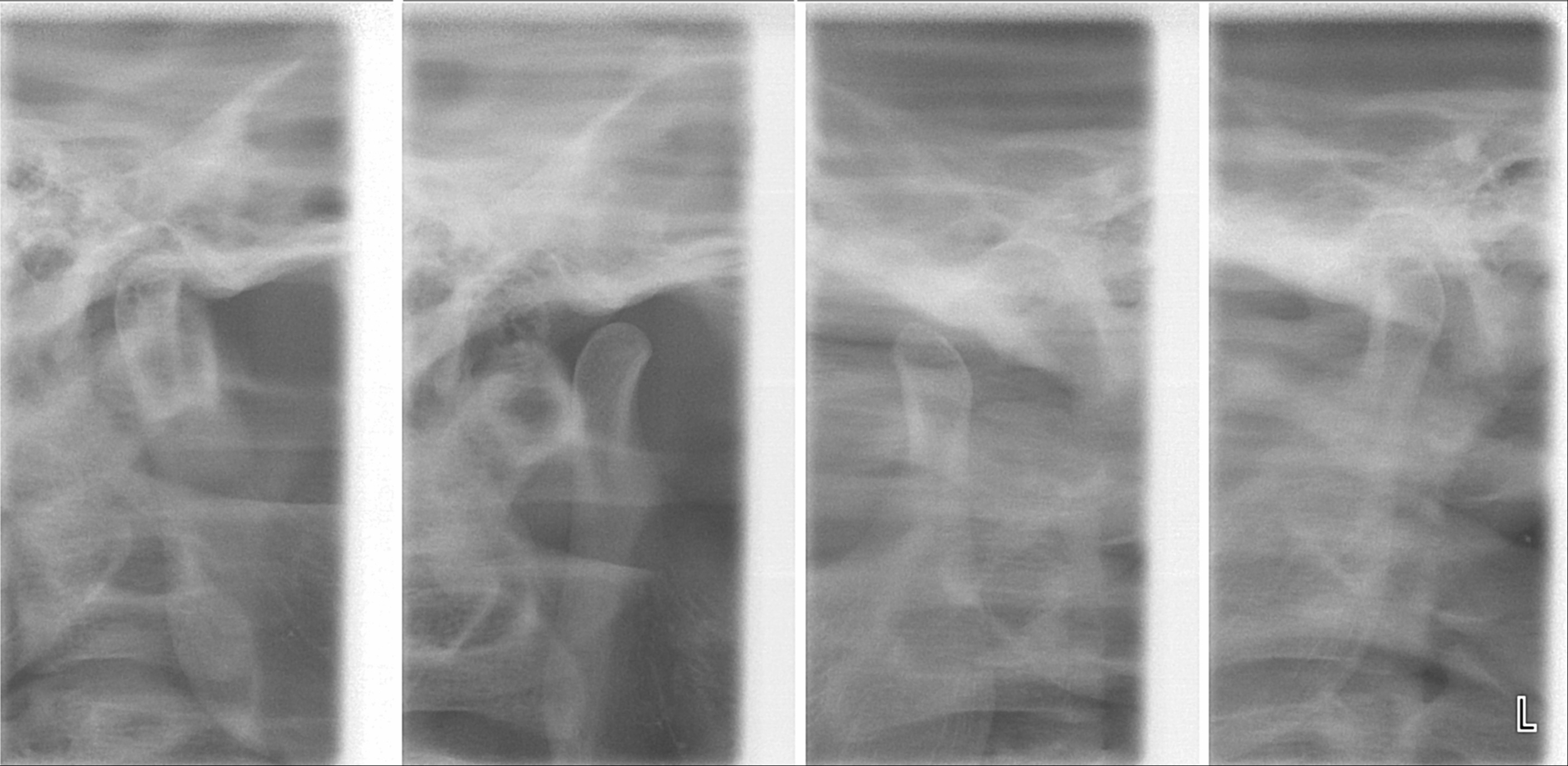
Idiopathic Condylar Resorption
Condylar resorption, also called idiopathic condylar resorption, ICR, and condylysis, is a temporomandibular joint disorder in which one or both of the mandibular condyles are broken down in a bone resorption process. This disorder is nine times more likely to be present in females than males, and is more common among teenagers. Symptoms and signs Symptoms that may be associated with condylar resorption are both aesthetic and functional. These include: *Occlusion (dentistry), Occlusion *Anterior open bite *Receding chin *Loss of Mandible#Ramus, ramus height *Antegonial notching *Hyperplasia of the coronoid process of the mandible *Clicking or popping when opening or closing the jaw *Pain when opening or closing the jaw *Limited jaw mobility Causes The cause of condylar resorption is unknown, but there are theories. Because condylar resorption is much more likely to occur in young females, hormonal mediation may be involved. Strain on the temporomandibular joint from orthodontics ... [...More Info...] [...Related Items...] OR: [Wikipedia] [Google] [Baidu] |
Temporomandibular Joint Disorder
Temporomandibular joint dysfunction (TMD, TMJD) is an umbrella term covering pain and dysfunction of the muscles of mastication (the muscles that move the jaw) and the temporomandibular joints (the joints which connect the mandible to the skull). The most important feature is pain, followed by restricted mandibular movement, and noises from the temporomandibular joints (TMJ) during jaw movement. Although TMD is not life-threatening, it can be detrimental to quality of life; this is because the symptoms can become chronic and difficult to manage. In this article, the term ''temporomandibular disorder'' is taken to mean any disorder that affects the temporomandibular joint, and ''temporomandibular joint dysfunction'' (here also abbreviated to TMD) is taken to mean symptomatic (e.g. pain, limitation of movement, clicking) dysfunction of the temporomandibular joint. However, there is no single, globally accepted term or definition concerning this topic. TMDs have a range of cause ... [...More Info...] [...Related Items...] OR: [Wikipedia] [Google] [Baidu] |
Single-photon Emission Computed Tomography
Single-photon emission computed tomography (SPECT, or less commonly, SPET) is a nuclear medicine tomographic imaging technique using gamma rays. It is very similar to conventional nuclear medicine planar imaging using a gamma camera (that is, scintigraphy), but is able to provide true 3D information. This information is typically presented as cross-sectional slices through the patient, but can be freely reformatted or manipulated as required. The technique needs delivery of a gamma-emitting radioisotope (a radionuclide) into the patient, normally through injection into the bloodstream. On occasion, the radioisotope is a simple soluble dissolved ion, such as an isotope of gallium(III). Most of the time, though, a marker radioisotope is attached to a specific ligand to create a radioligand, whose properties bind it to certain types of tissues. This marriage allows the combination of ligand and radiopharmaceutical to be carried and bound to a place of interest in the body, where ... [...More Info...] [...Related Items...] OR: [Wikipedia] [Google] [Baidu] |
Idiopathic Diseases
An idiopathic disease is any disease with an unknown cause or mechanism of apparent spontaneous origin. From Greek ἴδιος ''idios'' "one's own" and πάθος ''pathos'' "suffering", ''idiopathy'' means approximately "a disease of its own kind". For some medical conditions, one or more causes are somewhat understood, but in a certain percentage of people with the condition, the cause may not be readily apparent or characterized. In these cases, the origin of the condition is said to be idiopathic. With some other medical conditions, the root cause for a large percentage of all cases have not been established—for example, focal segmental glomerulosclerosis or ankylosing spondylitis; the majority of these cases are deemed idiopathic. Medical advances and this term Advances in medical science improve the understanding of causes of diseases and the classification of diseases; thus, regarding any particular condition or disease, as more root causes are discovered and as events t ... [...More Info...] [...Related Items...] OR: [Wikipedia] [Google] [Baidu] |
Musculoskeletal Disorders
Musculoskeletal disorders (MSDs) are injuries or pain in the human musculoskeletal system, including the joints, ligaments, muscles, nerves, tendons, and structures that support limbs, neck and back. MSDs can arise from a sudden exertion (e.g., lifting a heavy object), or they can arise from making the same motions repeatedly repetitive strain, or from repeated exposure to force, vibration, or awkward posture. Injuries and pain in the musculoskeletal system caused by acute traumatic events like a car accident or fall are not considered musculoskeletal disorders. MSDs can affect many different parts of the body including upper and lower back, neck, shoulders and extremities (arms, legs, feet, and hands). Examples of MSDs include carpal tunnel syndrome, epicondylitis, tendinitis, back pain, tension neck syndrome, and hand-arm vibration syndrome. Causes MSDs can arise from the interaction of physical factors with ergonomic, psychological, social, and occupational factors. Biomechanic ... [...More Info...] [...Related Items...] OR: [Wikipedia] [Google] [Baidu] |
Condylar Hyperplasia
Condylar hyperplasia (mandibular hyperplasia) is over-enlargement of the mandible bone in the skull. It was first described by Robert Adams in 1836 who related it to the overdevelopment of mandible. In humans, mandibular bone has two condyles which are known as growth centers of the mandible. When growth at the condyle exceeds its normal time span, it is referred to as condylar hyperplasia. The most common form of condylar hyperplasia is ''unilateral condylar hyperplasia'' where one condyle overgrows the other condyle leading to facial asymmetry. Hugo Obwegeser et al. classified condylar hyperplasia into two categories: ''hemimandibular hyperplasia'' and ''hemimandibular elongation''. It is estimated that about 30% of people with facial asymmetry express condylar hyperplasia. In 1986, Obwegeser and Makek specifically detailed two hemimandibular anomalies, hemimandibular hyperplasia and hemimandibular elongation. These anomalies can be clinically present in a pure form or in ... [...More Info...] [...Related Items...] OR: [Wikipedia] [Google] [Baidu] |
TMJ Disorder
Temporomandibular joint dysfunction (TMD, TMJD) is an umbrella term covering pain and dysfunction of the muscles of mastication (the muscles that move the jaw) and the temporomandibular joints (the joints which connect the mandible to the skull). The most important feature is pain, followed by restricted mandibular movement, and noises from the temporomandibular joints (TMJ) during jaw movement. Although TMD is not life-threatening, it can be detrimental to quality of life; this is because the symptoms can become chronic and difficult to manage. In this article, the term ''temporomandibular disorder'' is taken to mean any disorder that affects the temporomandibular joint, and ''temporomandibular joint dysfunction'' (here also abbreviated to TMD) is taken to mean symptomatic (e.g. pain, limitation of movement, clicking) dysfunction of the temporomandibular joint. However, there is no single, globally accepted term or definition concerning this topic. TMDs have a range of cause ... [...More Info...] [...Related Items...] OR: [Wikipedia] [Google] [Baidu] |
Surgery For Temporomandibular Joint Dysfunction
Attempts in the last decade to develop surgical treatments based on MRI and CAT scans now receive less attention. These techniques are reserved for the most difficult cases where other therapeutic modalities have failed. The American Society of Maxillofacial Surgeons recommends a conservative/non-surgical approach first. Only 20% of patients need to proceed to surgery. Examples of surgical procedures that are used in TMD, some more commonly than others, include arthrocentesis, arthroscopy, meniscectomy, disc repositioning, condylotomy or joint replacement. Invasive surgical procedures in TMD may cause symptoms to worsen. Menisectomy, also termed discectomy refers to the surgical removal of the articular disc. This is rarely carried out in TMD, it may have some benefits for pain, but dysfunction may persist and overall there it leads to degeneration or remodeling of the TMJ. Arthrocentesis TMJ arthrocentesis refers to lavage (flushing out) of the upper joint space (where most of ... [...More Info...] [...Related Items...] OR: [Wikipedia] [Google] [Baidu] |
Arthroscopic
Arthroscopy (also called arthroscopic or keyhole surgery) is a minimally invasive surgical procedure on a joint in which an examination and sometimes treatment of damage is performed using an arthroscope, an endoscope that is inserted into the joint through a small incision. Arthroscopic procedures can be performed during ACL reconstruction. The advantage over traditional open surgery is that the joint does not have to be opened up fully. For knee arthroscopy only two small incisions are made, one for the arthroscope and one for the surgical instruments to be used in the knee cavity. This reduces recovery time and may increase the rate of success due to less trauma to the connective tissue. It has gained popularity due to evidence of faster recovery times with less scarring, because of the smaller incisions. Irrigation fluid (most commonly 'normal' saline) is used to distend the joint and make a surgical space. The surgical instruments are smaller than traditional instruments. ... [...More Info...] [...Related Items...] OR: [Wikipedia] [Google] [Baidu] |
Arthrocentesis
Arthrocentesis, or joint aspiration, is the clinical procedure performed to diagnose and, in some cases, treat musculoskeletal conditions. The procedure entails using a syringe to collect synovial fluid from or inject medication into the joint capsule. Laboratory analysis of synovial fluid can further help characterize the diseased joint and distinguish between gout, arthritis, and synovial infections such as septic arthritis. Uses In general, arthrocentesis should be strongly considered if there is suspected trauma, infection, or effusion of the joint. Diagnostic Arthrocentesis can be used to diagnose septic arthritis or crystal arthropathy. In the case of a septic joint, arthrocentesis should preferably be performed prior to starting treatment with antibiotics, in order to ensure a proper sample of synovial fluid is obtained. Synovial Fluid Analysis Patients with a fever, suspected flare of existing arthritis, or unknown cause of joint effusion should undergo arthrocente ... [...More Info...] [...Related Items...] OR: [Wikipedia] [Google] [Baidu] |
Inferior Alveolar Nerve
The inferior alveolar nerve (IAN) (also the inferior dental nerve) is a branch of the mandibular nerve, which is itself the third branch of the trigeminal nerve. The inferior alveolar nerves supply sensation to the lower teeth. Structure The inferior alveolar nerve is a branch of the mandibular nerve. After branching from the mandibular nerve, the inferior alveolar nerve travels behind the lateral pterygoid muscle. It gives off a branch, the mylohyoid nerve, and then enters the mandibular foramen. While in the mandibular canal within the mandible, it supplies the lower teeth (molars and second premolar) with sensory branches that form into the inferior dental plexus and give off small gingival and dental nerves to the teeth. Anteriorly, the nerve gives off the mental nerve at about the level of the mandibular 2nd premolars, which exits the mandible via the mental foramen and supplies sensory branches to the chin and lower lip. The inferior alveolar nerve continues anteriorl ... [...More Info...] [...Related Items...] OR: [Wikipedia] [Google] [Baidu] |
Orthognathic Surgery
Orthognathic surgery (), also known as corrective jaw surgery or simply jaw surgery, is surgery designed to correct conditions of the jaw and lower face related to structure, growth, airway issues including sleep apnea, TMJ disorders, malocclusion problems primarily arising from skeletal disharmonies, other orthodontic dental bite problems that cannot be easily treated with braces, as well as the broad range of facial imbalances, disharmonies, asymmetries and malproportions where correction can be considered to improve facial aesthetics and self esteem. The origins of orthognathic surgery belong in oral surgery, and the basic operations related to the surgical removal of impacted or displaced teeth – especially where indicated by orthodontics to enhance dental treatments of malocclusion and dental crowding. One of the first published cases of orthognathic surgery was the one from Dr. Simon P. Hullihen in 1849. Originally coined by Harold Hargis, it was more widely popularised ... [...More Info...] [...Related Items...] OR: [Wikipedia] [Google] [Baidu] |
Orthodontics
Orthodontics is a dentistry specialty that addresses the diagnosis, prevention, management, and correction of mal-positioned teeth and jaws, and misaligned bite patterns. It may also address the modification of facial growth, known as dentofacial orthopedics. Abnormal alignment of the teeth and jaws is very common. Nearly 50% of the developed world's population, according to the American Association of Orthodontics, has malocclusions severe enough to benefit from orthodontic treatment: although this figure decreases to less than 10% according to the same AAO statement when referring to medically necessary orthodontics. However, conclusive scientific evidence for the health benefits of orthodontic treatment is lacking, although patients with completed orthodontic treatment have reported a higher quality of life than that of untreated patients undergoing orthodontic treatment. Treatment may require several months to a few years, and entails using dental braces and other appliances t ... [...More Info...] [...Related Items...] OR: [Wikipedia] [Google] [Baidu] |


