|
Hypertrophic Cardiomyopathy Screening
Hypertrophic cardiomyopathy screening is an assessment and testing to detect hypertrophic cardiomyopathy (HCM). It is a way of identifying HCM in immediate relatives of family members diagnosed with HCM, and athletes as part of a sports medical. It aims to detect HCM early, so that interventions can be commenced to prevent complications and sudden cardiac death. __TOC__ Purpose HCM is a heart disease in which a portion of the heart becomes thickened without an obvious cause. It affects up to one in 200 people and runs in families. A significant number of people with the condition have no symptoms. Screening is a way of identifying HCM in immediate relatives of family members diagnosed with hypertrophic cardiomyopathy (HCM), and athletes as part of a sports medical. Additional tests may also be performed in those who faint or have exertional chest pain. It aims to detect HCM early, so that interventions can be commenced to prevent complications and sudden cardiac death. ... [...More Info...] [...Related Items...] OR: [Wikipedia] [Google] [Baidu] |
Hypertrophic Cardiomyopathy
Hypertrophic cardiomyopathy (HCM, or HOCM when obstructive) is a condition in which the heart becomes thickened without an obvious cause. The parts of the heart most commonly affected are the interventricular septum and the ventricles. This results in the heart being less able to pump blood effectively and also may cause electrical conduction problems. People who have HCM may have a range of symptoms. People may be asymptomatic, or may have fatigue, leg swelling, and shortness of breath. It may also result in chest pain or fainting. Symptoms may be worse when the person is dehydrated. Complications may include heart failure, an irregular heartbeat, and sudden cardiac death. HCM is most commonly inherited from a person's parents in an autosomal dominant pattern. It is often due to mutations in certain genes involved with making heart muscle proteins. Other inherited causes of left ventricular hypertrophy may include Fabry disease, Friedreich's ataxia, and certain medica ... [...More Info...] [...Related Items...] OR: [Wikipedia] [Google] [Baidu] |
Cardiac MRI
Cardiac magnetic resonance imaging (cardiac MRI), also known as cardiovascular MRI, is a magnetic resonance imaging (MRI) medical imaging, technology used for non-invasive assessment of the function and structure of the cardiovascular system. Conditions in which it is performed include congenital heart disease, cardiomyopathies and valvular heart disease, diseases of the aorta such as aortic dissection, dissection, aortic aneurysm, aneurysm and coarctation of aorta, coarctation, coronary heart disease and it can be used to look at pulmonary veins. It is contraindicated if there is a permanent pacemaker or defibrillator, intracerebral clipping (medicine), clips or claustrophobia. Conventional MRI sequences are adapted for cardiac imaging by using Electrocardiography, ECG gating and high temporal resolution protocols. The development of cardiac MRI is an active field of research and continues to see a rapid expansion of new and emerging techniques. Uses Cardiovascular MRI is complem ... [...More Info...] [...Related Items...] OR: [Wikipedia] [Google] [Baidu] |
Cardiomyopathy
Cardiomyopathy is a group of diseases that affect the heart muscle. Early on there may be few or no symptoms. As the disease worsens, shortness of breath, feeling tired, and swelling of the legs may occur, due to the onset of heart failure. An irregular heart beat and fainting may occur. Those affected are at an increased risk of sudden cardiac death. Types of cardiomyopathy include hypertrophic cardiomyopathy, dilated cardiomyopathy, restrictive cardiomyopathy, arrhythmogenic right ventricular dysplasia, and Takotsubo cardiomyopathy (broken heart syndrome). In hypertrophic cardiomyopathy the heart muscle enlarges and thickens. In dilated cardiomyopathy the ventricles enlarge and weaken. In restrictive cardiomyopathy the ventricle stiffens. In many cases, the cause cannot be determined. Hypertrophic cardiomyopathy is usually inherited, whereas dilated cardiomyopathy is inherited in about one third of cases. Dilated cardiomyopathy may also result from alcohol, heavy m ... [...More Info...] [...Related Items...] OR: [Wikipedia] [Google] [Baidu] |
False Positives And False Negatives
A false positive is an error in binary classification in which a test result incorrectly indicates the presence of a condition (such as a disease when the disease is not present), while a false negative is the opposite error, where the test result incorrectly indicates the absence of a condition when it is actually present. These are the two kinds of errors in a binary test, in contrast to the two kinds of correct result (a and a ). They are also known in medicine as a false positive (or false negative) diagnosis, and in statistical classification as a false positive (or false negative) error. In statistical hypothesis testing the analogous concepts are known as type I and type II errors, where a positive result corresponds to rejecting the null hypothesis, and a negative result corresponds to not rejecting the null hypothesis. The terms are often used interchangeably, but there are differences in detail and interpretation due to the differences between medical testing and statist ... [...More Info...] [...Related Items...] OR: [Wikipedia] [Google] [Baidu] |
Preparticipation Physical Evaluation
In sports medicine, a preparticipation physical evaluation (PPE) is a physical examination of an athlete. PPEs screen for a variety of conditions, including athletic heart syndrome and risk of sudden cardiac death. PPEs are required for athletic participation according to the laws of some jurisdictions and the rules of many sports governing bodies. PPE is known by a variety of other names, such as preparticipation evaluation, preparticipation physical examination, preparticipation screening, sports physical, sports physical exam, examination for participation in sport, and similar. As of 2019, the latest PPE recommendation published by several US physician organizations is the 5th edition, called PPE5. PPE5 was published by American Academy of Pediatrics, American Academy of Family Physicians, American College of Sports Medicine, American Medical Society for Sports Medicine, American Orthopaedic Society for Sports Medicine, and American Osteopathic Academy of Sports Medicine ... [...More Info...] [...Related Items...] OR: [Wikipedia] [Google] [Baidu] |
Overdiagnosis
Overdiagnosis is the diagnosis of disease that will never cause symptoms or death during a patient's ordinarily expected lifetime and thus presents no practical threat regardless of being pathologic. Overdiagnosis is a side effect of screening for early forms of disease. Although screening saves lives in some cases, in others it may turn people into patients unnecessarily and may lead to treatments that do no good and perhaps do harm. Given the tremendous variability that is normal in biology, it is inherent that the more one screens, the more incidental findings will generally be found. For a large percentage of them, the most appropriate medical response is to recognize them as something that does not require intervention; but determining which action a particular finding warrants ("ignoring", watchful waiting, or intervention) can be very difficult, whether because the differential diagnosis is uncertain or because the risk ratio is uncertain (risks posed by intervention, namel ... [...More Info...] [...Related Items...] OR: [Wikipedia] [Google] [Baidu] |
Misdiagnosis
A medical error is a preventable adverse effect of care ("iatrogenesis"), whether or not it is evident or harmful to the patient. This might include an inaccurate or incomplete diagnosis or treatment of a disease, injury, syndrome, behavior, infection, or other ailment. Definitions The word ''error'' in medicine is used as a label for nearly all of the clinical incidents that harm patients. Medical errors are often described as human errors in healthcare. Whether the label is a medical error or human error, one definition used in medicine says that it occurs when a healthcare provider chooses an inappropriate method of care, improperly executes an appropriate method of care, or reads the wrong CT scan. It has been said that the definition should be the subject of more debate. For instance, studies of hand hygiene compliance of physicians in an ICU show that compliance varied from 19% to 85%. The deaths that result from infections caught as a result of treatment providers imp ... [...More Info...] [...Related Items...] OR: [Wikipedia] [Google] [Baidu] |
Left Ventricular Hypertrophy
Left ventricular hypertrophy (LVH) is thickening of the heart muscle of the left ventricle of the heart, that is, left-sided ventricular hypertrophy and resulting increased left ventricular mass. Causes While ventricular hypertrophy occurs naturally as a reaction to aerobic exercise and strength training, it is most frequently referred to as a pathological reaction to cardiovascular disease, or high blood pressure. It is one aspect of ventricular remodeling. While LVH itself is not a disease, it is usually a marker for disease involving the heart. Disease processes that can cause LVH include any disease that increases the afterload that the heart has to contract against, and some primary diseases of the muscle of the heart. Causes of increased afterload that can cause LVH include aortic stenosis, aortic insufficiency and hypertension. Primary disease of the muscle of the heart that cause LVH are known as hypertrophic cardiomyopathies, which can lead into heart failure. Lon ... [...More Info...] [...Related Items...] OR: [Wikipedia] [Google] [Baidu] |
QRS Complex
The QRS complex is the combination of three of the graphical deflections seen on a typical electrocardiogram (ECG or EKG). It is usually the central and most visually obvious part of the tracing. It corresponds to the depolarization of the right and left ventricles of the heart and contraction of the large ventricular muscles. In adults, the QRS complex normally lasts ; in children it may be shorter. The Q, R, and S waves occur in rapid succession, do not all appear in all leads, and reflect a single event and thus are usually considered together. A Q wave is any downward deflection immediately following the P wave. An R wave follows as an upward deflection, and the S wave is any downward deflection after the R wave. The T wave follows the S wave, and in some cases, an additional U wave follows the T wave. To measure the QRS interval start at the end of the PR interval (or beginning of the Q wave) to the end of the S wave. Normally this interval is 0.08 to 0.10 seconds. When ... [...More Info...] [...Related Items...] OR: [Wikipedia] [Google] [Baidu] |
ST Depression
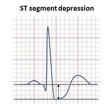
ST depression refers to a finding on an electrocardiogram, wherein the trace in the ST segment is abnormally low below the baseline. Causes It is often a sign of myocardial ischemia, of which coronary insufficiency is a major cause. Other ischemic heart diseases causing ST depression include: * Subendocardial ischemia or even infarction. Subendocardial means non full thickness ischemia. In contrast, ST elevation is transmural (or full thickness) ischemia * Non Q-wave myocardial infarction * Reciprocal changes in acute Q-wave myocardial infarction (e.g., ST depression in leads I & aVL with acute inferior myocardial infarction) * ST segment depression and T-wave changes may be seen in patients with unstable angina Depressed but ''upsloping'' ST segment generally rules out ischemia as a cause. Also, it can be a normal variant or artifacts, such as: * Pseudo-ST-depression, which is a wandering baseline due to poor skin contact of the electrode MicroEKG ManualRetrieved September 2 ... [...More Info...] [...Related Items...] OR: [Wikipedia] [Google] [Baidu] |
T Wave
In electrocardiography, the T wave represents the repolarization of the ventricles. The interval from the beginning of the QRS complex to the apex of the T wave is referred to as the ''absolute refractory period''. The last half of the T wave is referred to as the ''relative refractory period'' or ''vulnerable period''. The T wave contains more information than the QT interval. The T wave can be described by its symmetry, skewness, slope of ascending and descending limbs, amplitude and subintervals like the Tpeak–Tend interval. In most leads, the T wave is positive. This is due to the repolarization of the membrane. During ventricle contraction (QRS complex), the heart depolarizes. Repolarization of the ventricle happens in the opposite direction of depolarization and is negative current, signifying the relaxation of the cardiac muscle of the ventricles. But this negative flow causes a positive T wave; although the cell becomes more negatively charged, the net effect is in t ... [...More Info...] [...Related Items...] OR: [Wikipedia] [Google] [Baidu] |
12-lead ECG
Electrocardiography is the process of producing an electrocardiogram (ECG or EKG), a recording of the heart's electrical activity. It is an electrogram of the heart which is a graph of voltage versus time of the electrical activity of the heart using electrodes placed on the skin. These electrodes detect the small electrical changes that are a consequence of cardiac muscle depolarization followed by repolarization during each cardiac cycle (heartbeat). Changes in the normal ECG pattern occur in numerous cardiac abnormalities, including cardiac rhythm disturbances (such as atrial fibrillation and ventricular tachycardia), inadequate coronary artery blood flow (such as myocardial ischemia and myocardial infarction), and electrolyte disturbances (such as hypokalemia and hyperkalemia). Traditionally, "ECG" usually means a 12-lead ECG taken while lying down as discussed below. However, other devices can record the electrical activity of the heart such as a Holter monitor but also s ... [...More Info...] [...Related Items...] OR: [Wikipedia] [Google] [Baidu] |


