|
Human Rib Cage
The rib cage, as an enclosure that comprises the ribs, vertebral column and sternum in the thorax of most vertebrates, protects vital organs such as the heart, lungs and great vessels. The sternum, together known as the thoracic cage, is a semi-rigid bony and cartilaginous structure which surrounds the thoracic cavity and supports the shoulder girdle to form the core part of the human skeleton. A typical human thoracic cage consists of 12 pairs of ribs and the adjoining costal cartilages, the sternum (along with the manubrium and xiphoid process), and the 12 thoracic vertebrae articulating with the ribs. Together with the skin and associated fascia and muscles, the thoracic cage makes up the thoracic wall and provides attachments for extrinsic skeletal muscles of the neck, upper limbs, upper abdomen and back. The rib cage intrinsically holds the muscles of respiration ( diaphragm, intercostal muscles, etc.) that are crucial for active inhalation and forced exhalation, and the ... [...More Info...] [...Related Items...] OR: [Wikipedia] [Google] [Baidu] |
Gray's Anatomy
''Gray's Anatomy'' is a reference book of human anatomy written by Henry Gray, illustrated by Henry Vandyke Carter, and first published in London in 1858. It has gone through multiple revised editions and the current edition, the 42nd (October 2020), remains a standard reference, often considered "the doctors' bible". Earlier editions were called ''Anatomy: Descriptive and Surgical'', ''Anatomy of the Human Body'' and ''Gray's Anatomy: Descriptive and Applied'', but the book's name is commonly shortened to, and later editions are titled, ''Gray's Anatomy''. The book is widely regarded as an extremely influential work on the subject. Publication history Origins The English anatomist Henry Gray was born in 1827. He studied the development of the endocrine glands and spleen and in 1853 was appointed Lecturer on Anatomy at St George's Hospital Medical School in London. In 1855, he approached his colleague Henry Vandyke Carter with his idea to produce an inexpensive and ac ... [...More Info...] [...Related Items...] OR: [Wikipedia] [Google] [Baidu] |
Xiphoid Process
The xiphoid process , or xiphisternum or metasternum, is a small cartilaginous process (extension) of the inferior (lower) part of the sternum, which is usually ossified in the adult human. It may also be referred to as the ensiform process. Both the Greek-derived ''xiphoid'' and its Latin equivalent ''ensiform'' mean 'swordlike' or 'sword-shaped' Structure The xiphoid process is considered to be at the level of the 9th thoracic vertebra and the T7 dermatome. Development In newborns and young (especially small) infants, the tip of the xiphoid process may be both seen and felt as a lump just below the sternal notch. At 15 to 29 years old, the xiphoid usually fuses to the body of the sternum with a fibrous joint. Unlike the synovial articulation of major joints, this is non-movable. Ossification of the xiphoid process occurs around age 40. Variation The xiphoid process can be naturally bifurcated or sometimes perforated (xiphoidal foramen). These variances in morphology are inher ... [...More Info...] [...Related Items...] OR: [Wikipedia] [Google] [Baidu] |
Intercostal Muscles
Intercostal muscles are many different groups of muscles that run between the ribs, and help form and move the chest wall. The intercostal muscles are mainly involved in the mechanical aspect of breathing by helping expand and shrink the size of the chest cavity. Structure There are three principal layers; # External intercostal muscles also known as intercostalis externus aid in quiet and forced inhalation. They originate on ribs 1–11 and have their insertion on ribs 2–12. The external intercostals are responsible for the elevation of the ribs and bending them more open, thus expanding the transverse dimensions of the thoracic cavity. The muscle fibers are directed downwards, forwards and medially in the anterior part. # Internal intercostal muscles also known as intercostalis internus aid in forced expiration (quiet expiration is a passive process). They originate on ribs 2–12 and have their insertions on ribs 1–11.Their fibers pass anterior and supe ... [...More Info...] [...Related Items...] OR: [Wikipedia] [Google] [Baidu] |
Thoracic Diaphragm
The thoracic diaphragm, or simply the diaphragm ( grc, διάφραγμα, diáphragma, partition), is a sheet of internal Skeletal striated muscle, skeletal muscle in humans and other mammals that extends across the bottom of the thoracic cavity. The diaphragm is the most important Muscles of respiration, muscle of respiration, and separates the thoracic cavity, containing the heart and lungs, from the abdominal cavity: as the diaphragm contracts, the volume of the thoracic cavity increases, creating a negative pressure there, which draws air into the lungs. Its high oxygen consumption is noted by the many mitochondria and capillaries present; more than in any other skeletal muscle. The term ''diaphragm'' in anatomy, created by Gerard of Cremona, can refer to other flat structures such as the urogenital diaphragm or Pelvic floor, pelvic diaphragm, but "the diaphragm" generally refers to the thoracic diaphragm. In humans, the diaphragm is slightly asymmetric—its right half is h ... [...More Info...] [...Related Items...] OR: [Wikipedia] [Google] [Baidu] |
Muscles Of Respiration
The muscles of respiration are the muscles that contribute to inhalation and exhalation, by aiding in the expansion and contraction of the thoracic cavity. The diaphragm and, to a lesser extent, the intercostal muscles drive respiration during quiet breathing. The elasticity of these muscles is crucial to the health of the respiratory system and to maximize its functional capabilities. Diaphragm The diaphragm is the major muscle responsible for breathing. It is a thin, dome-shaped muscle that separates the abdominal cavity from the thoracic cavity. During inhalation, the diaphragm contracts, so that its center moves caudally (downward) and its edges move cranially (upward). This compresses the abdominal cavity, raises the ribs upward and outward and thus expands the thoracic cavity. This expansion draws air into the lungs. When the diaphragm relaxes, elastic recoil of the lungs causes the thoracic cavity to contract, forcing air out of the lungs, and returning to its dome-shap ... [...More Info...] [...Related Items...] OR: [Wikipedia] [Google] [Baidu] |
Back
The human back, also called the dorsum, is the large posterior area of the human body, rising from the top of the buttocks to the back of the neck. It is the surface of the body opposite from the chest and the abdomen. The vertebral column runs the length of the back and creates a central area of recession. The breadth of the back is created by the shoulders at the top and the pelvis at the bottom. Back pain is a common medical condition, generally benign in origin. Structure The central feature of the human back is the vertebral column, specifically the length from the top of the thoracic vertebrae to the bottom of the lumbar vertebrae, which houses the spinal cord in its spinal canal, and which generally has some curvature that gives shape to the back. The ribcage extends from the spine at the top of the back (with the top of the ribcage corresponding to the T1 vertebra), more than halfway down the length of the back, leaving an area with less protection between the bottom ... [...More Info...] [...Related Items...] OR: [Wikipedia] [Google] [Baidu] |
Upper Abdomen
In anatomy, the epigastrium (or epigastric region) is the upper central region of the abdomen. It is located between the costal margins and the subcostal plane. Pain may be referred to the epigastrium from damage to structures derived from the foregut. Structure The epigastrium is one of the nine regions of the abdomen, along with the right and left hypochondria, right and left lateral regions (lumbar areas or flanks), right and left inguinal regions (or fossae), and the umbilical and pubic regions. It is located between the costal margins and the subcostal plane. During breathing, the diaphragm contracts and flattens, displacing the viscera and producing an outward movement of the upper abdominal wall (epigastric region). It is a convergence of the diaphragm and the abdominals, so that "when both sets of muscles (diaphragm and abdominals) tense, the epigastrium pushes forward". Therefore, the epigastric region is not a muscle nor is it an organ, but it is a zone of activit ... [...More Info...] [...Related Items...] OR: [Wikipedia] [Google] [Baidu] |
Upper Limb
The upper limbs or upper extremities are the forelimbs of an upright-postured tetrapod vertebrate, extending from the scapulae and clavicles down to and including the digits, including all the musculatures and ligaments involved with the shoulder, elbow, wrist and knuckle joints. In humans, each upper limb is divided into the arm, forearm and hand, and is primarily used for climbing, lifting and manipulating objects. Definition In formal usage, the term "arm" only refers to the structures from the shoulder to the elbow, explicitly excluding the forearm, and thus "upper limb" and "arm" are not synonymous. However, in casual usage, the terms are often used interchangeably. The term "upper arm" is redundant in anatomy, but in informal usage is used to distinguish between the two terms. Structure In the human body the muscles of the upper limb can be classified by origin, topography, function, or innervation. While a grouping by innervation reveals embryological and phylogenet ... [...More Info...] [...Related Items...] OR: [Wikipedia] [Google] [Baidu] |
Neck
The neck is the part of the body on many vertebrates that connects the head with the torso. The neck supports the weight of the head and protects the nerves that carry sensory and motor information from the brain down to the rest of the body. In addition, the neck is highly flexible and allows the head to turn and flex in all directions. The structures of the human neck are anatomically grouped into four compartments; vertebral, visceral and two vascular compartments. Within these compartments, the neck houses the cervical vertebrae and cervical part of the spinal cord, upper parts of the respiratory and digestive tracts, endocrine glands, nerves, arteries and veins. Muscles of the neck are described separately from the compartments. They bound the neck triangles. In anatomy, the neck is also called by its Latin names, or , although when used alone, in context, the word ''cervix'' more often refers to the uterine cervix, the neck of the uterus. Thus the adjective ''cervical'' ma ... [...More Info...] [...Related Items...] OR: [Wikipedia] [Google] [Baidu] |
Skeletal Muscle
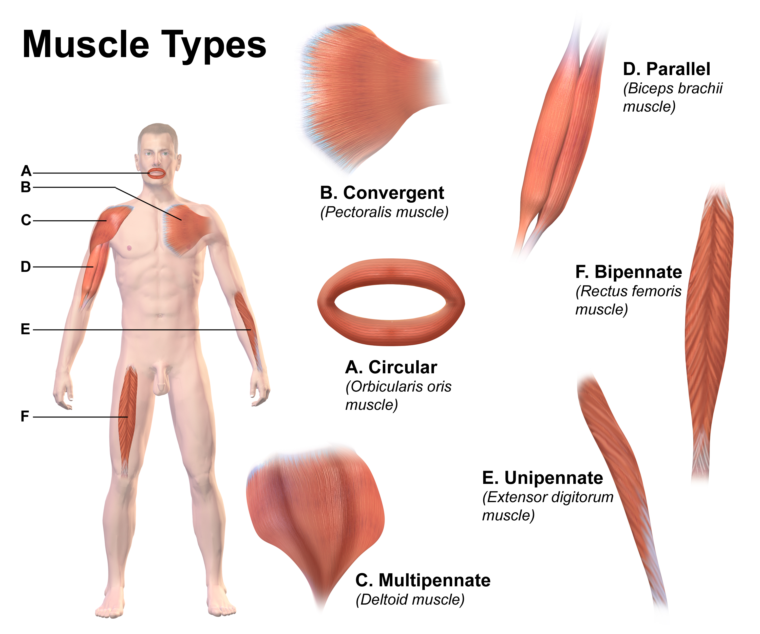
Skeletal muscles (commonly referred to as muscles) are organs of the vertebrate muscular system and typically are attached by tendons to bones of a skeleton. The muscle cells of skeletal muscles are much longer than in the other types of muscle tissue, and are often known as muscle fibers. The muscle tissue of a skeletal muscle is striated – having a striped appearance due to the arrangement of the sarcomeres. Skeletal muscles are voluntary muscles under the control of the somatic nervous system. The other types of muscle are cardiac muscle which is also striated and smooth muscle which is non-striated; both of these types of muscle tissue are classified as involuntary, or, under the control of the autonomic nervous system. A skeletal muscle contains multiple muscle fascicle, fascicles – bundles of muscle fibers. Each individual fiber, and each muscle is surrounded by a type of connective tissue layer of fascia. Muscle fibers are formed from the cell fusion, fusion of ... [...More Info...] [...Related Items...] OR: [Wikipedia] [Google] [Baidu] |
Thoracic Wall
The thoracic wall or chest wall is the boundary of the thoracic cavity. Structure The bony skeletal part of the thoracic wall is the rib cage, and the rest is made up of muscle, skin, and fasciae. The chest wall has 10 layers, namely (from superficial to deep) skin (epidermis and dermis), superficial fascia, deep fascia and the invested extrinsic muscles (from the upper limbs), intrinsic muscles associated with the ribs (three layers of intercostal muscles), endothoracic fascia and parietal pleura. However, the extrinsic muscular layers vary according to the region of the chest wall. For example, the front and back sides may include attachments of large upper limb muscles like pectoralis major or latissimus dorsi, while the sides only have serratus anterior. Function Diving reflex When not breathing for long and dangerous periods of time in cold water, a person's body undergoes great temporary changes to try to prevent death. It achieves this through the activation of ... [...More Info...] [...Related Items...] OR: [Wikipedia] [Google] [Baidu] |
Muscle
Skeletal muscles (commonly referred to as muscles) are organs of the vertebrate muscular system and typically are attached by tendons to bones of a skeleton. The muscle cells of skeletal muscles are much longer than in the other types of muscle tissue, and are often known as muscle fibers. The muscle tissue of a skeletal muscle is striated – having a striped appearance due to the arrangement of the sarcomeres. Skeletal muscles are voluntary muscles under the control of the somatic nervous system. The other types of muscle are cardiac muscle which is also striated and smooth muscle which is non-striated; both of these types of muscle tissue are classified as involuntary, or, under the control of the autonomic nervous system. A skeletal muscle contains multiple fascicles – bundles of muscle fibers. Each individual fiber, and each muscle is surrounded by a type of connective tissue layer of fascia. Muscle fibers are formed from the fusion of developmental myoblasts in ... [...More Info...] [...Related Items...] OR: [Wikipedia] [Google] [Baidu] |


