|
Hounsfield Unit
The Hounsfield scale , named after Sir Godfrey Hounsfield, is a quantitative scale for describing radiodensity. It is frequently used in CT scans, where its value is also termed CT number. Definition The Hounsfield unit (HU) scale is a linear transformation of the original linear attenuation coefficient measurement into one in which the radiodensity of distilled water at standard pressure and temperature (STP) is defined as 0 Hounsfield units (HU), while the radiodensity of air at STP is defined as −1000 HU. In a voxel with average linear attenuation coefficient \mu, the corresponding HU value is therefore given by: HU = 1000\times\frac where \mu_ and \mu_ are respectively the linear attenuation coefficients of water and air. Thus, a change of one Hounsfield unit (HU) represents a change of 0.1% of the attenuation coefficient of water since the attenuation coefficient of air is nearly zero. Calibration tests of HU with reference to water and other materials may be done to ensu ... [...More Info...] [...Related Items...] OR: [Wikipedia] [Google] [Baidu] |
Godfrey Hounsfield
Sir Godfrey Newbold Hounsfield (28 August 1919 – 12 August 2004) was an English electrical engineer who shared the 1979 Nobel Prize for Physiology or Medicine with Allan MacLeod Cormack for his part in developing the diagnostic technique of X-ray computed tomography (CT). His name is immortalised in the Hounsfield scale, a quantitative measure of radiodensity used in evaluating CT scans. The scale is defined in Hounsfield units (symbol HU), running from air at −1000 HU, through water at 0 HU, and up to dense cortical bone at +1000 HU and more. Early life Hounsfield was born in Sutton-on-Trent, Nottinghamshire, England on 28 August 1919. He was the youngest of five children (two brothers, two sisters). His father, Thomas Hounsfield was a farmer from Beighton, and was linked to the prominent Hounsfield and Newbold families of Hackenthorpe Hall, his mother was Blanche Dilcock. As a child he was fascinated by the electrical gadgets and machinery found all over his parents' f ... [...More Info...] [...Related Items...] OR: [Wikipedia] [Google] [Baidu] |
Exudate
An exudate is a fluid emitted by an organism through pores or a wound, a process known as exuding or exudation. ''Exudate'' is derived from ''exude'' 'to ooze' from Latin ''exsūdāre'' 'to (ooze out) sweat' (''ex-'' 'out' and ''sūdāre'' 'to sweat'). Medicine An exudate is any fluid that filters from the circulatory system into lesions or areas of inflammation. It can be a pus-like or clear fluid. When an injury occurs, leaving skin exposed, it leaks out of the blood vessels and into nearby tissues. The fluid is composed of serum, fibrin, and leukocytes. Exudate may ooze from cuts or from areas of infection or inflammation. Types * Purulent or suppurative exudate consists of plasma with both active and dead neutrophils, fibrinogen, and necrotic parenchymal cells. This kind of exudate is consistent with more severe infections, and is commonly referred to as pus. * Fibrinous exudate is composed mainly of fibrinogen and fibrin. It is characteristic of rheumatic carditis, bu ... [...More Info...] [...Related Items...] OR: [Wikipedia] [Google] [Baidu] |
Transudate
Transudate is extravascular fluid with low protein content and a low specific gravity (< 1.012). It has low nucleated cell counts (less than 500 to 1000 /microliter) and the primary cell types are mononuclear cells: s, s and l cells. For instance, an of |
Pleural Effusion
A pleural effusion is accumulation of excessive fluid in the pleural space, the potential space that surrounds each lung. Under normal conditions, pleural fluid is secreted by the parietal pleural capillaries at a rate of 0.6 millilitre per kilogram weight per hour, and is cleared by lymphatic absorption leaving behind only 5–15 millilitres of fluid, which helps to maintain a functional vacuum between the parietal and visceral pleurae. Excess fluid within the pleural space can impair inspiration by upsetting the functional vacuum and hydrostatically increasing the resistance against lung expansion, resulting in a fully or partially collapsed lung. Various kinds of fluid can accumulate in the pleural space, such as serous fluid (hydrothorax), blood (hemothorax), pus (pyothorax, more commonly known as pleural empyema), chyle ( chylothorax), or very rarely urine (urinothorax). When unspecified, the term "pleural effusion" normally refers to hydrothorax. A pleural effusion can a ... [...More Info...] [...Related Items...] OR: [Wikipedia] [Google] [Baidu] |
Coagulation
Coagulation, also known as clotting, is the process by which blood changes from a liquid to a gel, forming a blood clot. It potentially results in hemostasis, the cessation of blood loss from a damaged vessel, followed by repair. The mechanism of coagulation involves activation, adhesion and aggregation of platelets, as well as deposition and maturation of fibrin. Coagulation begins almost instantly after an injury to the endothelium lining a blood vessel. Exposure of blood to the subendothelial space initiates two processes: changes in platelets, and the exposure of subendothelial tissue factor to plasma factor VII, which ultimately leads to cross-linked fibrin formation. Platelets immediately form a plug at the site of injury; this is called ''primary hemostasis. Secondary hemostasis'' occurs simultaneously: additional coagulation (clotting) factors beyond factor VII ( listed below) respond in a cascade to form fibrin strands, which strengthen the platelet plug. Disorders of ... [...More Info...] [...Related Items...] OR: [Wikipedia] [Google] [Baidu] |
Blood
Blood is a body fluid in the circulatory system of humans and other vertebrates that delivers necessary substances such as nutrients and oxygen to the cells, and transports metabolic waste products away from those same cells. Blood in the circulatory system is also known as ''peripheral blood'', and the blood cells it carries, ''peripheral blood cells''. Blood is composed of blood cells suspended in blood plasma. Plasma, which constitutes 55% of blood fluid, is mostly water (92% by volume), and contains proteins, glucose, mineral ions, hormones, carbon dioxide (plasma being the main medium for excretory product transportation), and blood cells themselves. Albumin is the main protein in plasma, and it functions to regulate the colloidal osmotic pressure of blood. The blood cells are mainly red blood cells (also called RBCs or erythrocytes), white blood cells (also called WBCs or leukocytes) and platelets (also called thrombocytes). The most abundant cells in vertebrate blo ... [...More Info...] [...Related Items...] OR: [Wikipedia] [Google] [Baidu] |
Radiopaedia
Radiopaedia is a wiki-based international collaborative educational web resource containing a radiology encyclopedia and imaging case repository. It is currently the largest freely available radiology related resource in the world with more than 50,000 patient cases and over 16,000 reference articles on radiology-related topics. The open edit nature of articles allows radiologists, radiology trainees, radiographers, sonographers, and other healthcare professionals interested in medical imaging to refine most content through time. An editorial board peer reviews all contributions. Background Radiopaedia was started as a past-time project to store radiology notes and cases online by the Australian neuroradiologist Associate Professor Frank Gaillard in December 2005, while he was a radiology resident. He later became passionate in building the website and decided to release it on the web, advocating free dissemination of knowledge. The domain name for radiopaedia.org was registered ... [...More Info...] [...Related Items...] OR: [Wikipedia] [Google] [Baidu] |
Subdural Hematoma
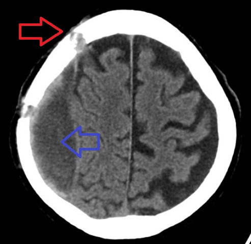
A subdural hematoma (SDH) is a type of bleeding in which a Hematoma, collection of blood—usually but not always associated with a traumatic brain injury—gathers between the inner layer of the dura mater and the arachnoid mater of the meninges surrounding the brain. It usually results from tears in bridging veins that cross the subdural space. Subdural hematomas may cause an increase in the intracranial pressure, pressure inside the skull, which in turn can cause compression of and damage to delicate brain tissue. Acute subdural hematomas are often life-threatening. Chronic subdural hematomas have a better prognosis if properly managed. In contrast, epidural hematomas are usually caused by tears in arteries, resulting in a build-up of blood between the dura mater and the skull. The third type of brain hemorrhage, known as a subarachnoid hemorrhage, causes bleeding into the subarachnoid space between the arachnoid mater and the pia mater. __TOC__ Signs and symptoms The sympt ... [...More Info...] [...Related Items...] OR: [Wikipedia] [Google] [Baidu] |
Bone
A bone is a Stiffness, rigid Organ (biology), organ that constitutes part of the skeleton in most vertebrate animals. Bones protect the various other organs of the body, produce red blood cell, red and white blood cells, store minerals, provide structure and support for the body, and enable animal locomotion, mobility. Bones come in a variety of shapes and sizes and have complex internal and external structures. They are lightweight yet strong and hard and serve multiple Function (biology), functions. Bone tissue (osseous tissue), which is also called bone in the mass noun, uncountable sense of that word, is hard tissue, a type of specialized connective tissue. It has a honeycomb-like matrix (biology), matrix internally, which helps to give the bone rigidity. Bone tissue is made up of different types of bone cells. Osteoblasts and osteocytes are involved in the formation and mineralization (biology), mineralization of bone; osteoclasts are involved in the bone resorption, resor ... [...More Info...] [...Related Items...] OR: [Wikipedia] [Google] [Baidu] |
Contrast CT
Contrast CT, or contrast enhanced computed tomography (CECT), is X-ray computed tomography (CT) using radiocontrast. Radiocontrasts for X-ray CT are generally iodine-based types. This is useful to highlight structures such as blood vessels that otherwise would be difficult to delineate from their surroundings. Using contrast material can also help to obtain functional information about tissues. Often, images are taken both with and without radiocontrast. CT images are called ''precontrast'' or ''native-phase'' images before any radiocontrast has been administrated, and ''postcontrast'' after radiocontrast administration. Bolus tracking Bolus tracking is a technique to optimize timing of the imaging. A small bolus of radio-opaque contrast media is injected into a patient via a peripheral intravenous cannula. Depending on the vessel being imaged, the volume of contrast is tracked using a region of interest (abbreviated "R.O.I.") at a certain level and then followed by the CT ... [...More Info...] [...Related Items...] OR: [Wikipedia] [Google] [Baidu] |
Cone Beam Computed Tomography
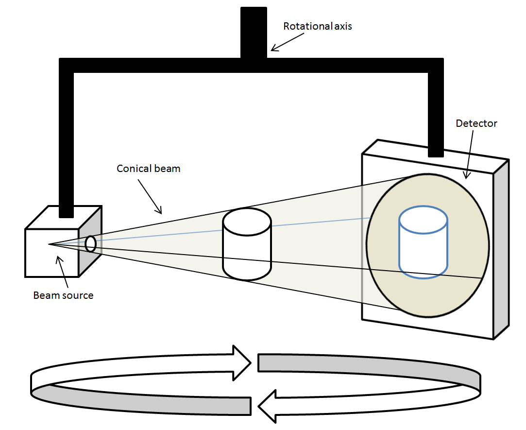
Cone beam computed tomography (or CBCT, also referred to as C-arm CT, cone beam volume CT, flat panel CT or Digital Volume Tomography (DVT)) is a medical imaging technique consisting of X-ray computed tomography where the X-rays are divergent, forming a cone. CBCT has become increasingly important in treatment planning and diagnosis in implant dentistry, ENT, orthopedics, and interventional radiology (IR), among other things. Perhaps because of the increased access to such technology, CBCT scanners are now finding many uses in dentistry, such as in the fields of oral surgery, endodontics and orthodontics. Integrated CBCT is also an important tool for patient positioning and verification in image-guided radiation therapy (IGRT). During dental/orthodontic imaging, the CBCT scanner rotates around the patient's head, obtaining up to nearly 600 distinct images. For interventional radiology, the patient is positioned offset to the table so that the region of interest is centered in ... [...More Info...] [...Related Items...] OR: [Wikipedia] [Google] [Baidu] |


