|
Holmes Tremor
Holmes tremor, first identified by Gordon Holmes in 1904, can be described as a wing-beating movement localized in the upper body that is caused by cerebellar damage. Holmes tremor is a combination of rest, action, and postural tremors. Tremor frequency ranges from 2 to 5 Hertz and is aggravated with posture and movement. It may arise from various underlying structural disorders including stroke, tumors, trauma, and other cerebellar lesions. Because Holmes tremor is rare, much of the research is based on individual cases. The formation of tremors is due to two main factors: the over-excited rhythmic movement of neuronal loops and permanent structural changes from neurodegeneration. Two major neuronal networks, the corticostriatothalamocortical hap and the inferior olivary nucleus (ION) specifically target the development of the tremors. When diagnosing a patient with Holmes tremor, one must look at the neurological signs and symptoms, as well as the possibility that the tremor is c ... [...More Info...] [...Related Items...] OR: [Wikipedia] [Google] [Baidu] |
Tremor
A tremor is an involuntary, somewhat rhythmic, muscle contraction and relaxation involving oscillations or twitching movements of one or more body parts. It is the most common of all involuntary movements and can affect the hands, arms, eyes, face, head, vocal folds, trunk, and legs. Most tremors occur in the hands. In some people, a tremor is a symptom of another neurological disorder. A very common tremor is the teeth chattering, usually induced by cold temperatures or by fear. Types Tremor is most commonly classified by clinical features and cause or origin. Some of the better-known forms of tremor, with their symptoms, include the following: * Cerebellar tremor (also known as intention tremor) is a slow, broad tremor of the extremities that occurs at the end of a purposeful movement, such as trying to press a button or touching a finger to the tip of one's nose. Cerebellar tremor is caused by lesions in or damage to the cerebellum resulting from stroke, tumor, or disease such ... [...More Info...] [...Related Items...] OR: [Wikipedia] [Google] [Baidu] |
Brainstem Stroke
A brainstem stroke syndrome falls under the broader category of stroke syndromes, or specific symptoms caused by vascular injury to an area of brain (for example, the lacunar syndromes). As the brainstem contains numerous cranial nuclei and white matter tracts, a stroke in this area can have a number of unique symptoms depending on the particular blood vessel that was injured and the group of cranial nerves and tracts that are no longer perfused. Symptoms of a brainstem stroke frequently include sudden vertigo and ataxia, with or without weakness. Brainstem stroke can also cause diplopia, slurred speech and decreased level of consciousness. A more serious outcome is locked-in syndrome. Syndromes * The midbrain syndromes (Significant overlap between these three syndromes) ** Superior alternating hemiplegia or Weber's syndrome ** Paramedian midbrain syndrome or Benedikt's syndrome ** Claude's syndrome * Medial pontine syndrome or Middle alternating hemiplegia or Foville's syndrom ... [...More Info...] [...Related Items...] OR: [Wikipedia] [Google] [Baidu] |
Coping Strategies
Coping refers to conscious strategies used to reduce unpleasant emotions. Coping strategies can be cognitions or behaviours and can be individual or social. Theories of coping Hundreds of coping strategies have been proposed in an attempt to understand how people cope. Classification of these strategies into a broader architecture has not been agreed upon. Researchers try to group coping responses rationally, empirically by factor analysis, or through a blend of both techniques. In the early days, Folkman and Richard Lazarus, Lazarus split the coping strategies into four groups, namely problem-focused, emotion-focused, support-seeking, and meaning-making coping. Weiten has identified four types of coping strategies:Weiten, W. & Lloyd, M.A. (2008) ''Psychology Applied to Modern Life (9th ed.)''. Wadsworth Cengage Learning. . appraisal-focused (adaptive cognitive), problem-focused (adaptive behavioral), emotion-focused, and occupation-focused coping. Billings and Moos added avoid ... [...More Info...] [...Related Items...] OR: [Wikipedia] [Google] [Baidu] |
Substantia Nigra
The substantia nigra (SN) is a basal ganglia structure located in the midbrain that plays an important role in reward and movement. ''Substantia nigra'' is Latin for "black substance", reflecting the fact that parts of the substantia nigra appear darker than neighboring areas due to high levels of neuromelanin in dopaminergic neurons. Parkinson's disease is characterized by the loss of dopaminergic neurons in the substantia nigra pars compacta. Although the substantia nigra appears as a continuous band in brain sections, anatomical studies have found that it actually consists of two parts with very different connections and functions: the pars compacta (SNpc) and the pars reticulata (SNpr). The pars compacta serves mainly as a projection to the basal ganglia circuit, supplying the striatum with dopamine. The pars reticulata conveys signals from the basal ganglia to numerous other brain structures. Structure The substantia nigra, along with four other nuclei, is part ... [...More Info...] [...Related Items...] OR: [Wikipedia] [Google] [Baidu] |
Midbrain Tegmentum
The midbrain is anatomically delineated into the tectum (roof) and the tegmentum (floor). The midbrain tegmentum extends from the substantia nigra to the cerebral aqueduct in a horizontal section of the midbrain. It forms the floor of the midbrain that surrounds below the cerebral aqueduct as well as the floor of the fourth ventricle while the midbrain tectum forms the roof of the fourth ventricle. The tegmentum contains a collection of tracts and nuclei with movement-related, species-specific, and pain-perception functions. The general structures of midbrain tegmentum include red nucleus and the periaqueductal grey matter. Unlike the midbrain tectum (which is a sensory structure located posteriorly), the midbrain tegmentum, which locates anteriorly, is related to a number of motor functions. Within the tegmentum, the red nucleus is in charge of motor coordination (specifically for limb movements) and the periaqueductal gray matter (PAG) contains critical circuits for modulating b ... [...More Info...] [...Related Items...] OR: [Wikipedia] [Google] [Baidu] |
Thalamus
The thalamus (from Greek θάλαμος, "chamber") is a large mass of gray matter located in the dorsal part of the diencephalon (a division of the forebrain). Nerve fibers project out of the thalamus to the cerebral cortex in all directions, allowing hub-like exchanges of information. It has several functions, such as the relaying of sensory signals, including motor signals to the cerebral cortex and the regulation of consciousness, sleep, and alertness. Anatomically, it is a paramedian symmetrical structure of two halves (left and right), within the vertebrate brain, situated between the cerebral cortex and the midbrain. It forms during embryonic development as the main product of the diencephalon, as first recognized by the Swiss embryologist and anatomist Wilhelm His Sr. in 1893. Anatomy The thalamus is a paired structure of gray matter located in the forebrain which is superior to the midbrain, near the center of the brain, with nerve fibers projecting out to the ... [...More Info...] [...Related Items...] OR: [Wikipedia] [Google] [Baidu] |
Thyroid
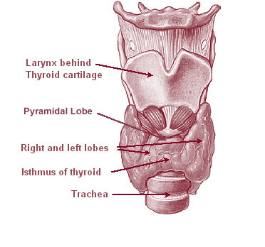
The thyroid, or thyroid gland, is an endocrine gland in vertebrates. In humans it is in the neck and consists of two connected lobes. The lower two thirds of the lobes are connected by a thin band of tissue called the thyroid isthmus. The thyroid is located at the front of the neck, below the Adam's apple. Microscopically, the functional unit of the thyroid gland is the spherical thyroid follicle, lined with follicular cells (thyrocytes), and occasional parafollicular cells that surround a lumen containing colloid. The thyroid gland secretes three hormones: the two thyroid hormones triiodothyronine (T3) and thyroxine (T4)and a peptide hormone, calcitonin. The thyroid hormones influence the metabolic rate and protein synthesis, and in children, growth and development. Calcitonin plays a role in calcium homeostasis. Secretion of the two thyroid hormones is regulated by thyroid-stimulating hormone (TSH), which is secreted from the anterior pituitary gland. TSH is regula ... [...More Info...] [...Related Items...] OR: [Wikipedia] [Google] [Baidu] |
Thyroid Stimulating Hormone
Thyroid-stimulating hormone (also known as thyrotropin, thyrotropic hormone, or abbreviated TSH) is a pituitary hormone that stimulates the thyroid gland to produce thyroxine (T4), and then triiodothyronine (T3) which stimulates the metabolism of almost every tissue in the body. It is a glycoprotein hormone produced by thyrotrope cells in the anterior pituitary gland, which regulates the endocrine function of the thyroid. Physiology Hormone levels TSH (with a half-life of about an hour) stimulates the thyroid gland to secrete the hormone thyroxine (T4), which has only a slight effect on metabolism. T4 is converted to triiodothyronine (T3), which is the active hormone that stimulates metabolism. About 80% of this conversion is in the liver and other organs, and 20% in the thyroid itself. [...More Info...] [...Related Items...] OR: [Wikipedia] [Google] [Baidu] |
Dentate Nucleus
The dentate nucleus is a cluster of neurons, or nerve cells, in the central nervous system that has a dentate – tooth-like or serrated – edge. It is located within the deep white matter of each cerebellar hemisphere, and it is the largest single structure linking the cerebellum to the rest of the brain.Sultan, F., Hamodeh, S., & Baizer, J. S. (2010). THE HUMAN DENTATE NUCLEUS: A COMPLEX SHAPE UNTANGLED. rticle Neuroscience, 167(4), 965–968. It is the largest and most lateral, or farthest from the midline, of the four pairs of deep cerebellar nuclei, the others being the globose and emboliform nuclei, which together are referred to as the interposed nucleus, and the fastigial nucleus. The dentate nucleus is responsible for the planning, initiation and control of voluntary movements. The dorsal region of the dentate nucleus contains output channels involved in motor function, which is the movement of skeletal muscle, while the ventral region contains output channels involv ... [...More Info...] [...Related Items...] OR: [Wikipedia] [Google] [Baidu] |
Red Nucleus
The red nucleus or nucleus ruber is a structure in the rostral midbrain involved in motor coordination. The red nucleus is pale pink, which is believed to be due to the presence of iron in at least two different forms: hemoglobin and ferritin. The structure is located in the tegmentum of the midbrain next to the substantia nigra and comprises caudal magnocellular and rostral parvocellular components. The red nucleus and substantia nigra are subcortical centers of the extrapyramidal motor system. Function In a vertebrate without a significant corticospinal tract, gait is mainly controlled by the red nucleus. However, in primates, where the corticospinal tract is dominant, the rubrospinal tract may be regarded as vestigial in motor function. Therefore, the red nucleus is less important in primates than in many other mammals. Nevertheless, the crawling of babies is controlled by the red nucleus, as is arm swinging in typical walking. The red nucleus may play an additional role ... [...More Info...] [...Related Items...] OR: [Wikipedia] [Google] [Baidu] |
Myoclonic Triangle
The myoclonic triangle (also known by its eponym Triangle of Guillain-Mollaret or dentato-rubro-olivary pathway) is an important feedback circuit of the brainstem and deep cerebellar nuclei which is responsible for modulating spinal cord motor activity. The circuit is thus composed: # Fibers of the rubro-olivary tract project from the parvocellular red nucleus via the central tegmental tract to the ipsilateral inferior olivary nucleus. # The inferior olivary nucleus sends its afferents via climbing fibers in the inferior cerebellar peduncle to Purkinje cells of the contralateral cerebellar cortex. # The Purkinje cells send their afferents to the ipsilateral dentate nucleus. # Dentatorubral tract fibers: the dentate nucleus afferents travel via the superior cerebellar peduncle to the contralateral red nucleus, thus completing the circuit. Of note, this circuit contains a double decussation, implying that a lesion in this tract ... [...More Info...] [...Related Items...] OR: [Wikipedia] [Google] [Baidu] |
Neuronal Network
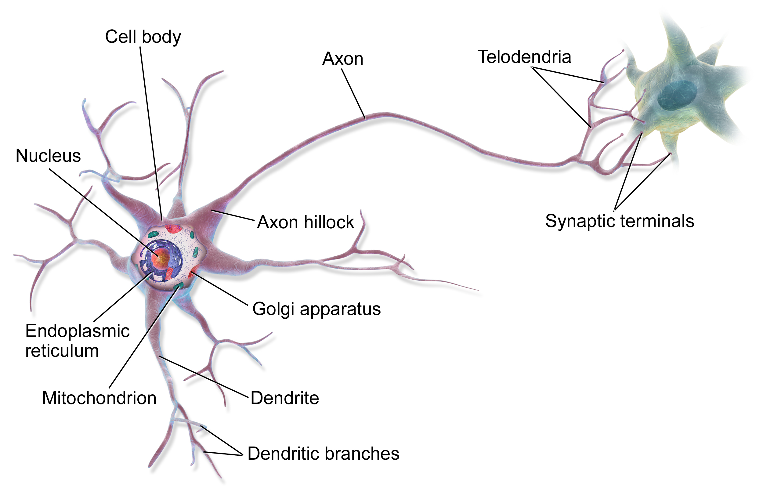
A neural circuit is a population of neurons interconnected by synapses to carry out a specific function when activated. Neural circuits interconnect to one another to form large scale brain networks. Biological neural networks have inspired the design of artificial neural networks, but artificial neural networks are usually not strict copies of their biological counterparts. Early study Early treatments of neural networks can be found in Herbert Spencer's ''Principles of Psychology'', 3rd edition (1872), Theodor Meynert's ''Psychiatry'' (1884), William James' ''Principles of Psychology'' (1890), and Sigmund Freud's Project for a Scientific Psychology (composed 1895). The first rule of neuronal learning was described by Hebb in 1949, in the Hebbian theory. Thus, Hebbian pairing of pre-synaptic and post-synaptic activity can substantially alter the dynamic characteristics of the synaptic connection and therefore either facilitate or inhibit signal transmission. In 1959, the neur ... [...More Info...] [...Related Items...] OR: [Wikipedia] [Google] [Baidu] |


_(14779159452).jpg)
