|
Hofbauer Cells
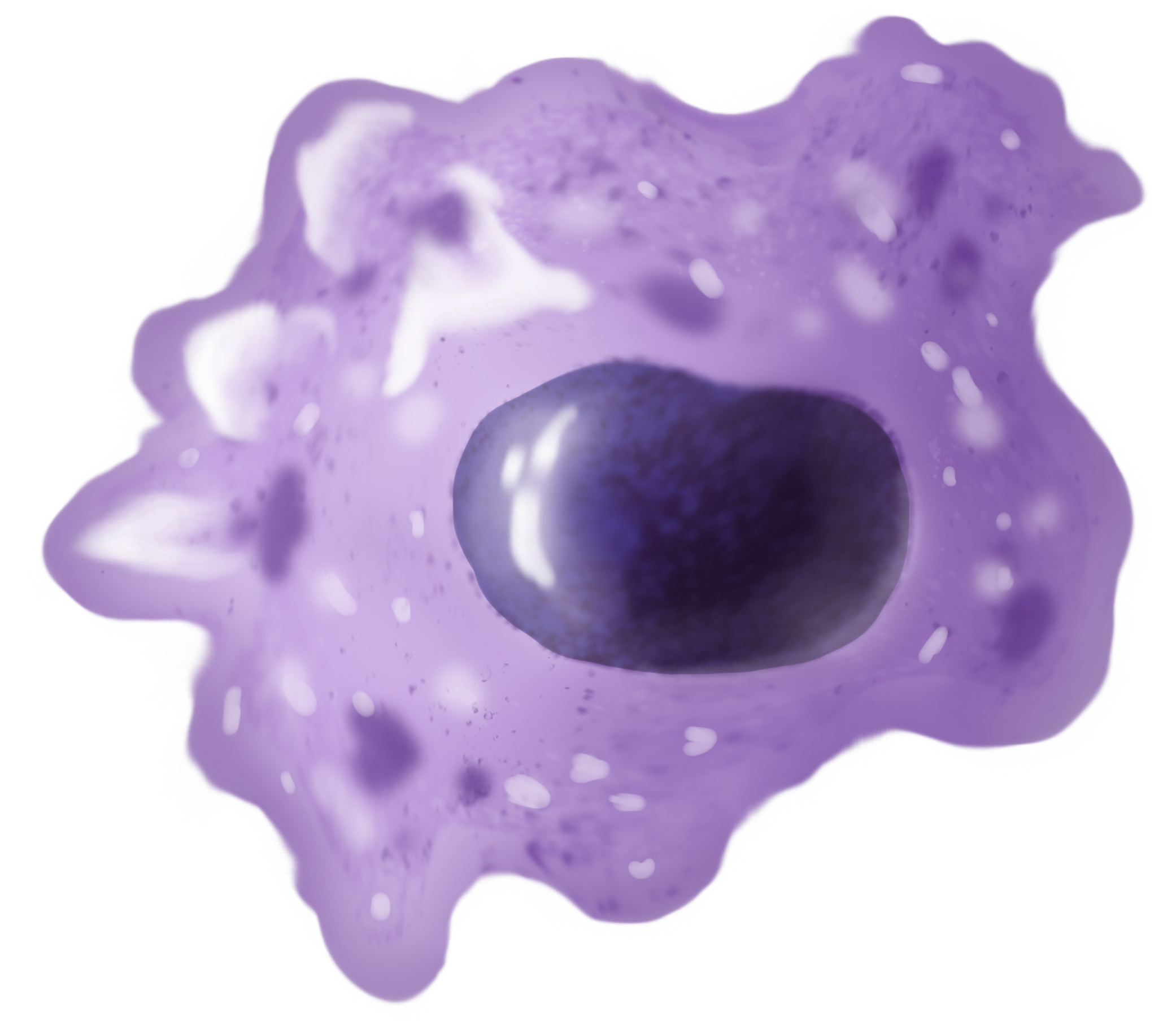
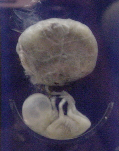
Hofbauer cells are oval eosinophilic histiocytes with granules and vacuoles found in the placenta, which are of mesenchymal origin, in mesoderm of the chorionic villus, particularly numerous in early pregnancy. Etymology They are named after J. Isfred Isidore Hofbauer (1871-1961), a German-American gynecologist who described the cell type in his book (''Biology of the Human Placenta with a special emphasis on the question of fetal nourishment''). Function They are believed to be a type of macrophage and are most likely involved in preventing the transmission of pathogens from the mother to the fetus (vertical transmission). Although there are many studies concerning placental vasculogenesis and angiogenesis, there has been a lack of evidence on the possible roles of Hofbauer cells in these processes. According to a systems level single-cell transcriptomics based study of human placental cell-cell communication, Hofbauer cells produce HBEGF, an EGFR ligand, which drives differentiat ... [...More Info...] [...Related Items...] OR: [Wikipedia] [Google] [Baidu] |
Eosinophilic
Eosinophilic (Greek suffix -phil-, meaning ''loves eosin'') is the staining of tissues, cells, or organelles after they have been washed with eosin, a dye. Eosin is an acidic dye for staining cell cytoplasm, collagen, and muscle fibers. ''Eosinophilic'' describes the appearance of cells and structures seen in histological sections that take up the staining dye eosin. Such eosinophilic structures are, in general, composed of protein. Eosin is usually combined with a stain called hematoxylin to produce a hematoxylin- and eosin-stained section (also called an H&E stain, HE or H+E section). It is the most widely used histological stain for a medical diagnosis. When a pathologist examines a biopsy of a suspected cancer, they will stain the biopsy with H&E. Some structures seen inside cells are described as being eosinophilic; for example, Lewy and Mallory bodies. [...More Info...] [...Related Items...] OR: [Wikipedia] [Google] [Baidu] |
Histiocyte
A histiocyte is a vertebrate cell that is part of the mononuclear phagocyte system (also known as the reticuloendothelial system or lymphoreticular system). The mononuclear phagocytic system is part of the organism's immune system. The histiocyte is a tissue macrophage or a dendritic cell (histio, diminutive of histo, meaning ''tissue'', and cyte, meaning ''cell''). Part of their job is to clear out neutrophils once they've reached the end of their lifespan. Development Histiocytes are derived from the bone marrow by multiplication from a stem cell. The derived cells migrate from the bone marrow to the blood as monocytes. They circulate through the body and enter various organs, where they undergo differentiation into histiocytes, which are part of the mononuclear phagocytic system (MPS). However, the term ''histiocyte'' has been used for multiple purposes in the past, and some cells called "histocytes" do not appear to derive from monocytic-macrophage lines. The term Histioc ... [...More Info...] [...Related Items...] OR: [Wikipedia] [Google] [Baidu] |
Vacuole
A vacuole () is a membrane-bound organelle which is present in plant and fungal cells and some protist, animal, and bacterial cells. Vacuoles are essentially enclosed compartments which are filled with water containing inorganic and organic molecules including enzymes in solution, though in certain cases they may contain solids which have been engulfed. Vacuoles are formed by the fusion of multiple membrane vesicles and are effectively just larger forms of these. The organelle has no basic shape or size; its structure varies according to the requirements of the cell. Discovery Contractile vacuoles ("stars") were first observed by Spallanzani (1776) in protozoa, although mistaken for respiratory organs. Dujardin (1841) named these "stars" as ''vacuoles''. In 1842, Schleiden applied the term for plant cells, to distinguish the structure with cell sap from the rest of the protoplasm. In 1885, de Vries named the vacuole membrane as tonoplast. Function The function and signifi ... [...More Info...] [...Related Items...] OR: [Wikipedia] [Google] [Baidu] |
Placenta
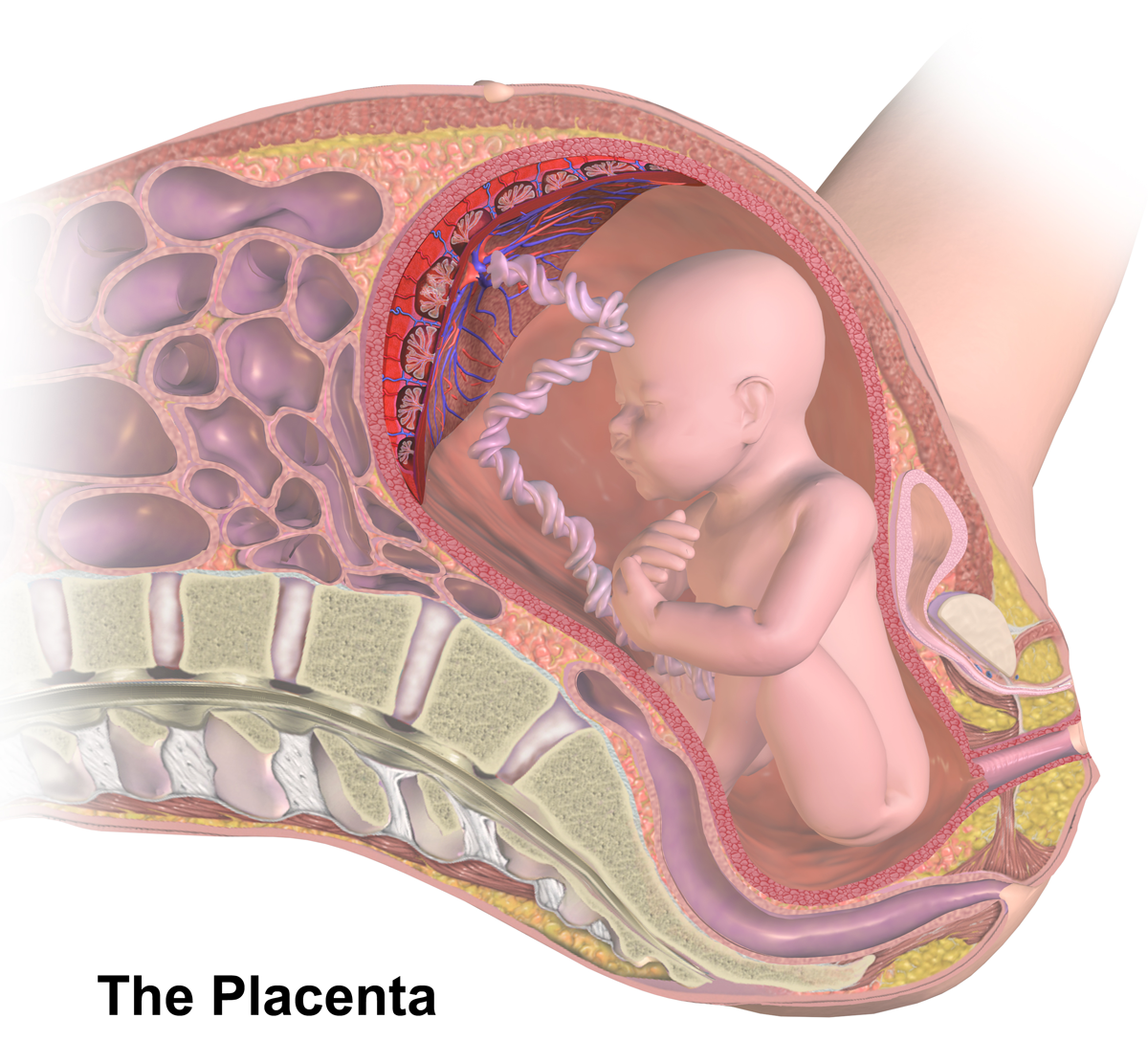
The placenta is a temporary embryonic and later fetal organ that begins developing from the blastocyst shortly after implantation. It plays critical roles in facilitating nutrient, gas and waste exchange between the physically separate maternal and fetal circulations, and is an important endocrine organ, producing hormones that regulate both maternal and fetal physiology during pregnancy. The placenta connects to the fetus via the umbilical cord, and on the opposite aspect to the maternal uterus in a species-dependent manner. In humans, a thin layer of maternal decidual (endometrial) tissue comes away with the placenta when it is expelled from the uterus following birth (sometimes incorrectly referred to as the 'maternal part' of the placenta). Placentas are a defining characteristic of placental mammals, but are also found in marsupials and some non-mammals with varying levels of development. Mammalian placentas probably first evolved about 150 million to 200 million years ... [...More Info...] [...Related Items...] OR: [Wikipedia] [Google] [Baidu] |
Mesenchyme
Mesenchyme () is a type of loosely organized animal embryonic connective tissue of undifferentiated cells that give rise to most tissues, such as skin, blood or bone. The interactions between mesenchyme and epithelium help to form nearly every organ in the developing embryo. Vertebrates Structure Mesenchyme is characterized morphologically by a prominent ground substance matrix containing a loose aggregate of reticular fibers and unspecialized mesenchymal stem cells. Mesenchymal cells can migrate easily (in contrast to epithelial cells, which lack mobility), are organized into closely adherent sheets, and are polarized in an apical-basal orientation. Development The mesenchyme originates from the mesoderm. From the mesoderm, the mesenchyme appears as an embryologically primitive "soup". This "soup" exists as a combination of the mesenchymal cells plus serous fluid plus the many different tissue proteins. Serous fluid is typically stocked with the many serous elements, such a ... [...More Info...] [...Related Items...] OR: [Wikipedia] [Google] [Baidu] |
Mesoderm
The mesoderm is the middle layer of the three germ layers that develops during gastrulation in the very early development of the embryo of most animals. The outer layer is the ectoderm, and the inner layer is the endoderm.Langman's Medical Embryology, 11th edition. 2010. The mesoderm forms mesenchyme, mesothelium, non-epithelial blood cells and coelomocytes. Mesothelium lines coeloms. Mesoderm forms the muscles in a process known as myogenesis, septa (cross-wise partitions) and mesenteries (length-wise partitions); and forms part of the gonads (the rest being the gametes). Myogenesis is specifically a function of mesenchyme. The mesoderm differentiates from the rest of the embryo through intercellular signaling, after which the mesoderm is polarized by an organizing center. The position of the organizing center is in turn determined by the regions in which beta-catenin is protected from degradation by GSK-3. Beta-catenin acts as a co-factor that alters the activity of ... [...More Info...] [...Related Items...] OR: [Wikipedia] [Google] [Baidu] |
Chorionic Villi
Chorionic villi are villi that sprout from the chorion to provide maximal contact area with maternal blood. They are an essential element in pregnancy from a histomorphologic perspective, and are, by definition, a product of conception. Branches of the umbilical arteries carry embryonic blood to the villi. After circulating through the capillaries of the villi, blood returns to the embryo through the umbilical vein. Thus, villi are part of the border between maternal and fetal blood during pregnancy. Structure Villi can also be classified by their relations: * Floating villi float freely in the intervillous space. They exhibit a bi-layered epithelium consisting of cytotrophoblasts with overlaying syncytium ( syncytiotrophoblast). * Anchoring (stem) villi stabilize mechanical integrity of the placental-maternal interface. Development The chorion undergoes rapid proliferation and forms numerous processes, the chorionic villi, which invade and destroy the uterine decidua and a ... [...More Info...] [...Related Items...] OR: [Wikipedia] [Google] [Baidu] |
Pregnancy
Pregnancy is the time during which one or more offspring develops ( gestates) inside a woman's uterus (womb). A multiple pregnancy involves more than one offspring, such as with twins. Pregnancy usually occurs by sexual intercourse, but can also occur through assisted reproductive technology procedures. A pregnancy may end in a live birth, a miscarriage, an induced abortion, or a stillbirth. Childbirth typically occurs around 40 weeks from the start of the last menstrual period (LMP), a span known as the gestational age. This is just over nine months. Counting by fertilization age, the length is about 38 weeks. Pregnancy is "the presence of an implanted human embryo or fetus in the uterus"; implantation occurs on average 8–9 days after fertilization. An '' embryo'' is the term for the developing offspring during the first seven weeks following implantation (i.e. ten weeks' gestational age), after which the term ''fetus'' is used until birth. Signs an ... [...More Info...] [...Related Items...] OR: [Wikipedia] [Google] [Baidu] |
Macrophage
Macrophages (abbreviated as M φ, MΦ or MP) ( el, large eaters, from Greek ''μακρός'' (') = large, ''φαγεῖν'' (') = to eat) are a type of white blood cell of the immune system that engulfs and digests pathogens, such as cancer cells, microbes, cellular debris, and foreign substances, which do not have proteins that are specific to healthy body cells on their surface. The process is called phagocytosis, which acts to defend the host against infection and injury. These large phagocytes are found in essentially all tissues, where they patrol for potential pathogens by amoeboid movement. They take various forms (with various names) throughout the body (e.g., histiocytes, Kupffer cells, alveolar macrophages, microglia, and others), but all are part of the mononuclear phagocyte system. Besides phagocytosis, they play a critical role in nonspecific defense (innate immunity) and also help initiate specific defense mechanisms (adaptive immunity) by recruiting other immune ... [...More Info...] [...Related Items...] OR: [Wikipedia] [Google] [Baidu] |
Pathogens
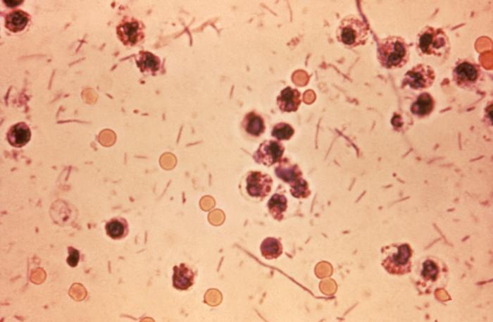
In biology, a pathogen ( el, πάθος, "suffering", "passion" and , "producer of") in the oldest and broadest sense, is any organism or agent that can produce disease. A pathogen may also be referred to as an infectious agent, or simply a germ. The term ''pathogen'' came into use in the 1880s. Typically, the term ''pathogen'' is used to describe an ''infectious'' microorganism or agent, such as a virus, bacterium, protozoan, prion, viroid, or fungus. Small animals, such as helminths and insects, can also cause or transmit disease. However, these animals are usually referred to as parasites rather than pathogens. The scientific study of microscopic organisms, including microscopic pathogenic organisms, is called microbiology, while parasitology refers to the scientific study of parasites and the organisms that host them. There are several pathways through which pathogens can invade a host. The principal pathways have different episodic time frames, but soil has the longest ... [...More Info...] [...Related Items...] OR: [Wikipedia] [Google] [Baidu] |
Fetus
A fetus or foetus (; plural fetuses, feti, foetuses, or foeti) is the unborn offspring that develops from an animal embryo. Following embryonic development the fetal stage of development takes place. In human prenatal development, fetal development begins from the ninth week after fertilization (or eleventh week gestational age) and continues until birth. Prenatal development is a continuum, with no clear defining feature distinguishing an embryo from a fetus. However, a fetus is characterized by the presence of all the major body organs, though they will not yet be fully developed and functional and some not yet situated in their final anatomical location. Etymology The word ''fetus'' (plural ''fetuses'' or '' feti'') is related to the Latin '' fētus'' ("offspring", "bringing forth", "hatching of young") and the Greek "φυτώ" to plant. The word "fetus" was used by Ovid in Metamorphoses, book 1, line 104. The predominant British, Irish, and Commonwealth spelling is '' ... [...More Info...] [...Related Items...] OR: [Wikipedia] [Google] [Baidu] |
Vertical Transmission
Vertical transmission of symbionts is the transfer of a microbial symbiont from the parent directly to the offspring. Many metazoan species carry symbiotic bacteria which play a mutualistic, commensal, or parasitic role. A symbiont is acquired by a host via horizontal, vertical, or mixed transmission. Implications Complex interdependence occurs between host and symbiont. The genetic pool of the symbiont is generally smaller and more subject to genetic drift. In true vertical transmission, the evolutionary outcomes of the host and symbiont are linked. If there is mixed transmission, new genetic material may be introduced. Generally, symbionts settle into specific niches and can even transfer part of their genome into the host nucleus. Evolutionary consequences Benefits The mechanism promotes tightly coupled evolutionary pressure, which causes the host and symbiont to function as a holobiont. Disadvantages Evolutionary bottlenecks lead to less symbiont diversity, and ... [...More Info...] [...Related Items...] OR: [Wikipedia] [Google] [Baidu] |

