|
Hypomyces Hyalinus
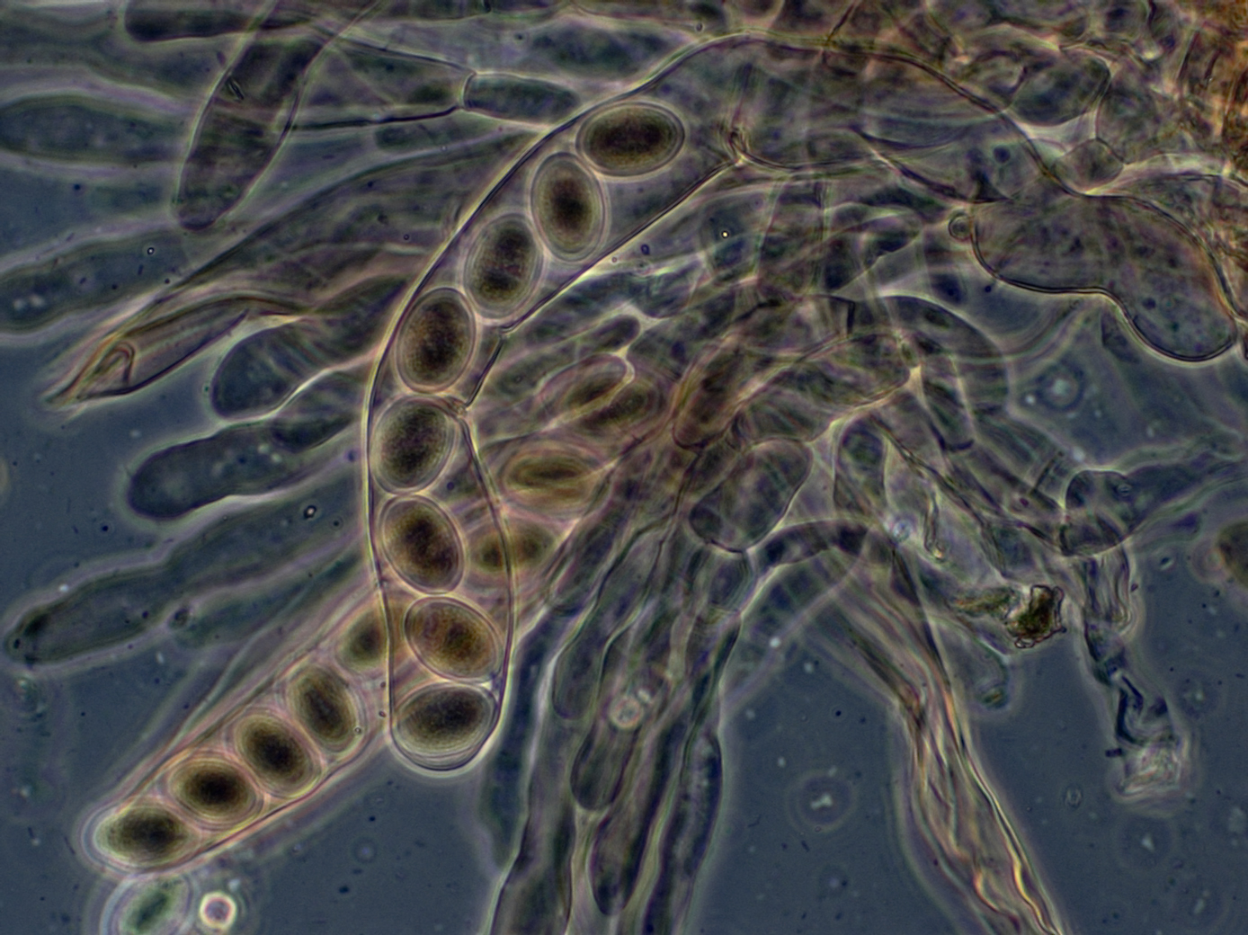
''Hypomyces hyalinus'' is a species of parasitic fungi that attacks fungi of the genus ''Amanita''. The earliest recording of this parasite was in 1822 in Salem, North Carolina, but microscopic descriptions of ''H. hyalinus'' do not appear in the literature until 1886. Host ''Hypomyces hyalinus'' is a host-specific pathogen which exclusively attacks species of the genus ''Amanita'', which is famous for containing some of the most toxic mushrooms in the world. ''Hypomyces hyalinus'' specifically attaches to the basidiocarp on the Sporocarp (fungi), sporocarp (fruiting body) of the fungus.Kadri Põldmaa, Põldmaa, K., Farr, D.F., & McCray, E.B. Hypomyces Online, Systematic Mycology and Microbiology Laboratory, ARS, USDA. Effects The parasitic effects of ''H. hyalinus'' thoroughly disfigures its host and in the absence of a nearby healthy specimen it can be impossible to determine the identity of the host in the field. Infection often covers the host mushroom preventing the expans ... [...More Info...] [...Related Items...] OR: [Wikipedia] [Google] [Baidu] |
Parasitic
Parasitism is a close relationship between species, where one organism, the parasite, lives on or inside another organism, the host, causing it some harm, and is adapted structurally to this way of life. The entomologist E. O. Wilson has characterised parasites as "predators that eat prey in units of less than one". Parasites include single-celled protozoans such as the agents of malaria, sleeping sickness, and amoebic dysentery; animals such as hookworms, lice, mosquitoes, and vampire bats; fungi such as honey fungus and the agents of ringworm; and plants such as mistletoe, dodder, and the broomrapes. There are six major parasitic strategies of exploitation of animal hosts, namely parasitic castration, directly transmitted parasitism (by contact), trophicallytransmitted parasitism (by being eaten), vector-transmitted parasitism, parasitoidism, and micropredation. One major axis of classification concerns invasiveness: an endoparasite lives inside the host's body; an e ... [...More Info...] [...Related Items...] OR: [Wikipedia] [Google] [Baidu] |
Anamorph
In mycology, the terms teleomorph, anamorph, and holomorph apply to portions of the life cycles of fungi in the phyla Ascomycota and Basidiomycota: *Teleomorph: the sexual reproductive stage (morph), typically a fruiting body. *Anamorph: an asexual reproductive stage (morph), often mold-like. When a single fungus produces multiple morphologically distinct anamorphs, these are called synanamorphs. *Holomorph: the whole fungus, including anamorphs and teleomorph. Dual naming of fungi Fungi are classified primarily based on the structures associated with sexual reproduction, which tend to be evolutionarily conserved. However, many fungi reproduce only asexually, and cannot easily be classified based on sexual characteristics; some produce both asexual and sexual states. These problematic species are often members of the Ascomycota, but a few of them belong to the Basidiomycota. Even among fungi that reproduce both sexually and asexually, often only one method of reproduction can be ... [...More Info...] [...Related Items...] OR: [Wikipedia] [Google] [Baidu] |
Hypocreaceae
The Hypocreaceae are a family (biology), family within the class Sordariomycetes. Species of Hypocreaceae are usually recognized by their brightly colored, perithecial Ascocarp, ascomata, typically yellow, orange or red. The family was proposed by Giuseppe De Notaris in 1844. According to the ''Dictionary of the Fungi'' (10th edition, 2008), the family has 22 genera and 454 species. Genera *''Acrostalagmus'' *''Aphysiostroma'' *''Cladobotryum'' *''Gliocladium'' *''Hypocrea'' *''Hypocreopsis'' *''Hypomyces'' *''Mycogone'' *''Podostroma'' *''Protocrea'' *''Rogersonia'' *''Sarawakus'' *''Sepedonium'' *''Sphaerostilbella'' *''Sporophagomyces'' *''Stephanoma'' *''Trichoderma'' References {{Taxonbar, from=Q3144255 Hypocreaceae, Ascomycota families Taxa named by Giuseppe De Notaris Taxa described in 1844 ... [...More Info...] [...Related Items...] OR: [Wikipedia] [Google] [Baidu] |
Phase Contrast Microscopy
__NOTOC__ Phase-contrast microscopy (PCM) is an optical microscopy technique that converts phase shifts in light passing through a transparent specimen to brightness changes in the image. Phase shifts themselves are invisible, but become visible when shown as brightness variations. When light waves travel through a medium other than a vacuum, interaction with the medium causes the wave amplitude and phase to change in a manner dependent on properties of the medium. Changes in amplitude (brightness) arise from the scattering and absorption of light, which is often wavelength-dependent and may give rise to colors. Photographic equipment and the human eye are only sensitive to amplitude variations. Without special arrangements, phase changes are therefore invisible. Yet, phase changes often convey important information. Phase-contrast microscopy is particularly important in biology. It reveals many cellular structures that are invisible with a bright-field microscope, as exemplif ... [...More Info...] [...Related Items...] OR: [Wikipedia] [Google] [Baidu] |
Differential Interference Contrast Microscopy
Differential interference contrast (DIC) microscopy, also known as Nomarski interference contrast (NIC) or Nomarski microscopy, is an optical microscopy technique used to enhance the contrast in unstained, transparent samples. DIC works on the principle of interferometry to gain information about the optical path length of the sample, to see otherwise invisible features. A relatively complex optical system produces an image with the object appearing black to white on a grey background. This image is similar to that obtained by phase contrast microscopy but without the bright diffraction halo. The technique was developed by Polish physicist Georges Nomarski in 1952. DIC works by separating a polarized light source into two orthogonally polarized mutually coherent parts which are spatially displaced (sheared) at the sample plane, and recombined before observation. The interference of the two parts at recombination is sensitive to their optical path difference (i.e. the product ... [...More Info...] [...Related Items...] OR: [Wikipedia] [Google] [Baidu] |
Fluorescence Microscopy
A fluorescence microscope is an optical microscope that uses fluorescence instead of, or in addition to, scattering, reflection, and attenuation or absorption, to study the properties of organic or inorganic substances. "Fluorescence microscope" refers to any microscope that uses fluorescence to generate an image, whether it is a simple set up like an epifluorescence microscope or a more complicated design such as a confocal microscope, which uses optical sectioning to get better resolution of the fluorescence image. Principle The specimen is illuminated with light of a specific wavelength (or wavelengths) which is absorbed by the fluorophores, causing them to emit light of longer wavelengths (i.e., of a different color than the absorbed light). The illumination light is separated from the much weaker emitted fluorescence through the use of a spectral emission filter. Typical components of a fluorescence microscope are a light source (xenon arc lamp or mercury-vapor lamp are ... [...More Info...] [...Related Items...] OR: [Wikipedia] [Google] [Baidu] |
Bright-field Microscopy
Bright-field microscopy (BF) is the simplest of all the optical microscopy illumination techniques. Sample illumination is transmitted (i.e., illuminated from below and observed from above) white light, and contrast in the sample is caused by attenuation of the transmitted light in dense areas of the sample. Bright-field microscopy is the simplest of a range of techniques used for illumination of samples in light microscopes, and its simplicity makes it a popular technique. The typical appearance of a bright-field microscopy image is a dark sample on a bright background, hence the name. Light path The light path of a bright-field microscope is extremely simple, no additional components are required beyond the normal light-microscope setup. The light path therefore consists of: * a transillumination light source, commonly a halogen lamp in the microscope stand; * a condenser lens, which focuses light from the light source onto the sample; * an objective lens, which collects light ... [...More Info...] [...Related Items...] OR: [Wikipedia] [Google] [Baidu] |
Microscopy
Microscopy is the technical field of using microscopes to view objects and areas of objects that cannot be seen with the naked eye (objects that are not within the resolution range of the normal eye). There are three well-known branches of microscopy: optical, electron, and scanning probe microscopy, along with the emerging field of X-ray microscopy. Optical microscopy and electron microscopy involve the diffraction, reflection, or refraction of electromagnetic radiation/electron beams interacting with the specimen, and the collection of the scattered radiation or another signal in order to create an image. This process may be carried out by wide-field irradiation of the sample (for example standard light microscopy and transmission electron microscopy) or by scanning a fine beam over the sample (for example confocal laser scanning microscopy and scanning electron microscopy). Scanning probe microscopy involves the interaction of a scanning probe with the surface of the objec ... [...More Info...] [...Related Items...] OR: [Wikipedia] [Google] [Baidu] |
Dextrose
Glucose is a simple sugar with the molecular formula . Glucose is overall the most abundant monosaccharide, a subcategory of carbohydrates. Glucose is mainly made by plants and most algae during photosynthesis from water and carbon dioxide, using energy from sunlight, where it is used to make cellulose in cell walls, the most abundant carbohydrate in the world. In energy metabolism, glucose is the most important source of energy in all organisms. Glucose for metabolism is stored as a polymer, in plants mainly as starch and amylopectin, and in animals as glycogen. Glucose circulates in the blood of animals as blood sugar. The naturally occurring form of glucose is -glucose, while -glucose is produced synthetically in comparatively small amounts and is less biologically active. Glucose is a monosaccharide containing six carbon atoms and an aldehyde group, and is therefore an aldohexose. The glucose molecule can exist in an open-chain (acyclic) as well as ring (cyclic) form. Gluco ... [...More Info...] [...Related Items...] OR: [Wikipedia] [Google] [Baidu] |
Agar
Agar ( or ), or agar-agar, is a jelly-like substance consisting of polysaccharides obtained from the cell walls of some species of red algae, primarily from ogonori (''Gracilaria'') and "tengusa" (''Gelidiaceae''). As found in nature, agar is a mixture of two components, the linear polysaccharide agarose and a heterogeneous mixture of smaller molecules called agaropectin. It forms the supporting structure in the cell walls of certain species of algae and is released on boiling. These algae are known as agarophytes, belonging to the Rhodophyta (red algae) phylum. The processing of food-grade agar removes the agaropectin, and the commercial product is essentially pure agarose. Agar has been used as an ingredient in desserts throughout Asia and also as a solid substrate to contain culture media for microbiological work. Agar can be used as a laxative; an appetite suppressant; a vegan substitute for gelatin; a thickener for soups; in fruit preserves, ice cream, and other desser ... [...More Info...] [...Related Items...] OR: [Wikipedia] [Google] [Baidu] |
Ascospore
An ascus (; ) is the sexual spore-bearing cell produced in ascomycete fungi. Each ascus usually contains eight ascospores (or octad), produced by meiosis followed, in most species, by a mitotic cell division. However, asci in some genera or species can occur in numbers of one (e.g. ''Monosporascus cannonballus''), two, four, or multiples of four. In a few cases, the ascospores can bud off conidia that may fill the asci (e.g. ''Tympanis'') with hundreds of conidia, or the ascospores may fragment, e.g. some ''Cordyceps'', also filling the asci with smaller cells. Ascospores are nonmotile, usually single celled, but not infrequently may be coenocytic (lacking a septum), and in some cases coenocytic in multiple planes. Mitotic divisions within the developing spores populate each resulting cell in septate ascospores with nuclei. The term ocular chamber, or oculus, refers to the epiplasm (the portion of cytoplasm not used in ascospore formation) that is surrounded by the "bourrelet ... [...More Info...] [...Related Items...] OR: [Wikipedia] [Google] [Baidu] |
Perithecia
An ascocarp, or ascoma (), is the fruiting body ( sporocarp) of an ascomycete phylum fungus. It consists of very tightly interwoven hyphae and millions of embedded asci, each of which typically contains four to eight ascospores. Ascocarps are most commonly bowl-shaped (apothecia) but may take on a spherical or flask-like form that has a pore opening to release spores (perithecia) or no opening (cleistothecia). Classification The ascocarp is classified according to its placement (in ways not fundamental to the basic taxonomy). It is called ''epigeous'' if it grows above ground, as with the morels, while underground ascocarps, such as truffles, are termed ''hypogeous''. The structure enclosing the hymenium is divided into the types described below (apothecium, cleistothecium, etc.) and this character ''is'' important for the taxonomic classification of the fungus. Apothecia can be relatively large and fleshy, whereas the others are microscopic—about the size of flecks of ... [...More Info...] [...Related Items...] OR: [Wikipedia] [Google] [Baidu] |

