|
Hypofrontality
Hypofrontality is a state of decreased cerebral blood flow (CBF) in the prefrontal cortex of the brain. Hypofrontality is symptomatic of several neurological medical conditions, such as schizophrenia, attention deficit hyperactivity disorder (ADHD), bipolar disorder, and major depressive disorder. This condition was initially described by Ingvar and Franzén in 1974, through the use of xenon blood flow technique with 32 detectors to image the brains of patients with schizophrenia. This finding was confirmed in subsequent studies using the improved spatial resolution of positron emission tomography with the fluorodeoxyglucose (18F-FDG) tracer. Subsequent neuroimaging work has shown that the decreases in prefrontal CBF are localized to the medial, lateral, and orbital portions of the prefrontal cortex. Hypofrontality is thought to contribute to the negative symptoms of schizophrenia. Definition Hypofrontality is a symptom of numerous neurological diseases defined as reduced u ... [...More Info...] [...Related Items...] OR: [Wikipedia] [Google] [Baidu] |
Prefrontal Cortex
In mammalian brain anatomy, the prefrontal cortex (PFC) covers the front part of the frontal lobe of the cerebral cortex. The PFC contains the Brodmann areas BA8, BA9, BA10, BA11, BA12, BA13, BA14, BA24, BA25, BA32, BA44, BA45, BA46, and BA47. The basic activity of this brain region is considered to be orchestration of thoughts and actions in accordance with internal goals. Many authors have indicated an integral link between a person's will to live, personality, and the functions of the prefrontal cortex. This brain region has been implicated in executive functions, such as planning, decision making, short-term memory, personality expression, moderating social behavior and controlling certain aspects of speech and language. Executive function relates to abilities to differentiate among conflicting thoughts, determine good and bad, better and best, same and different, future consequences of current activities, working toward a defined goal, prediction of outcomes, e ... [...More Info...] [...Related Items...] OR: [Wikipedia] [Google] [Baidu] |
Frontal Lobe
The frontal lobe is the largest of the four major lobes of the brain in mammals, and is located at the front of each cerebral hemisphere (in front of the parietal lobe and the temporal lobe). It is parted from the parietal lobe by a groove between tissues called the central sulcus and from the temporal lobe by a deeper groove called the lateral sulcus (Sylvian fissure). The most anterior rounded part of the frontal lobe (though not well-defined) is known as the frontal pole, one of the three poles of the cerebrum. The frontal lobe is covered by the frontal cortex. The frontal cortex includes the premotor cortex, and the primary motor cortex – parts of the motor cortex. The front part of the frontal cortex is covered by the prefrontal cortex. There are four principal gyri in the frontal lobe. The precentral gyrus is directly anterior to the central sulcus, running parallel to it and contains the primary motor cortex, which controls voluntary movements of specific body parts ... [...More Info...] [...Related Items...] OR: [Wikipedia] [Google] [Baidu] |
Clonidine
Clonidine, sold under the brand name Catapres among others, is an α2-adrenergic agonist medication used to treat high blood pressure, ADHD, drug withdrawal ( alcohol, opioids, or nicotine), menopausal flushing, diarrhea, spasticity, and certain pain conditions. It is used by mouth, by injection, or as a skin patch. Onset of action is typically within an hour with the effects on blood pressure lasting for up to eight hours. Common side effect include dry mouth, dizziness, headaches, hypotension, and sleepiness. Severe side effects may include hallucinations, heart arrhythmias, and confusion. If rapidly stopped, withdrawal effects may occur. Use during pregnancy or breastfeeding is not recommended. Clonidine lowers blood pressure by stimulating α2 receptors in the brain, which results in relaxation of many arteries. Clonidine was patented in 1961 and came into medical use in 1966. It is available as a generic medication. In 2020, it was the 75th most commonly prescri ... [...More Info...] [...Related Items...] OR: [Wikipedia] [Google] [Baidu] |
Haloperidol
Haloperidol, sold under the brand name Haldol among others, is a typical antipsychotic medication. Haloperidol is used in the treatment of schizophrenia, tics in Tourette syndrome, mania in bipolar disorder, delirium, agitation, acute psychosis, and hallucinations from alcohol withdrawal. It may be used by mouth or injection into a muscle or a vein. Haloperidol typically works within 30 to 60 minutes. A long-acting formulation may be used as an injection every four weeks by people with schizophrenia or related illnesses, who either forget or refuse to take the medication by mouth. Haloperidol may result in a movement disorder known as tardive dyskinesia which may be permanent. Neuroleptic malignant syndrome and QT interval prolongation may occur. In older people with psychosis due to dementia it results in an increased risk of death. When taken during pregnancy it may result in problems in the infant. It should not be used by people with Parkinson's disease. Haloperidol was ... [...More Info...] [...Related Items...] OR: [Wikipedia] [Google] [Baidu] |
Clozapine
Clozapine is a psychiatric medication and is the first atypical antipsychotic (also called second-generation antipsychotic). It is primarily used to treat people with schizophrenia and schizoaffective disorders who have had an inadequate response to other antipsychotics or who have been unable to tolerate other drugs due to extrapyramidal side effects. It is also used for the treatment of psychosis in Parkinson's disease. Clozapine is regarded as the gold-standard treatment when other medication has been insufficiently effective and its use is recommended by multiple international treatment guidelines, after resistance to earlier neuroleptic treatment is established. The role of clozapine in treatment-resistant schizophrenia was established by a 1988 landmark study in which clozapine showed marked benefits compared to chlorpromazine in a group of patients with protracted psychosis who had already shown an inadequate response to other antipsychotics. While there are significan ... [...More Info...] [...Related Items...] OR: [Wikipedia] [Google] [Baidu] |
PET Scanner
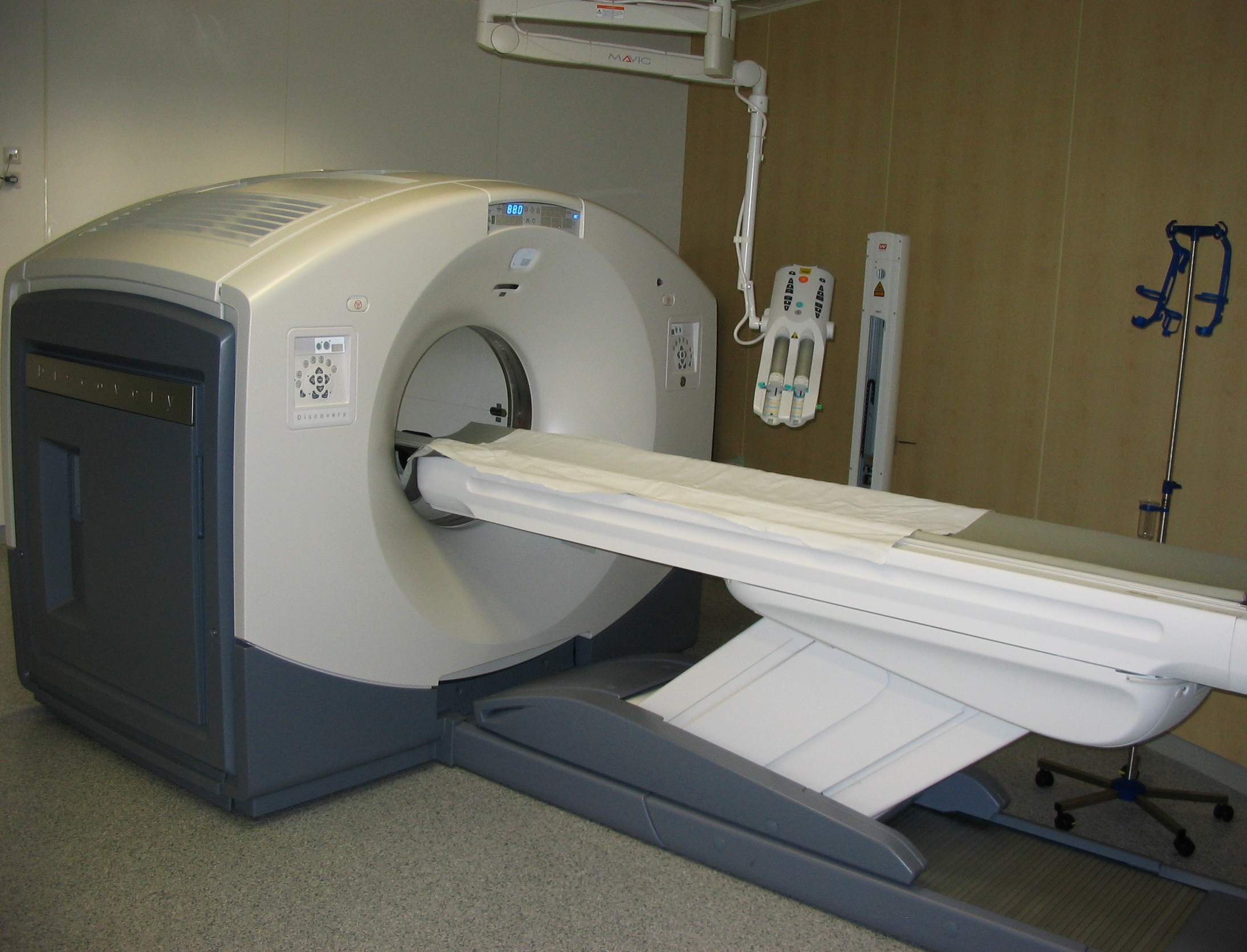
Positron emission tomography (PET) is a functional imaging technique that uses radioactive substances known as radiotracers to visualize and measure changes in metabolic processes, and in other physiological activities including blood flow, regional chemical composition, and absorption. Different tracers are used for various imaging purposes, depending on the target process within the body. For example, -FDG is commonly used to detect cancer, NaF is widely used for detecting bone formation, and oxygen-15 is sometimes used to measure blood flow. PET is a common imaging technique, a medical scintillography technique used in nuclear medicine. A radiopharmaceutical — a radioisotope attached to a drug — is injected into the body as a tracer. When the radiopharmaceutical undergoes beta plus decay, a positron is emitted, and when the positron collides with an ordinary electron, the two particles annihilate and gamma rays are emitted. These gamma rays are detected ... [...More Info...] [...Related Items...] OR: [Wikipedia] [Google] [Baidu] |
FMRI
Functional magnetic resonance imaging or functional MRI (fMRI) measures brain activity by detecting changes associated with blood flow. This technique relies on the fact that cerebral blood flow and neuronal activation are coupled. When an area of the brain is in use, blood flow to that region also increases. The primary form of fMRI uses the blood-oxygen-level dependent (BOLD) contrast, discovered by Seiji Ogawa in 1990. This is a type of specialized brain and body scan used to map neural activity in the brain or spinal cord of humans or other animals by imaging the change in blood flow (hemodynamic response) related to energy use by brain cells. Since the early 1990s, fMRI has come to dominate brain mapping research because it does not involve the use of injections, surgery, the ingestion of substances, or exposure to ionizing radiation. This measure is frequently corrupted by noise from various sources; hence, statistical procedures are used to extract the underlying signal. T ... [...More Info...] [...Related Items...] OR: [Wikipedia] [Google] [Baidu] |
Interneuron
Interneurons (also called internuncial neurons, relay neurons, association neurons, connector neurons, intermediate neurons or local circuit neurons) are neurons that connect two brain regions, i.e. not direct motor neurons or sensory neurons. Interneurons are the central nodes of neural circuits, enabling communication between sensory or motor neurons and the central nervous system (CNS). They play vital roles in reflexes, neuronal oscillations, and neurogenesis in the adult mammalian brain. Interneurons can be further broken down into two groups: local interneurons and relay interneurons. Local interneurons have short axons and form circuits with nearby neurons to analyze small pieces of information. Relay interneurons have long axons and connect circuits of neurons in one region of the brain with those in other regions. However, interneurons are generally considered to operate mainly within local brain areas. The interaction between interneurons allow the brain to perform c ... [...More Info...] [...Related Items...] OR: [Wikipedia] [Google] [Baidu] |
Mediodorsal Thalamic Nucleus
The medial dorsal nucleus (or dorsomedial nucleus of thalamus) is a large nucleus in the thalamus. It is believed to play a role in memory. Structure It relays inputs from the amygdala and olfactory cortex and projects to the prefrontal cortex and the limbic system and in turn relays them to the prefrontal association cortex. As a result, it plays a crucial role in attention, planning, organization, abstract thinking, multi-tasking, and active memory. The connections of the medial dorsal nucleus have even been used to delineate the prefrontal cortex of the Göttingen minipig brain. By stereology the number of brain cells in the region has been estimated to around 6.43 million neurons in the adult human brain and 36.3 million glial cells, and with the newborn having quite different numbers: around 11.2 million neurons and 10.6 million glial cells. Function Pain processing While both the ventral and medial dorsal nuclei process pain, the medial dorsal nucleus bypasses pri ... [...More Info...] [...Related Items...] OR: [Wikipedia] [Google] [Baidu] |
Dopamine
Dopamine (DA, a contraction of 3,4-dihydroxyphenethylamine) is a neuromodulatory molecule that plays several important roles in cells. It is an organic compound, organic chemical of the catecholamine and phenethylamine families. Dopamine constitutes about 80% of the catecholamine content in the brain. It is an amine synthesized by removing a carboxyl group from a molecule of its precursor (chemistry), precursor chemical, L-DOPA, which is biosynthesis, synthesized in the brain and kidneys. Dopamine is also synthesized in plants and most animals. In the brain, dopamine functions as a neurotransmitter—a chemical released by neurons (nerve cells) to send signals to other nerve cells. Neurotransmitters are synthesized in specific regions of the brain, but affect many regions systemically. The brain includes several distinct dopaminergic pathway, dopamine pathways, one of which plays a major role in the motivational component of reward system, reward-motivated behavior. The anticipa ... [...More Info...] [...Related Items...] OR: [Wikipedia] [Google] [Baidu] |


