|
Hospital Nacional
Hospital Nacional ( en, National Hospital) is a private hospital in Panama City, Panama. It is located at Avenida Cuba and 38th Street in Bella Vista. History Clinica Nacional (National Clinic), a small 10-bed facility with one operating room, one delivery room, and a small newborn unit, opened on 9 July 1973 at Justo Arosemena Avenue and 38th Street in Panama City, Panama.It exclusively provided OB/GYN services and became known as "Clinica de la Mujer" (The Women's Clinic). To accommodate its growth to 25 beds, it moved to a larger location across the street in January 1980 In September 1994, Centro Médico Nacional (National Medical Center) opened, housing 110 specialists. By that time, the smaller clinic's fame made further expansion feasible. On 20 August 1998, a new building was inaugurated on Avenida Cuba, between 38th and 39th Streets in Panama City, where the clinic was renamed Hospital Nacional. Facilities Hospital Nacional is an 80-bed facility offering all major medi ... [...More Info...] [...Related Items...] OR: [Wikipedia] [Google] [Baidu] |
Panama City
Panama City ( es, Ciudad de Panamá, links=no; ), also known as Panama (or Panamá in Spanish), is the capital and largest city of Panama. It has an urban population of 880,691, with over 1.5 million in its metropolitan area. The city is located at the Pacific entrance of the Panama Canal, in the province of Panama. The city is the political and administrative center of the country, as well as a hub for banking and commerce. The city of Panama was founded on 15 August 1519, by Spanish conquistador Pedro Arias Dávila. The city was the starting point for expeditions that conquered the Inca Empire in Peru. It was a stopover point on one of the most important trade routes in the American continent, leading to the fairs of Nombre de Dios and Portobelo, through which passed most of the gold and silver that Spain mined from the Americas. On 28 January 1671, the original city was destroyed by a fire when the privateer Henry Morgan sacked and set fire to it. The city was formally ... [...More Info...] [...Related Items...] OR: [Wikipedia] [Google] [Baidu] |
Pathology
Pathology is the study of the causes and effects of disease or injury. The word ''pathology'' also refers to the study of disease in general, incorporating a wide range of biology research fields and medical practices. However, when used in the context of modern medical treatment, the term is often used in a narrower fashion to refer to processes and tests that fall within the contemporary medical field of "general pathology", an area which includes a number of distinct but inter-related medical specialties that diagnose disease, mostly through analysis of tissue, cell, and body fluid samples. Idiomatically, "a pathology" may also refer to the predicted or actual progression of particular diseases (as in the statement "the many different forms of cancer have diverse pathologies", in which case a more proper choice of word would be " pathophysiologies"), and the affix ''pathy'' is sometimes used to indicate a state of disease in cases of both physical ailment (as in cardiomy ... [...More Info...] [...Related Items...] OR: [Wikipedia] [Google] [Baidu] |
Physical Therapy
Physical therapy (PT), also known as physiotherapy, is one of the allied health professions. It is provided by physical therapists who promote, maintain, or restore health through physical examination, diagnosis, management, prognosis, patient education, physical intervention, rehabilitation, disease prevention, and health promotion. Physical therapists are known as physiotherapists in many countries. In addition to clinical practice, other aspects of physical therapist practice include research, education, consultation, and health administration. Physical therapy is provided as a primary care treatment or alongside, or in conjunction with, other medical services. In some jurisdictions, such as the United Kingdom, physical therapists have the authority to prescribe medication. Overview Physical therapy addresses the illnesses or injuries that limit a person's abilities to move and perform functional activities in their daily lives. PTs use an individual's history and physic ... [...More Info...] [...Related Items...] OR: [Wikipedia] [Google] [Baidu] |
Endoscopy
An endoscopy is a procedure used in medicine to look inside the body. The endoscopy procedure uses an endoscope to examine the interior of a hollow organ or cavity of the body. Unlike many other medical imaging techniques, endoscopes are inserted directly into the organ. There are many types of endoscopies. Depending on the site in the body and type of procedure, an endoscopy may be performed by either a doctor or a surgeon. A patient may be fully conscious or anaesthesia, anaesthetised during the procedure. Most often, the term ''endoscopy'' is used to refer to an examination of the upper part of the human gastrointestinal tract, gastrointestinal tract, known as an esophagogastroduodenoscopy. For nonmedical use, similar instruments are called borescopes. History Adolf Kussmaul was fascinated by sword swallowers who would insert a sword down their throat without gagging. This drew inspiration to insert a camera, the next problem to solve was how to insert a source of light, as ... [...More Info...] [...Related Items...] OR: [Wikipedia] [Google] [Baidu] |
DEXA
Dual-energy X-ray absorptiometry (DXA, or DEXA) is a means of measuring bone mineral density (BMD) using spectral imaging. Two X-ray beams, with different energy levels, are aimed at the patient's bones. When soft tissue absorption is subtracted out, the bone mineral density (BMD) can be determined from the absorption of each beam by bone. Dual-energy X-ray absorptiometry is the most widely used and most thoroughly studied bone density measurement technology. The DXA scan is typically used to diagnose and follow osteoporosis, as contrasted to the nuclear bone scan, which is sensitive to certain metabolic diseases of bones in which bones are attempting to heal from infections, fractures, or tumors. It is also sometimes used to assess body composition. Physics Soft tissue and bone have different attenuation coefficients to X-rays. A single X-ray beam passing through the body will be attenuated by both soft tissue and bone, and it is not possible to determine, from a single beam, ... [...More Info...] [...Related Items...] OR: [Wikipedia] [Google] [Baidu] |
Mammography
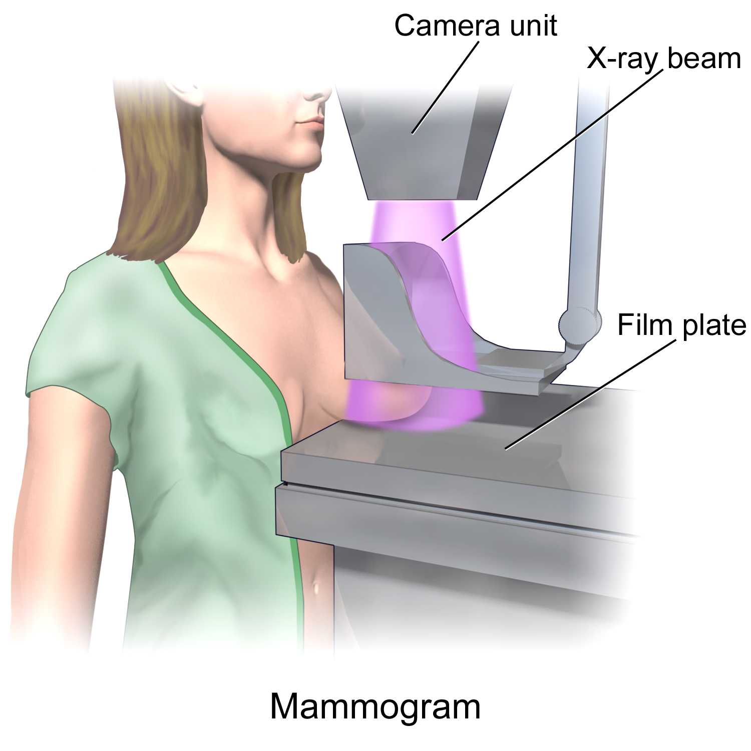
Mammography (also called mastography) is the process of using low-energy X-rays (usually around 30 kVp) to examine the human breast for diagnosis and screening. The goal of mammography is the early detection of breast cancer, typically through detection of characteristic masses or microcalcifications. As with all X-rays, mammograms use doses of ionizing radiation to create images. These images are then analyzed for abnormal findings. It is usual to employ lower-energy X-rays, typically Mo (K-shell X-ray energies of 17.5 and 19.6 keV) and Rh (20.2 and 22.7 keV) than those used for radiography of bones. Mammography may be 2D or 3D (tomosynthesis), depending on the available equipment and/or purpose of the examination. Ultrasound, ductography, positron emission mammography (PEM), and magnetic resonance imaging (MRI) are adjuncts to mammography. Ultrasound is typically used for further evaluation of masses found on mammography or palpable masses that may or may not be seen on mammogr ... [...More Info...] [...Related Items...] OR: [Wikipedia] [Google] [Baidu] |
Angiography
Angiography or arteriography is a medical imaging technique used to visualize the inside, or lumen, of blood vessels and organs of the body, with particular interest in the arteries, veins, and the heart chambers. Modern angiography is performed by injecting a radio-opaque contrast agent into the blood vessel and imaging using X-ray based techniques such as fluoroscopy. The word itself comes from the Greek words ἀγγεῖον ''angeion'' 'vessel' and γράφειν ''graphein'' 'to write, record'. The film or image of the blood vessels is called an ''angiograph'', or more commonly an ''angiogram''. Though the word can describe both an arteriogram and a venogram, in everyday usage the terms angiogram and arteriogram are often used synonymously, whereas the term venogram is used more precisely. The term angiography has been applied to radionuclide angiography and newer vascular imaging techniques such as CO2 angiography, CT angiography and MR angiography. The term ''isotope a ... [...More Info...] [...Related Items...] OR: [Wikipedia] [Google] [Baidu] |
Fluoroscopy
Fluoroscopy () is an imaging technique that uses X-rays to obtain real-time moving images of the interior of an object. In its primary application of medical imaging, a fluoroscope () allows a physician to see the internal structure and function of a patient, so that the pumping action of the heart or the motion of swallowing, for example, can be watched. This is useful for both diagnosis and therapy and occurs in general radiology, interventional radiology, and image-guided surgery. In its simplest form, a fluoroscope consists of an X-ray source and a fluorescent screen, between which a patient is placed. However, since the 1950s most fluoroscopes have included X-ray image intensifiers and cameras as well, to improve the image's visibility and make it available on a remote display screen. For many decades, fluoroscopy tended to produce live pictures that were not recorded, but since the 1960s, as technology improved, recording and playback became the norm. Fluoroscopy is s ... [...More Info...] [...Related Items...] OR: [Wikipedia] [Google] [Baidu] |
Nuclear Medicine
Nuclear medicine or nucleology is a medical specialty involving the application of radioactive substances in the diagnosis and treatment of disease. Nuclear imaging, in a sense, is "radiology done inside out" because it records radiation emitting from within the body rather than radiation that is generated by external sources like X-rays. In addition, nuclear medicine scans differ from radiology, as the emphasis is not on imaging anatomy, but on the function. For such reason, it is called a physiological imaging modality. Single photon emission computed tomography (SPECT) and positron emission tomography (PET) scans are the two most common imaging modalities in nuclear medicine. Diagnostic medical imaging Diagnostic In nuclear medicine imaging, radiopharmaceuticals are taken internally, for example, through inhalation, intravenously or orally. Then, external detectors (gamma cameras) capture and form images from the radiation emitted by the radiopharmaceuticals. This process ... [...More Info...] [...Related Items...] OR: [Wikipedia] [Google] [Baidu] |
Medical Ultrasonography
Medical ultrasound includes diagnostic techniques (mainly medical imaging, imaging techniques) using ultrasound, as well as therapeutic ultrasound, therapeutic applications of ultrasound. In diagnosis, it is used to create an image of internal body structures such as tendons, muscles, joints, blood vessels, and internal organs, to measure some characteristics (e.g. distances and velocities) or to generate an informative audible sound. Its aim is usually to find a source of disease or to exclude pathology. The usage of ultrasound to produce visual images for medicine is called medical ultrasonography or simply sonography. The practice of examining pregnant women using ultrasound is called obstetric ultrasonography, and was an early development of clinical ultrasonography. Ultrasound is composed of sound waves with frequency, frequencies which are significantly higher than the range of human hearing (>20,000 Hz). Ultrasonic images, also known as sonograms, are created by se ... [...More Info...] [...Related Items...] OR: [Wikipedia] [Google] [Baidu] |
Computed Tomography
A computed tomography scan (CT scan; formerly called computed axial tomography scan or CAT scan) is a medical imaging technique used to obtain detailed internal images of the body. The personnel that perform CT scans are called radiographers or radiology technologists. CT scanners use a rotating X-ray tube and a row of detectors placed in a gantry to measure X-ray attenuations by different tissues inside the body. The multiple X-ray measurements taken from different angles are then processed on a computer using tomographic reconstruction algorithms to produce tomographic (cross-sectional) images (virtual "slices") of a body. CT scans can be used in patients with metallic implants or pacemakers, for whom magnetic resonance imaging (MRI) is contraindicated. Since its development in the 1970s, CT scanning has proven to be a versatile imaging technique. While CT is most prominently used in medical diagnosis, it can also be used to form images of non-living objects. The 1979 Nob ... [...More Info...] [...Related Items...] OR: [Wikipedia] [Google] [Baidu] |
Magnetic Resonance Imaging
Magnetic resonance imaging (MRI) is a medical imaging technique used in radiology to form pictures of the anatomy and the physiological processes of the body. MRI scanners use strong magnetic fields, magnetic field gradients, and radio waves to generate images of the organs in the body. MRI does not involve X-rays or the use of ionizing radiation, which distinguishes it from CT and PET scans. MRI is a medical application of nuclear magnetic resonance (NMR) which can also be used for imaging in other NMR applications, such as NMR spectroscopy. MRI is widely used in hospitals and clinics for medical diagnosis, staging and follow-up of disease. Compared to CT, MRI provides better contrast in images of soft-tissues, e.g. in the brain or abdomen. However, it may be perceived as less comfortable by patients, due to the usually longer and louder measurements with the subject in a long, confining tube, though "Open" MRI designs mostly relieve this. Additionally, implants and oth ... [...More Info...] [...Related Items...] OR: [Wikipedia] [Google] [Baidu] |
_(14597444780).jpg)
.jpg)


