|
Holdsworth Fracture
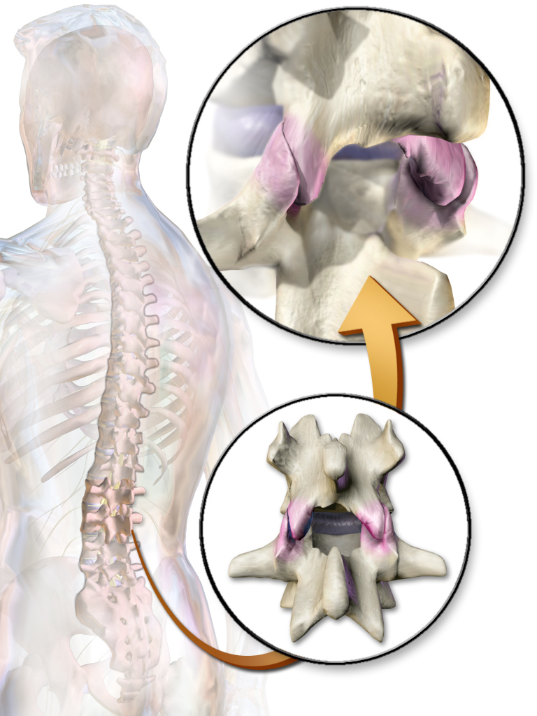
In medicine the Holdsworth fracture is an unstable fracture dislocation of the thoraco lumbar junction of the spine. The injury comprises a fracture through a vertebral body, rupture of the posterior spinal ligaments and fractures of the facet joints. The injury was described by Frank Wild Holdsworth in 1963. He described the mechanism of this injury as a flexion-rotation injury, and said that the unstable fracture dislocation should be treated by fusion Fusion, or synthesis, is the process of combining two or more distinct entities into a new whole. Fusion may also refer to: Science and technology Physics *Nuclear fusion, multiple atomic nuclei combining to form one or more different atomic nucl ... of the two affected vertebrae. See also * Busch fracture References Bone fractures {{injury-stub ... [...More Info...] [...Related Items...] OR: [Wikipedia] [Google] [Baidu] |
Medicine
Medicine is the science and practice of caring for a patient, managing the diagnosis, prognosis, prevention, treatment, palliation of their injury or disease, and promoting their health. Medicine encompasses a variety of health care practices evolved to maintain and restore health by the prevention and treatment of illness. Contemporary medicine applies biomedical sciences, biomedical research, genetics, and medical technology to diagnose, treat, and prevent injury and disease, typically through pharmaceuticals or surgery, but also through therapies as diverse as psychotherapy, external splints and traction, medical devices, biologics, and ionizing radiation, amongst others. Medicine has been practiced since prehistoric times, and for most of this time it was an art (an area of skill and knowledge), frequently having connections to the religious and philosophical beliefs of local culture. For example, a medicine man would apply herbs and say prayers for healing, o ... [...More Info...] [...Related Items...] OR: [Wikipedia] [Google] [Baidu] |
Bone Fracture
A bone fracture (abbreviated FRX or Fx, Fx, or #) is a medical condition in which there is a partial or complete break in the continuity of any bone in the body. In more severe cases, the bone may be broken into several fragments, known as a ''comminuted fracture''. A bone fracture may be the result of high force impact or stress, or a minimal trauma injury as a result of certain medical conditions that weaken the bones, such as osteoporosis, osteopenia, bone cancer, or osteogenesis imperfecta, where the fracture is then properly termed a pathologic fracture. Signs and symptoms Although bone tissue contains no pain receptors, a bone fracture is painful for several reasons: * Breaking in the continuity of the periosteum, with or without similar discontinuity in endosteum, as both contain multiple pain receptors. * Edema and hematoma of nearby soft tissues caused by ruptured bone marrow evokes pressure pain. * Involuntary muscle spasms trying to hold bone fragments in place. D ... [...More Info...] [...Related Items...] OR: [Wikipedia] [Google] [Baidu] |
Joint Dislocation
A joint dislocation, also called luxation, occurs when there is an abnormal separation in the joint, where two or more bones meet.Dislocations. Lucile Packard Children’s Hospital at Stanford. Retrieved 3 March 2013 A partial dislocation is referred to as a subluxation. Dislocations are often caused by sudden trauma on the joint like an impact or fall. A joint dislocation can cause damage to the surrounding ligaments, tendons, muscles, and nerves. Dislocations can occur in any major joint (shoulder, knees, etc.) or minor joint (toes, fingers, etc.). The most common joint dislocation is a shoulder dislocation. Treatment for joint dislocation is usually by closed reduction, that is, skilled manipulation to return the bones to their normal position. Reduction should only be performed by trained medical professionals, because it can cause injury to soft tissue and/or the nerves and vascular structures around the dislocation. Symptoms and signs The following symptoms are common with ... [...More Info...] [...Related Items...] OR: [Wikipedia] [Google] [Baidu] |
Thoracic Vertebrae
In vertebrates, thoracic vertebrae compose the middle segment of the vertebral column, between the cervical vertebrae and the lumbar vertebrae. In humans, there are twelve thoracic vertebra (anatomy), vertebrae and they are intermediate in size between the cervical and lumbar vertebrae; they increase in size going towards the lumbar vertebrae, with the lower ones being much larger than the upper. They are distinguished by the presence of Zygapophysial joint, facets on the sides of the bodies for Articulation (anatomy), articulation with the head of rib, heads of the ribs, as well as facets on the transverse processes of all, except the eleventh and twelfth, for articulation with the tubercle (rib), tubercles of the ribs. By convention, the human thoracic vertebrae are numbered T1–T12, with the first one (T1) located closest to the skull and the others going down the spine toward the lumbar region. General characteristics These are the general characteristics of the second throu ... [...More Info...] [...Related Items...] OR: [Wikipedia] [Google] [Baidu] |
Lumbar Vertebrae
The lumbar vertebrae are, in human anatomy, the five vertebrae between the rib cage and the pelvis. They are the largest segments of the vertebral column and are characterized by the absence of the foramen transversarium within the transverse process (since it is only found in the cervical region) and by the absence of facets on the sides of the body (as found only in the thoracic region). They are designated L1 to L5, starting at the top. The lumbar vertebrae help support the weight of the body, and permit movement. Human anatomy General characteristics The adjacent figure depicts the general characteristics of the first through fourth lumbar vertebrae. The fifth vertebra contains certain peculiarities, which are detailed below. As with other vertebrae, each lumbar vertebra consists of a ''vertebral body'' and a ''vertebral arch''. The vertebral arch, consisting of a pair of ''pedicles'' and a pair of ''laminae'', encloses the ''vertebral foramen'' (opening) and sup ... [...More Info...] [...Related Items...] OR: [Wikipedia] [Google] [Baidu] |
Vertebral Column
The vertebral column, also known as the backbone or spine, is part of the axial skeleton. The vertebral column is the defining characteristic of a vertebrate in which the notochord (a flexible rod of uniform composition) found in all chordata, chordates has been replaced by a segmented series of bone: vertebrae separated by intervertebral discs. Individual vertebrae are named according to their region and position, and can be used as anatomical landmarks in order to guide procedures such as Lumbar puncture, lumbar punctures. The vertebral column houses the spinal canal, a cavity that encloses and protects the spinal cord. There are about 50,000 species of animals that have a vertebral column. The human vertebral column is one of the most-studied examples. Many different diseases in humans can affect the spine, with spina bifida and scoliosis being recognisable examples. The general structure of human vertebrae is fairly typical of that found in mammals, reptiles, and birds. Th ... [...More Info...] [...Related Items...] OR: [Wikipedia] [Google] [Baidu] |
Vertebra
The spinal column, a defining synapomorphy shared by nearly all vertebrates,Hagfish are believed to have secondarily lost their spinal column is a moderately flexible series of vertebrae (singular vertebra), each constituting a characteristic irregular bone whose complex structure is composed primarily of bone, and secondarily of hyaline cartilage. They show variation in the proportion contributed by these two tissue types; such variations correlate on one hand with the cerebral/caudal rank (i.e., location within the backbone), and on the other with phylogenetic differences among the vertebrate taxa. The basic configuration of a vertebra varies, but the bone is its ''body'', with the central part of the body constituting the ''centrum''. The upper (closer to) and lower (further from), respectively, the cranium and its central nervous system surfaces of the vertebra body support attachment to the intervertebral discs. The posterior part of a vertebra forms a vertebral arch ... [...More Info...] [...Related Items...] OR: [Wikipedia] [Google] [Baidu] |
Zygapophysial Joint
The facet joints (or zygapophysial joints, zygapophyseal, apophyseal, or Z-joints) are a set of synovial joint, synovial, plane joints between the articular processes of two adjacent vertebrae. There are two facet joints in each functional spinal unit, spinal motion segment and each facet joint is innervated by the Meningeal branches of spinal nerve, recurrent meningeal nerves. Innervation Innervation to the facet joints vary between segments of the spinal, but they are generally innervated by medial branch nerves that come off the dorsal rami. It is thought that these nerves are for primary sensory input, though there is some evidence that they have some motor input local musculature. Within the cervical spine, most joints are innervated by the medial branch nerve (a branch of the dorsal rami) from the same levels. In other words, the facet joint between C4 and C5 vertebral segments is innervated by the C4 and C5 medial branch nerves. However, there are two exceptions: # The ... [...More Info...] [...Related Items...] OR: [Wikipedia] [Google] [Baidu] |
Frank Wild Holdsworth
Sir Frank Wild Holdsworth (22 September 1904 – 11 December 1969) was an English orthopaedic surgeon remembered for pioneering work on rehabilitation of spinal injury patients. He described the Holdsworth fracture of the spine in 1963. Biography Holdsworth was born and brought up in Bradford, Yorkshire, the son of John William Holdsworth, a joiner and builder, and Martha Elizabeth Denison. He was baptised into the Wesleyan Methodist Church. He was educated at Bradford Grammar School. He studied medicine at Downing College, Cambridge, where he had won an exhibition, and St. George's Hospital Medical School. He qualified MRCS, LRCP in 1929, and was awarded FRCS in 1930 and M. Chir in 1935. On his return to Yorkshire, he worked at the Sheffield Royal Infirmary, becoming consultant orthopaedic surgeon there, and at the Sheffield Children's Hospital, in 1937. He established the orthopaedic and accident service in Sheffield, and later developed the registrar rotation ... [...More Info...] [...Related Items...] OR: [Wikipedia] [Google] [Baidu] |
Flexion
Motion, the process of movement, is described using specific anatomical terms. Motion includes movement of organs, joints, limbs, and specific sections of the body. The terminology used describes this motion according to its direction relative to the anatomical position of the body parts involved. Anatomists and others use a unified set of terms to describe most of the movements, although other, more specialized terms are necessary for describing unique movements such as those of the hands, feet, and eyes. In general, motion is classified according to the anatomical plane it occurs in. ''Flexion'' and ''extension'' are examples of ''angular'' motions, in which two axes of a joint are brought closer together or moved further apart. ''Rotational'' motion may occur at other joints, for example the shoulder, and are described as ''internal'' or ''external''. Other terms, such as ''elevation'' and ''depression'', describe movement above or below the horizontal plane. Many anatomica ... [...More Info...] [...Related Items...] OR: [Wikipedia] [Google] [Baidu] |
Arthrodesis
Arthrodesis, also known as artificial ankylosis or syndesis, is the artificial induction of joint ossification between two bones by surgery. This is done to relieve intractable pain in a joint which cannot be managed by pain medication, splints, or other normally indicated treatments. The typical causes of such pain are fractures which disrupt the joint, severe sprains, and arthritis. It is most commonly performed on joints in the spine, hand, ankle, and foot. Historically, knee and hip arthrodeses were also performed as pain-relieving procedures, but with the great successes achieved in hip and knee arthroplasty, arthrodesis of these large joints has fallen out of favour as a primary procedure, and now is only used as a procedure of last resort in some failed arthroplasties. Method Arthrodesis can be done in several ways: * A bone graft can be created between the two bones using a bone from elsewhere in the person's body (autograft) or using donor bone (allograft) from a ... [...More Info...] [...Related Items...] OR: [Wikipedia] [Google] [Baidu] |
Busch Fracture
In medicine a Busch fracture is a type of fracture of the base of the distal phalanx of the fingers, produced by the removal of the bone insertion ( avulsion) of the extensor tendon. Without the appropriate treatment, the finger becomes a hammer finger. It would correspond to the group B of the Albertoni classification. It is very common in motorcycle riders and soccer joggers, caused by hyperflexion when the tendon is exercising its maximum tension (the closed hand tightening the clutch lever or the brake lever). The Busch fracture is named after Friedrich Busch (1844–1916), who described this type of fracture in the 1860s. Busch's work was drawn on by Albert Hoffa in 1904, resulting in it sometimes being called a "Busch-Hoffa fracture". The mechanism of this injury can be described as an avulsion of the tendon fixed to the distal phalanx. See also * Holdsworth fracture * Galeazzi fracture The Galeazzi fracture is a fracture of the distal third of the radius with disloc ... [...More Info...] [...Related Items...] OR: [Wikipedia] [Google] [Baidu] |


