|
History Of Invasive And Interventional Cardiology
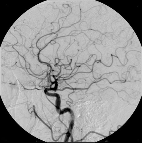
The history of invasive and interventional cardiology is complex, with multiple groups working independently on similar technologies. Invasive and interventional cardiology is currently closely associated with cardiologists (physicians who treat the diseases of the heart), though the development and most of its early research and procedures were performed by diagnostic and interventional radiologists. The birth of invasive cardiology The history of invasive cardiology begins with the development of cardiac catheterization in 1711, when Stephen Hales placed catheters into the right and left ventricles of a living horse. Variations on the technique were performed over the subsequent century, with formal study of cardiac physiology being performed by Claude Bernard in the 1840s. Catheterization of humans The technique of angiography itself was first developed in 1927 by the Portuguese physician Egas Moniz at the University of Lisbon for cerebral angiography, the viewing of brain vasc ... [...More Info...] [...Related Items...] OR: [Wikipedia] [Google] [Baidu] |
Cardiology
Cardiology () is a branch of medicine that deals with disorders of the heart and the cardiovascular system. The field includes medical diagnosis and treatment of congenital heart defects, coronary artery disease, heart failure, valvular heart disease and electrophysiology. Physicians who specialize in this field of medicine are called cardiologists, a specialty of internal medicine. Pediatric cardiologists are pediatricians who specialize in cardiology. Physicians who specialize in cardiac surgery are called cardiothoracic surgeons or cardiac surgeons, a specialty of general surgery. Specializations All cardiologists study the disorders of the heart, but the study of adult and child heart disorders each require different training pathways. Therefore, an adult cardiologist (often simply called "cardiologist") is inadequately trained to take care of children, and pediatric cardiologists are not trained to treat adult heart disease. Surgical aspects are not included in cardiology ... [...More Info...] [...Related Items...] OR: [Wikipedia] [Google] [Baidu] |
André Frédéric Cournand
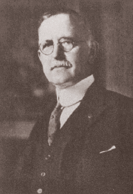
André Frédéric Cournand (September 24, 1895 – February 19, 1988) was a French-American physician and physiologist. Biography Cournand was awarded the Nobel Prize in Physiology or Medicine in 1956 along with Werner Forssmann and Dickinson W. Richards for the development of cardiac catheterization. Born in Paris, Cournand emigrated to the United States in 1930 and, in 1941, became a naturalized citizen. For most of his career, Cournand was a professor at the Columbia University College of Physicians and Surgeons and worked at Bellevue Hospital in New York City. Many seats of medical research have recognized his work, and he has received the Anders Retzius Silver Medal of the Swedish Society for Internal Medicine (1946), the Albert Lasker Award for Basic Medical Research (1949), the John Philipps Memorial Award of the American College of Physicians (1952), the Gold Medal of the Académie Royale de Médecine de Belgique and of the Académie Nationale de Médecine, Paris (19 ... [...More Info...] [...Related Items...] OR: [Wikipedia] [Google] [Baidu] |
Sven-Ivar Seldinger
Sven Ivar Seldinger (19 April 1921 – 21 February 1998), was a radiologist from Mora Municipality, Sweden. In 1953, he introduced the Seldinger technique to obtain safe access to blood vessels and other hollow organs. Biography Sven Ivar Seldinger was born on 19 April 1921 in Dalarna, Sweden. He was born to a family who had long run the local Mora Mechanical Workshop. He first began his medical training in 1940 at the Karolinska Institute. After graduating medical school in 1948, he went on to specialize in radiology. While attending at the Karolinska Hospital he came up with an idea of how to administer a catheter that would be able to reach every human artery. He was qualified with the title of Docent in Radiology in 1967 after successfully defending his thesis on percutaneous transhepatic cholangiography. He was later able to demonstrate, using "phantom experiments", how one could insert a catheter into the femoral artery and reach both the parathyroid and renal arteries. In ... [...More Info...] [...Related Items...] OR: [Wikipedia] [Google] [Baidu] |
Percutaneous
{{More citations needed, date=January 2021 In surgery, a percutaneous procedurei.e. Granger et al., 2012 is any medical procedure or method where access to inner organs or other tissue is done via needle-puncture of the skin, rather than by using an "open" approach where inner organs or tissue are exposed (typically with the use of a scalpel). The percutaneous approach is commonly used in vascular procedures such as angioplasty and stenting. This involves a needle catheter getting access to a blood vessel, followed by the introduction of a wire through the lumen (pathway) of the needle. It is over this wire that other catheters can be placed into the blood vessel. This technique is known as the modified Seldinger technique. More generally, "percutaneous", via its Latin roots means, 'by way of the skin'. An example would be percutaneous drug absorption from topical medications. More often, percutaneous is typically used in reference to placement of medical devices using a need ... [...More Info...] [...Related Items...] OR: [Wikipedia] [Google] [Baidu] |
Precordial Thump
Precordial thump is a medical procedure used in the treatment of ventricular fibrillation or pulseless ventricular tachycardia under certain conditions. The procedure has a very low success rate, but may be used in those with witnessed, monitored onset of one of the "shockable" cardiac rhythms if a defibrillator is not immediately available. It should not delay cardiopulmonary resuscitation (CPR) and defibrillation, nor should it be used in those with unwitnessed out-of-hospital cardiac arrest. Procedure In a precordial thump, a provider strikes at the middle of a person's sternum with the ulnar aspect of the fist. The intent is to interrupt a potentially life-threatening rhythm. The thump is thought to produce an electrical depolarization of 2 to 5 joules. Effectiveness Precordial thump may be effective only if used within seconds near the onset of ventricular fibrillation or pulseless ventricular tachycardia, and so should be used only when the arrest is witnessed and monitor ... [...More Info...] [...Related Items...] OR: [Wikipedia] [Google] [Baidu] |
Ventricular Fibrillation
Ventricular fibrillation (V-fib or VF) is an abnormal heart rhythm in which the ventricles of the heart quiver. It is due to disorganized electrical activity. Ventricular fibrillation results in cardiac arrest with loss of consciousness and no pulse. This is followed by sudden cardiac death in the absence of treatment. Ventricular fibrillation is initially found in about 10% of people with cardiac arrest. Ventricular fibrillation can occur due to coronary heart disease, valvular heart disease, cardiomyopathy, Brugada syndrome, long QT syndrome, electric shock, or intracranial hemorrhage. Diagnosis is by an electrocardiogram (ECG) showing irregular unformed QRS complexes without any clear P waves. An important differential diagnosis is torsades de pointes. Treatment is with cardiopulmonary resuscitation (CPR) and defibrillation. Biphasic defibrillation may be better than monophasic. The medication epinephrine or amiodarone may be given if initial treatments are not effect ... [...More Info...] [...Related Items...] OR: [Wikipedia] [Google] [Baidu] |
Radiocontrast
Radiocontrast agents are substances used to enhance the visibility of internal structures in X-ray-based imaging techniques such as computed tomography (contrast CT), projectional radiography, and fluoroscopy. Radiocontrast agents are typically iodine, or more rarely barium sulfate. The contrast agents absorb external X-rays, resulting in decreased exposure on the X-ray detector. This is different from radiopharmaceuticals used in nuclear medicine which emit radiation. Magnetic resonance imaging (MRI) functions through different principles and thus MRI contrast agents have a different mode of action. These compounds work by altering the magnetic properties of nearby hydrogen nuclei. Types and uses Radiocontrast agents used in X-ray examinations can be grouped in positive (iodinated agents, barium sulfate), and negative agents (air, carbon dioxide, methylcellulose). Iodine (circulatory system) Iodinated contrast contains iodine. It is the main type of radiocontrast used for intr ... [...More Info...] [...Related Items...] OR: [Wikipedia] [Google] [Baidu] |
Cleveland Clinic
Cleveland Clinic is a nonprofit American academic medical center based in Cleveland, Ohio. Owned and operated by the Cleveland Clinic Foundation, an Ohio nonprofit corporation established in 1921, it runs a 170-acre (69 ha) campus in Cleveland, as well as 11 affiliated hospitals, 19 family health centers in Northeast Ohio, and hospitals in Florida and Nevada. International operations include the Cleveland Clinic Abu Dhabi hospital in the United Arab Emirates and Cleveland Clinic Canada, which has two executive health and sports medicine clinics in Toronto."Facts & Figures" Cleveland Clinic. Another hospital campus in the United Kingdom, Cleveland Clinic London, opened to outpatients in 2021 and is scheduled to fully open in 2022. Tomislav Mihaljevic is the president and CEO. Cleveland Cl ... [...More Info...] [...Related Items...] OR: [Wikipedia] [Google] [Baidu] |
Mason Sones
F. Mason Sones, Jr. (October 28, 1918 – August 28, 1985) was an American physician whose pioneering work in cardiac catheterization was instrumental in the development of both coronary artery bypass surgery and interventional cardiology. Early life and career Sones was born in Noxapater, MississippiWestaby, p. 214 to Frank Mason and Myrtle (Bryan) Sones. He graduated from Western Maryland College (now McDaniel College) in 1940 and received his M.D. from the University of Maryland School of Medicine in 1943. In 1942, Sones married Geraldine Newton. The couple had four children, Frank Mason III, Geraldine Patricia, Steven, and David. From 1944 to 1946, he served in the United States Army Air Corps in the Pacific and would later serve as a national consultant to the Air Force. Sones served as an intern at University Hospital in Baltimore and was a Resident at Henry Ford Hospital in Detroit before joining the Cleveland Clinic Foundation in 1950. Discovery of coronary angiography ... [...More Info...] [...Related Items...] OR: [Wikipedia] [Google] [Baidu] |
Aortography
Aortography involves placement of a catheter in the aorta and injection of contrast material while taking X-rays of the aorta. The procedure is known as an aortogram. The diagnosis of aortic dissection can be made by visualization of the intimal flap and flow of contrast material in both the true lumen and the false lumen. The catheter has to be inserted through the right femoral artery, because in about two-thirds of cases the aortic dissection spreads into the left common iliac artery. The aortogram was previously considered the gold standard test for the diagnosis of aortic dissection, with a sensitivity of up to 80% and a specificity of about 94%. It is especially poor in the diagnosis of cases where the dissection is due to hemorrhage within the media without any initiating intimal tear. The advantage of the aortogram in the diagnosis of aortic dissection is that it can delineate the extent of involvement of the aorta and branch vessels and can diagnose aortic insuffic ... [...More Info...] [...Related Items...] OR: [Wikipedia] [Google] [Baidu] |
Charles Dotter
Charles Theodore Dotter (14 June 1920 – 15 February 1985) was a pioneering US vascular radiologist who is credited with developing interventional radiology. Dotter, with his trainee Dr Melvin P. Judkins, described angioplasty in 1964. Dotter received a bachelor of arts degree in 1941 from Duke University. He went to medical school at Cornell, where he met his future wife, Pamela Beattie, a head nurse at New York Hospital. They married in 1944. He completed his internship at the United States Naval Hospital in New York State, and his residency at New York Hospital. Dotter invented angioplasty and the catheter-delivered stent, which were first used to treat peripheral arterial disease. It was Dotter who, in 1950, developed an automatic X-Ray Roll-Film magazine capable of producing images at the rate of 2 per second. On January 16, 1964, at Oregon Health and Science University Dotter percutaneously dilated a tight, localized stenosis of the superficial femoral artery (SFA) in a ... [...More Info...] [...Related Items...] OR: [Wikipedia] [Google] [Baidu] |
Interventional Radiology
Interventional radiology (IR) is a medical specialty that performs various minimally-invasive procedures using medical imaging guidance, such as x-ray fluoroscopy, computed tomography, magnetic resonance imaging, or ultrasound. IR performs both diagnostic and therapeutic procedures through very small incisions or body orifices. Diagnostic IR procedures are those intended to help make a diagnosis or guide further medical treatment, and include image-guided biopsy of a tumor or injection of an imaging contrast agent into a hollow structure, such as a blood vessel or a duct. By contrast, therapeutic IR procedures provide direct treatment—they include catheter-based medicine delivery, medical device placement (e.g., stents), and angioplasty of narrowed structures. The main benefits of interventional radiology techniques are that they can reach the deep structures of the body through a body orifice or tiny incision using small needles and wires. That decreases risks, pain, an ... [...More Info...] [...Related Items...] OR: [Wikipedia] [Google] [Baidu] |


