|
Hiatus For Lesser Petrosal Nerve
The hiatus for lesser petrosal nerve is a hiatus in the petrous part of the temporal bone which transmits the lesser petrosal nerve. It is located posterior to the groove for the superior petrosal sinus and posterolateral to the jugular foramen. The hiatus for lesser petrosal nerve receives the lesser petrosal nerve as it branches from the glossopharyngeal nerve (CN IX) before the glossopharyngeal enters the posterior cranial fossa through the jugular foramen A jugular foramen is one of the two (left and right) large foramina (openings) in the base of the skull, located behind the carotid canal. It is formed by the temporal bone and the occipital bone. It allows many structures to pass, including the .... The lesser petrosal nerve then travels anteriorly from the hiatus toward the foramen ovale, through which it exits the cranial cavity.Frank H. Netter, MD; ''Atlas of Human Anatomy'', 4th Edition, References Foramina of the skull {{musculoskeletal-stub ... [...More Info...] [...Related Items...] OR: [Wikipedia] [Google] [Baidu] |
Hiatus (anatomy)
{{set index article In anatomy, a hiatus is a natural fissure in a structure. Examples include: * Adductor hiatus * Aortic hiatus * Esophageal hiatus, the opening in the diaphragm through which the oesophagus passes from the thorax into the abdomen * Greater petrosal nerve hiatus * Maxillary hiatus * Sacral hiatus * Semilunar hiatus The semilunar hiatus or hiatus semilunaris, is a crescent-shaped groove in the lateral wall of the nasal cavity just inferior to the ethmoid bulla. It is the location of the openings of the maxillary sinuses. It is bounded inferiorly and anterior ... Anatomy ... [...More Info...] [...Related Items...] OR: [Wikipedia] [Google] [Baidu] |
Petrous Part Of The Temporal Bone
The petrous part of the temporal bone is pyramid-shaped and is wedged in at the base of the skull between the sphenoid and occipital bones. Directed medially, forward, and a little upward, it presents a base, an apex, three surfaces, and three angles, and houses in its interior, the components of the inner ear. The petrous portion is among the most basal elements of the skull and forms part of the endocranium. Petrous comes from the Latin word ''petrosus'', meaning "stone-like, hard". It is one of the densest bones in the body. The petrous bone is important for studies of ancient DNA from skeletal remains, as it tends to contain extremely well-preserved DNA. Base The base is fused with the internal surfaces of the squamous and mastoid parts. Apex The apex, which is rough and uneven, is received into the angular interval between the posterior border of the great wing of the sphenoid bone and the basilar part of the occipital bone; it presents the anterior or internal openin ... [...More Info...] [...Related Items...] OR: [Wikipedia] [Google] [Baidu] |
Lesser Petrosal Nerve
The lesser petrosal nerve (also known as the small superficial petrosal nerve) is the general visceral efferent (GVE) component of the glossopharyngeal nerve (CN IX), carrying parasympathetic preganglionic fibers from the tympanic plexus to the parotid gland. It synapses in the otic ganglion, from where the postganglionic fibers emerge. Structure After arising in the tympanic plexus, the lesser petrosal nerve passes forward and then through the hiatus for lesser petrosal nerve on the anterior surface of the petrous part of the temporal bone into the middle cranial fossa. It travels across the floor of the middle cranial fossa, then exits the skull via canaliculus innominatus to reach the infratemporal fossa. The fibres synapse in the otic ganglion, and post-ganglionic fibres then travel briefly with the auriculotemporal nerve (a branch of V3) before entering the body of the parotid gland. The lesser petrosal nerve will distribute its parasympathetic post-ganglionic (GVE) fibers ... [...More Info...] [...Related Items...] OR: [Wikipedia] [Google] [Baidu] |
Glossopharyngeal Nerve
The glossopharyngeal nerve (), also known as the ninth cranial nerve, cranial nerve IX, or simply CN IX, is a cranial nerve that exits the brainstem from the sides of the upper Medulla oblongata, medulla, just anterior (closer to the nose) to the vagus nerve. Being a mixed nerve (sensorimotor), it carries afferent sensory and efferent motor information. The motor division of the glossopharyngeal nerve is derived from the Basal plate (neural tube), basal plate of the embryonic medulla oblongata, whereas the sensory division originates from the cranial neural crest. Structure From the anterior portion of the medulla oblongata, the glossopharyngeal nerve passes laterally across or below the Flocculus (cerebellar), flocculus, and leaves the skull through the central part of the jugular foramen. From the superior and inferior ganglia in jugular foramen, it has its own sheath of dura mater. The inferior ganglion on the inferior surface of petrous part of temporal is related with a tri ... [...More Info...] [...Related Items...] OR: [Wikipedia] [Google] [Baidu] |
Posterior Cranial Fossa
The posterior cranial fossa is part of the cranial cavity, located between the foramen magnum and tentorium cerebelli. It contains the brainstem and cerebellum. This is the most inferior of the fossae. It houses the cerebellum, medulla and pons. Anteriorly it extends to the apex of the petrous temporal. Posteriorly it is enclosed by the occipital bone. Laterally portions of the squamous temporal and mastoid part of the temporal bone form its walls. Features Foramen magnum The most conspicuous, large opening in the floor of the fossa. It transmits the medulla, the ascending portions of the spinal accessory nerve (XI), and the vertebral arteries. Internal acoustic meatus Lies in the anterior wall of the posterior cranial fossa. It transmits the facial (VII) and vestibulocochlear (VIII) cranial nerves into a canal in the petrous temporal bone. Jugular foramen Lies between the inferior edge of the petrous temporal bone and the adjacent occipital bone and transmits the internal ... [...More Info...] [...Related Items...] OR: [Wikipedia] [Google] [Baidu] |
Jugular Foramen
A jugular foramen is one of the two (left and right) large foramina (openings) in the base of the skull, located behind the carotid canal. It is formed by the temporal bone and the occipital bone. It allows many structures to pass, including the inferior petrosal sinus, three cranial nerves, the sigmoid sinus, and meningeal arteries. Structure The jugular foramen is formed in front by the petrous portion of the temporal bone, and behind by the occipital bone. It is generally slightly larger on the right side than on the left side. Contents The jugular foramen may be subdivided into three compartments, each with their own contents. * The ''anterior'' compartment transmits the inferior petrosal sinus. * The ''intermediate'' compartment transmits the glossopharyngeal nerve, the vagus nerve, and the accessory nerve. * The ''posterior'' compartment transmits the sigmoid sinus (becoming the internal jugular vein), and some meningeal branches from the occipital artery and ascending ... [...More Info...] [...Related Items...] OR: [Wikipedia] [Google] [Baidu] |
Foramen Ovale (skull)
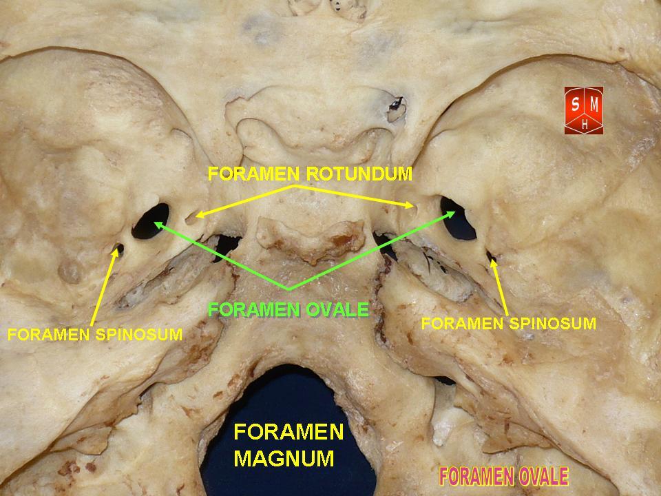
The foramen ovale (Latin: oval window) is a hole in the posterior part of the sphenoid bone, posterolateral to the foramen rotundum. It is one of the larger of the several holes (the foramina) in the skull. It transmits the mandibular nerve, a branch of the trigeminal nerve. Structure The foramen ovale is an opening in the greater wing of the sphenoid bone. The foramen ovale is one of two cranial foramina in the greater wing, the other being the foramen spinosum. The foramen ovale is posterolateral to the foramen rotundum and anteromedial to the foramen spinosum. Posterior and medial to the foramen is the opening for the carotid canal. Variation Similar to other foramina, the foramen ovale differs in shape and size throughout the natural life. The earliest perfect ring-shaped formation of the foramen ovale was observed in the 7th fetal month and the latest in 3 years after birth, in a study using over 350 skulls.In a study conducted on 100 skulls, the foramen ovale was d ... [...More Info...] [...Related Items...] OR: [Wikipedia] [Google] [Baidu] |
Cranial Cavity
The cranial cavity, also known as intracranial space, is the space within the skull that accommodates the brain. The skull minus the mandible is called the ''cranium''. The cavity is formed by eight cranial bones known as the neurocranium that in humans includes the skull cap and forms the protective case around the brain. The remainder of the skull is called the facial skeleton. Meninges are protective membranes that surround the brain to minimize damage of the brain when there is head trauma. Meningitis is the inflammation of meninges caused by bacterial or viral infections. Structure The capacity of an adult human cranial cavity is 1,200–1,700 cm3. The spaces between meninges and the brain are filled with a clear cerebrospinal fluid, increasing the protection of the brain. Facial bones of the skull are not included in the cranial cavity. There are only eight cranial bones: The occipital, sphenoid, frontal, ethmoid, two parietal, and two temporal bones are fused tog ... [...More Info...] [...Related Items...] OR: [Wikipedia] [Google] [Baidu] |
