|
Hepatic Portal Vein
The portal vein or hepatic portal vein (HPV) is a blood vessel that carries blood from the gastrointestinal tract, gallbladder, pancreas and spleen to the liver. This blood contains nutrients and toxins extracted from digested contents. Approximately 75% of total liver blood flow is through the portal vein, with the remainder coming from the hepatic artery proper. The blood leaves the liver to the heart in the hepatic veins. The portal vein is not a true vein, because it conducts blood to capillary beds in the liver and not directly to the heart. It is a major component of the hepatic portal system, one of only two portal venous systems in the body – with the hypophyseal portal system being the other. The portal vein is usually formed by the confluence of the superior mesenteric, splenic veins, inferior mesenteric, left, right gastric veins and the pancreatic vein. Conditions involving the portal vein cause considerable illness and death. An important example of such ... [...More Info...] [...Related Items...] OR: [Wikipedia] [Google] [Baidu] |
Superior Mesenteric Vein
In human anatomy, the superior mesenteric vein (SMV) is a blood vessel that drains blood from the small intestine ( jejunum and ileum). Behind the neck of the pancreas, the superior mesenteric vein combines with the splenic vein to form the hepatic portal vein. The superior mesenteric vein lies to the right of the similarly named artery, the superior mesenteric artery, which originates from the abdominal aorta. Structure Tributaries of the superior mesenteric vein drain the small intestine, large intestine, stomach, pancreas and appendix and include: * Right gastro-omental vein (also known as the right gastro-epiploic vein) * inferior pancreaticoduodenal veins * veins from jejunum * veins from ileum * middle colic vein – drains the transverse colon * right colic vein – drains the ascending colon * ileocolic vein The superior mesenteric vein combines with the splenic vein to form the portal vein. Clinical significance Thrombosis of the superior mesenteric vein is quit ... [...More Info...] [...Related Items...] OR: [Wikipedia] [Google] [Baidu] |
Hepatic Veins
In human anatomy, the hepatic veins are the veins that drain venous blood from the liver into the inferior vena cava (as opposed to the hepatic portal vein which conveys blood from the gastrointestinal organs to the liver). There are usually three large upper hepatic veins draining from the left, middle, and right parts of the liver, as well as a number (6-20) of lower hepatic veins. All hepatic veins are valveless. Structure All the hepatic veins drain into the inferior vena cava. The hepatic veins are divided into an upper and a lower group. Upper group The upper group consists of three hepatic veins - the right, middle, and left hepatic veins - draining the central veins from the right, middle, and left regions of the liver and are larger than the lower group of veins. The veins of the upper group drain into the suprahepatic part of the inferior vena cava (i.e. part superior to the liver). Right hepatic vein The right hepatic vein is the longest and largest of all the he ... [...More Info...] [...Related Items...] OR: [Wikipedia] [Google] [Baidu] |
Organ (biology)
In biology, an organ is a collection of Tissue (biology), tissues joined in a structural unit to serve a common function. In the biological organization, hierarchy of life, an organ lies between Tissue (biology), tissue and an organ system. Tissues are formed from same type Cell (biology), cells to act together in a function. Tissues of different types combine to form an organ which has a specific function. The Gastrointestinal tract, intestinal wall for example is formed by epithelial tissue and smooth muscle tissue. Two or more organs working together in the execution of a specific body function form an organ system, also called a biological system or body system. An organ's tissues can be broadly categorized as parenchyma, the functional tissue, and stroma (tissue), stroma, the structural tissue with supportive, connective, or ancillary functions. For example, the gland's tissue that makes the hormones is the parenchyma, whereas the stroma includes the nerve tissue, nerves tha ... [...More Info...] [...Related Items...] OR: [Wikipedia] [Google] [Baidu] |
Cytokine
Cytokines are a broad and loose category of small proteins (~5–25 kDa) important in cell signaling. Cytokines are peptides and cannot cross the lipid bilayer of cells to enter the cytoplasm. Cytokines have been shown to be involved in autocrine, paracrine and endocrine signaling as immunomodulating agents. Cytokines include chemokines, interferons, interleukins, lymphokines, and tumour necrosis factors, but generally not hormones or growth factors (despite some overlap in the terminology). Cytokines are produced by a broad range of cells, including immune cells like macrophages, B lymphocytes, T lymphocytes and mast cells, as well as endothelial cells, fibroblasts, and various stromal cells; a given cytokine may be produced by more than one type of cell. They act through cell surface receptors and are especially important in the immune system; cytokines modulate the balance between humoral and cell-based immune responses, and they regulate the maturati ... [...More Info...] [...Related Items...] OR: [Wikipedia] [Google] [Baidu] |
Abdominal Obesity
Abdominal obesity, also known as central obesity and truncal obesity, is a condition when excessive visceral fat around the stomach and abdomen has built up to the extent that it is likely to have a negative impact on health. Abdominal obesity has been strongly linked to cardiovascular disease, Alzheimer's disease, and other metabolic and vascular diseases. Visceral and central abdominal fat and waist circumference show a strong association with type 2 diabetes. Visceral fat, also known as organ fat or ''intra-abdominal fat'', is located inside the peritoneal cavity, packed in between internal organs and torso, as opposed to subcutaneous fat, which is found underneath the skin, and intramuscular fat, which is found interspersed in skeletal muscle. Visceral fat is composed of several adipose depots including mesenteric, epididymal white adipose tissue (EWAT), and perirenal fat. An excess of adipose visceral fat is known as central obesity, the "pot belly" or "beer bell ... [...More Info...] [...Related Items...] OR: [Wikipedia] [Google] [Baidu] |
Cirrhosis
Cirrhosis, also known as liver cirrhosis or hepatic cirrhosis, and end-stage liver disease, is the impaired liver function caused by the formation of scar tissue known as fibrosis due to damage caused by liver disease. Damage causes tissue repair and subsequent formation of scar tissue, which over time can replace normal parenchyma, functioning tissue, leading to the impaired liver function of cirrhosis. The disease typically develops slowly over months or years. Early symptoms may include Fatigue (medicine), tiredness, Asthenia, weakness, Anorexia (symptom), loss of appetite, weight loss, unexplained weight loss, nausea and vomiting, and discomfort in the right upper quadrant of the abdomen. As the disease worsens, symptoms may include Pruritus, itchiness, peripheral edema, swelling in the lower legs, ascites, fluid build-up in the abdomen, jaundice, coagulopathy, bruising easily, and the development of spider angioma, spider-like blood vessels in the skin. The fluid build-up in ... [...More Info...] [...Related Items...] OR: [Wikipedia] [Google] [Baidu] |
Portal Hypertension
Portal hypertension is abnormally increased portal venous pressure – blood pressure in the portal vein and its branches, that drain from most of the intestine to the liver. Portal hypertension is defined as a hepatic venous pressure gradient greater than 5 mmHg. Cirrhosis (a form of chronic liver failure) is the most common cause of portal hypertension; other, less frequent causes are therefore grouped as non-cirrhotic portal hypertension. When it becomes severe enough to cause symptoms or complications, treatment may be given to decrease portal hypertension itself or to manage its complications. Signs and symptoms Signs and symptoms of portal hypertension include: * Ascites (free fluid in the peritoneal cavity), ** Abdominal pain or tenderness (when bacteria infect the ascites, as in spontaneous bacterial peritonitis). * Increased spleen size (splenomegaly), which may lead to lower platelet counts (thrombocytopenia) * Anorectal varices * Swollen veins on the anterior abdomi ... [...More Info...] [...Related Items...] OR: [Wikipedia] [Google] [Baidu] |
Blood Pressure
Blood pressure (BP) is the pressure of circulating blood against the walls of blood vessels. Most of this pressure results from the heart pumping blood through the circulatory system. When used without qualification, the term "blood pressure" refers to the pressure in the large arteries. Blood pressure is usually expressed in terms of the systolic pressure (maximum pressure during one heartbeat) over diastolic pressure (minimum pressure between two heartbeats) in the cardiac cycle. It is measured in millimeters of mercury (mmHg) above the surrounding atmospheric pressure. Blood pressure is one of the vital signs—together with respiratory rate, heart rate, oxygen saturation, and body temperature—that healthcare professionals use in evaluating a patient's health. Normal resting blood pressure, in an adult is approximately systolic over diastolic, denoted as "120/80 mmHg". Globally, the average blood pressure, age standardized, has remained about the sam ... [...More Info...] [...Related Items...] OR: [Wikipedia] [Google] [Baidu] |
Right Gastric Vein
The right gastric vein (pyloric vein) drains blood from the lesser curvature of the stomach into the hepatic portal vein. It is part of the portal circulation. Structure The right gastric vein passes right along the lesser curvature of the stomach to the pylorus. Once there, it joins onto the portal vein before the duodenum The duodenum is the first section of the small intestine in most higher vertebrates, including mammals, reptiles, and birds. In fish, the divisions of the small intestine are not as clear, and the terms anterior intestine or proximal intestine .... The prepyloric vein is the last connecting branch onto the right gastric vein, marking the end of the stomach, and draining the proximal part of the duodenum. Function The right gastric vein drains deoxygenated blood from the lesser curvature of the stomach. See also * Left gastric vein References External links * () {{Authority control Veins of the torso Stomach ... [...More Info...] [...Related Items...] OR: [Wikipedia] [Google] [Baidu] |
Left Gastric Vein
The left gastric vein (or coronary vein) is a vein that derives from tributaries draining the lesser curvature of the stomach. Structure The left gastric vein runs from right to left along the lesser curvature of the stomach. It passes to the esophageal opening of the stomach, where it receives some esophageal veins. It then turns backward and passes from left to right behind the omental bursa. It drains into the portal vein near the superior border of the pancreas. Function The left gastric vein drains deoxygenated blood from the lesser curvature of the stomach. It also acts as collaterals between the portal vein and the systemic venous system of the lower esophagus (azygous vein). Clinical significance Esophageal and paraesophageal varices are supplied primarily by the left gastric vein (due to flow reversal) and typically drain into the azygos/hemiazygos venous system.Siegelman, E.: "Body MRI", page 47. Saunders, 2004 See also * Right gastric vein The right g ... [...More Info...] [...Related Items...] OR: [Wikipedia] [Google] [Baidu] |
Hypophyseal Portal System
The hypophyseal portal system is a system of blood vessels in the microcirculation at the base of the brain, connecting the hypothalamus with the anterior pituitary. Its main function is to quickly transport and exchange hormones between the hypothalamus arcuate nucleus and anterior pituitary gland. The capillaries in the portal system are fenestrated (have many small channels with high vascular permeability) which allows a rapid exchange between the hypothalamus and the pituitary. The main hormones transported by the system include gonadotropin-releasing hormone, corticotropin-releasing hormone, growth hormone–releasing hormone, and thyrotropin-releasing hormone. Structure The blood supply and direction of flow in the hypophyseal portal system has been studied over many years on laboratory animals and human cadaver specimens with injection and vascular corrosion casting methods. Short portal vessels between the neural and anterior pituitary lobes provide an avenue for rap ... [...More Info...] [...Related Items...] OR: [Wikipedia] [Google] [Baidu] |
Portal Venous System
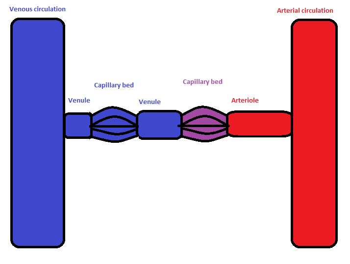
In the circulatory system of animals, a portal venous system occurs when a capillary bed pools into another capillary bed through veins, without first going through the heart. Both capillary beds and the blood vessels that connect them are considered part of the portal venous system. They are relatively uncommon as the majority of capillary beds drain into veins which then drain into the heart, not into another capillary bed. Portal venous systems are considered venous because the blood vessels that join the two capillary beds are either veins or venules. Examples of such systems include the hepatic portal system, the hypophyseal portal system, and (in non-mammals) the renal portal system. Unqualified, ''portal venous system'' often refers to the hepatic portal system. For this reason, ''portal vein'' most commonly refers to the hepatic portal vein. The functional significance of such a system is that it transports products of one region directly to another region in relati ... [...More Info...] [...Related Items...] OR: [Wikipedia] [Google] [Baidu] |


