|
Helicis Minor
The Helicis minor (musculus helicis minor or smaller muscle of helix) is a small skeletal muscle. The helicis minor is an intrinsic muscle of the outer ear. The muscle runs obliques and covers the helical crus, part of the helix located just above the tragus. The helicis minor originates from the base of the helical crus, runs obliques and inserts at the anterior aspect of the helical crus where it curves upward above the tragus. The function of the muscle is to assist in adjusting the shape of the anterior margin of the ear cartilage. While this is a potential action in some individuals, in the majority of individuals the muscle modifies auricular shape to a minimal degree. The helicis minor is developmentally derived from the second pharyngeal arch It seem that only in primates is the helicis major and minor two distinctive muscles. Additional images See also * Intrinsic muscles of external ear The outer ear, external ear, or auris externa is the external part o ... [...More Info...] [...Related Items...] OR: [Wikipedia] [Google] [Baidu] |
Helical Crus
Helical may refer to: * Helix, the mathematical concept for the shape * Helical engine, a proposed spacecraft propulsion drive * Helical spring, a coilspring * Helical plc, a British property company, once a maker of steel bar stock * Helicoil A threaded insert, also known as a threaded bushing, is a fastener element that is inserted into an object to add a threaded hole. They may be used to repair a stripped threaded hole, provide a durable threaded hole in a soft material, place a thr ..., a mechanical thread repairing insert * H-el-ical//, stage name for Hikaru, Japanese singer {{disambig ... [...More Info...] [...Related Items...] OR: [Wikipedia] [Google] [Baidu] |
Outer Ear
The outer ear, external ear, or auris externa is the external part of the ear, which consists of the auricle (also pinna) and the ear canal. It gathers sound energy and focuses it on the eardrum (tympanic membrane). Structure Auricle The visible part is called the auricle, also known as the pinna, especially in other animals. It is composed of a thin plate of yellow elastic cartilage, covered with integument, and connected to the surrounding parts by ligaments and muscles; and to the commencement of the ear canal by fibrous tissue. Many mammals can move the pinna (with the auriculares muscles) in order to focus their hearing in a certain direction in much the same way that they can turn their eyes. Most humans do not have this ability. Ear canal From the pinna, the sound waves move into the ear canal (also known as the ''external acoustic meatus'') a simple tube running through to the middle ear. This tube leads inward from the bottom of the auricula and conducts t ... [...More Info...] [...Related Items...] OR: [Wikipedia] [Google] [Baidu] |
Helicis Major
The helicis major (or large muscle of helix) is an intrinsic muscle of the outer ear. In human anatomy, it is the form of a narrow vertical band situated upon the anterior margin of the helix, at the point where the helix becomes transverse. It arises below, from the spina helicis, and is inserted into the anterior border of the helix, just where it is about to curve backward. The function of the muscle is to adjust the shape of the ear by depressing the anterior margin of the ear cartilage. While the muscle modifies the auricular shape only minimally in the majority of individuals, this action could increase the opening into the external acoustic meatus in some. The helicis major is developmentally derived from the second pharyngeal arch. It seem that only in primates is the helicis minor and major two distinctive muscles. Additional images See also * Intrinsic muscles of external ear * Helicis minor The Helicis minor (musculus helicis minor or smaller muscle of helix ... [...More Info...] [...Related Items...] OR: [Wikipedia] [Google] [Baidu] |
Primates
Primates are a diverse order of mammals. They are divided into the strepsirrhines, which include the lemurs, galagos, and lorisids, and the haplorhines, which include the tarsiers and the simians ( monkeys and apes, the latter including humans). Primates arose 85–55 million years ago first from small terrestrial mammals, which adapted to living in the trees of tropical forests: many primate characteristics represent adaptations to life in this challenging environment, including large brains, visual acuity, color vision, a shoulder girdle allowing a large degree of movement in the shoulder joint, and dextrous hands. Primates range in size from Madame Berthe's mouse lemur, which weighs , to the eastern gorilla, weighing over . There are 376–524 species of living primates, depending on which classification is used. New primate species continue to be discovered: over 25 species were described in the 2000s, 36 in the 2010s, and three in the 2020s. Primates have ... [...More Info...] [...Related Items...] OR: [Wikipedia] [Google] [Baidu] |
Pharyngeal Arch
The pharyngeal arches, also known as visceral arches'','' are structures seen in the embryonic development of vertebrates that are recognisable precursors for many structures. In fish, the arches are known as the branchial arches, or gill arches. In the human embryo, the arches are first seen during the fourth week of development. They appear as a series of outpouchings of mesoderm on both sides of the developing pharynx. The vasculature of the pharyngeal arches is known as the aortic arches. In fish, the branchial arches support the gills. Structure In vertebrates, the pharyngeal arches are derived from all three germ layers (the primary layers of cells that form during embryogenesis). Neural crest cells enter these arches where they contribute to features of the skull and facial skeleton such as bone and cartilage. However, the existence of pharyngeal structures before neural crest cells evolved is indicated by the existence of neural crest-independent mechanisms of pharyn ... [...More Info...] [...Related Items...] OR: [Wikipedia] [Google] [Baidu] |
Second Pharyngeal Arch
The pharyngeal arches, also known as visceral arches'','' are structures seen in the embryonic development of vertebrates that are recognisable precursors for many structures. In fish, the arches are known as the branchial arches, or gill arches. In the human embryo, the arches are first seen during the fourth week of development. They appear as a series of outpouchings of mesoderm on both sides of the developing pharynx. The vasculature of the pharyngeal arches is known as the aortic arches. In fish, the branchial arches support the gills. Structure In vertebrates, the pharyngeal arches are derived from all three germ layers (the primary layers of cells that form during embryogenesis). Neural crest cells enter these arches where they contribute to features of the skull and facial skeleton such as bone and cartilage. However, the existence of pharyngeal structures before neural crest cells evolved is indicated by the existence of neural crest-independent mechanisms of phar ... [...More Info...] [...Related Items...] OR: [Wikipedia] [Google] [Baidu] |
Tragus (ear)
The tragus is a small pointed eminence of the external ear, situated in front of the concha, and projecting backward over the meatus. It also is the name of hair growing at the entrance of the ear. Its name comes the Ancient Greek (), meaning 'goat', and is descriptive of its general covering on its under surface with a tuft of hair, resembling a goat's beard. The nearby antitragus projects forwards and upwards. Because the tragus faces rearwards, it aids in collecting sounds from behind. These sounds are delayed more than sounds arriving from the front, assisting the brain to sense front vs. rear sound sources. In a positive fistula test (for the presence of a fistula from cholesteatoma to the labyrinth), pressure on the tragus causes vertigo or eye deviation by inducing movement of perilymph. Other animals The tragus is a key feature in many bat species. As a piece of skin in front of the ear canal, it plays an important role in directing sounds into the ear for prey loc ... [...More Info...] [...Related Items...] OR: [Wikipedia] [Google] [Baidu] |
Helix (ear)
The helix is the prominent rim of the auricle. Where the helix turns downwards posteriorly, a small tubercle is sometimes seen, namely the '' auricular tubercle of Darwin''. Additional images File:Gray906.png, The muscles of the auricula. File:Darwin-s-tubercle.jpg, Left: Darwin's tubercle. Right: the homologous point in a macaque The macaques () constitute a genus (''Macaca'') of gregarious Old World monkeys of the subfamily Cercopithecinae. The 23 species of macaques inhabit ranges throughout Asia, North Africa, and (in one instance) Gibraltar. Macaques are principall .... File:Slide2COR.JPG, External ear. Right auricle.Lateral view. File:Slide3COR.JPG, External ear. Right auricle.Lateral view. File:Slide4COR.JPG, External ear. Right auricle.Lateral view. See also References Ear {{anatomy-stub ... [...More Info...] [...Related Items...] OR: [Wikipedia] [Google] [Baidu] |
Intrinsic Muscles Of External Ear
The outer ear, external ear, or auris externa is the external part of the ear, which consists of the auricle (also pinna) and the ear canal. It gathers sound energy and focuses it on the eardrum (tympanic membrane). Structure Auricle The visible part is called the auricle, also known as the pinna, especially in other animals. It is composed of a thin plate of yellow elastic cartilage, covered with integument, and connected to the surrounding parts by ligaments and muscles; and to the commencement of the ear canal by fibrous tissue. Many mammals can move the pinna (with the auriculares muscles) in order to focus their hearing in a certain direction in much the same way that they can turn their eyes. Most humans do not have this ability. Ear canal From the pinna, the sound waves move into the ear canal (also known as the ''external acoustic meatus'') a simple tube running through to the middle ear. This tube leads inward from the bottom of the auricula and conducts t ... [...More Info...] [...Related Items...] OR: [Wikipedia] [Google] [Baidu] |
Auricular Branch Of Posterior Auricular Artery
The auricular branch of posterior auricular artery is a small artery in the head. It branches off the posterior auricular artery and ascends behind the ear, beneath the posterior auricular muscle, and is distributed to the back of the auricula, upon which it ramifies minutely, some branches curving around the margin of the cartilage, others perforating it, to supply the anterior surface. It anastomoses with the parietal and anterior auricular branches of the superficial temporal artery In human anatomy, the superficial temporal artery is a major artery of the head. It arises from the external carotid artery when it splits into the superficial temporal artery and maxillary artery. Its pulse can be felt above the zygomatic a .... References Arteries of the head and neck {{circulatory-stub ... [...More Info...] [...Related Items...] OR: [Wikipedia] [Google] [Baidu] |
Muscle
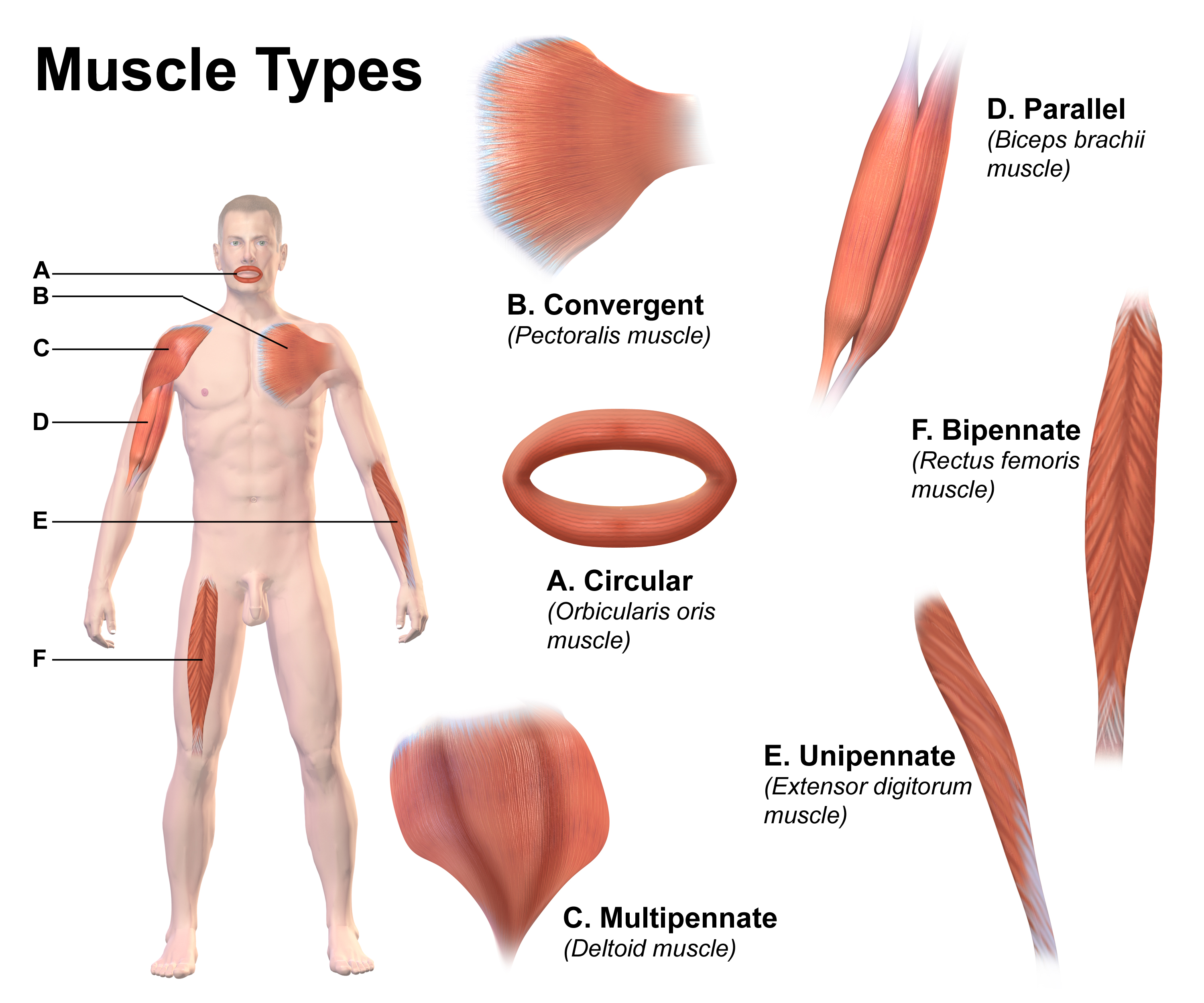
Skeletal muscles (commonly referred to as muscles) are organs of the vertebrate muscular system and typically are attached by tendons to bones of a skeleton. The muscle cells of skeletal muscles are much longer than in the other types of muscle tissue, and are often known as muscle fibers. The muscle tissue of a skeletal muscle is striated – having a striped appearance due to the arrangement of the sarcomeres. Skeletal muscles are voluntary muscles under the control of the somatic nervous system. The other types of muscle are cardiac muscle which is also striated and smooth muscle which is non-striated; both of these types of muscle tissue are classified as involuntary, or, under the control of the autonomic nervous system. A skeletal muscle contains multiple fascicles – bundles of muscle fibers. Each individual fiber, and each muscle is surrounded by a type of connective tissue layer of fascia. Muscle fibers are formed from the fusion of developmental myoblasts ... [...More Info...] [...Related Items...] OR: [Wikipedia] [Google] [Baidu] |
Skeletal Striated Muscle
Skeletal muscles (commonly referred to as muscles) are organs of the vertebrate muscular system and typically are attached by tendons to bones of a skeleton. The muscle cells of skeletal muscles are much longer than in the other types of muscle tissue, and are often known as muscle fibers. The muscle tissue of a skeletal muscle is striated – having a striped appearance due to the arrangement of the sarcomeres. Skeletal muscles are voluntary muscles under the control of the somatic nervous system. The other types of muscle are cardiac muscle which is also striated and smooth muscle which is non-striated; both of these types of muscle tissue are classified as involuntary, or, under the control of the autonomic nervous system. A skeletal muscle contains multiple fascicles – bundles of muscle fibers. Each individual fiber, and each muscle is surrounded by a type of connective tissue layer of fascia. Muscle fibers are formed from the fusion of developmental myoblasts in a pro ... [...More Info...] [...Related Items...] OR: [Wikipedia] [Google] [Baidu] |
.jpg)

