|
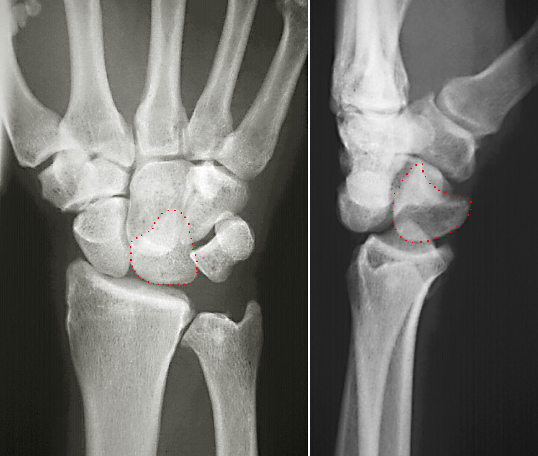
Hamate
The hamate bone (from Latin hamatus, "hooked"), or unciform bone (from Latin ''uncus'', "hook"), Latin os hamatum and occasionally abbreviated as just hamatum, is a bone in the human wrist readily distinguishable by its wedge shape and a hook-like process ("hamulus") projecting from its palmar surface. Structure The hamate is an irregularly shaped carpal bone found within the hand. The hamate is found within the distal row of carpal bones, and abuts the metacarpals of the little finger and ring finger. Adjacent to the hamate on the ulnar side, and slightly above it, is the pisiform bone. Adjacent on the radial side is the capitate, and proximal is the lunate bone. Surfaces The hamate bone has six surfaces: * The ''superior'', the apex of the wedge, is narrow, convex, smooth, and articulates with the lunate. * The ''inferior'' articulates with the fourth and fifth metacarpal bones, by concave facets which are separated by a ridge. * The ''dorsal'' is triangular and rough for l ... [...More Info...] [...Related Items...] OR: [Wikipedia] [Google] [Baidu] |
Hamulus Of Hamate (left Hand) - Animation02
The hamate bone (from Latin hamatus, "hooked"), or unciform bone (from Latin ''uncus'', "hook"), Latin os hamatum and occasionally abbreviated as just hamatum, is a bone in the human wrist readily distinguishable by its wedge shape and a hook-like process ("hamulus") projecting from its palmar surface. Structure The hamate is an irregularly shaped carpal bone found within the hand. The hamate is found within the distal row of carpal bones, and abuts the metacarpals of the little finger and ring finger. Adjacent to the hamate on the ulnar side, and slightly above it, is the pisiform bone. Adjacent on the radial side is the capitate, and proximal is the lunate bone. Surfaces The hamate bone has six surfaces: * The ''superior'', the apex of the wedge, is narrow, convex, smooth, and articulates with the lunate. * The ''inferior'' articulates with the fourth and fifth metacarpal bones, by concave facets which are separated by a ridge. * The ''dorsal'' is triangular and rough for l ... [...More Info...] [...Related Items...] OR: [Wikipedia] [Google] [Baidu] |
Hamulus Of Hamate Bone
The hamate bone (from Latin hamatus, "hooked"), or unciform bone (from Latin ''uncus'', "hook"), Latin os hamatum and occasionally abbreviated as just hamatum, is a bone in the human wrist readily distinguishable by its wedge shape and a hook-like process ("hamulus") projecting from its palmar surface. Structure The hamate is an irregularly shaped carpal bone found within the hand. The hamate is found within the distal row of carpal bones, and abuts the metacarpals of the little finger and ring finger. Adjacent to the hamate on the ulnar side, and slightly above it, is the pisiform bone. Adjacent on the radial side is the capitate, and proximal is the lunate bone. Surfaces The hamate bone has six surfaces: * The ''superior'', the apex of the wedge, is narrow, convex, smooth, and articulates with the lunate. * The ''inferior'' articulates with the fourth and fifth metacarpal bones, by concave facets which are separated by a ridge. * The ''dorsal'' is triangular and rough for l ... [...More Info...] [...Related Items...] OR: [Wikipedia] [Google] [Baidu] |
Carpal Bone
The carpal bones are the eight small bones that make up the wrist (or carpus) that connects the hand to the forearm. The term "carpus" is derived from the Latin wikt:carpus#Latin, carpus and the Greek language, Greek wikt:καρπός#Ancient Greek, καρπός (karpós), meaning "wrist". In human anatomy, the main role of the wrist is to facilitate effective positioning of the hand and powerful use of the Anatomical terms of motion, extensors and Anterior compartment of the forearm, flexors of the forearm, and the mobility of individual carpal bones increase the freedom of movements at the wrist.Kingston 2000, pp 126-127 In tetrapods, the carpus is the sole cluster of bones in the wrist between the radius (bone), radius and ulna and the metacarpus. The bones of the carpus do not belong to individual fingers (or toes in quadrupeds), whereas those of the metacarpus do. The corresponding part of the foot is the Tarsus (skeleton), tarsus. The carpal bones allow the wrist to move and ... [...More Info...] [...Related Items...] OR: [Wikipedia] [Google] [Baidu] |
Pisohamate Ligament
The pisohamate ligament is a ligament in the hand. It connects the pisiform to the hook of the hamate. It is a prolongation of the tendon of the flexor carpi ulnaris. It serves as part of the origin for the abductor digiti minimi. It also forms the floor of the ulnar canal, a canal that allows the ulnar nerve and ulnar artery The ulnar artery is the main blood vessel, with oxygenated blood, of the medial aspects of the forearm. It arises from the brachial artery and terminates in the superficial palmar arch, which joins with the superficial branch of the radial ar ... into the hand. References Ligaments of the upper limb {{ligament-stub ... [...More Info...] [...Related Items...] OR: [Wikipedia] [Google] [Baidu] |
Flexor Carpi Ulnaris
The flexor carpi ulnaris (FCU) is a muscle of the forearm that flexes and adducts at the wrist joint. Structure Origin The flexor carpi ulnaris has two heads; a humeral head and ulnar head. The humeral head originates from the medial epicondyle of the humerus via the common flexor tendon. The ulnar head originates from the medial margin of the olecranon of the ulnar and the upper two-thirds of the dorsal border of the ulnar by an aponeurosis. Between the two heads passes the ulnar nerve and ulnar artery. Insertion The flexor carpi ulnaris inserts onto the pisiform, hook of the hamate (via the pisohamate ligament) and the anterior surface of the base of the fifth metacarpal (via the pisometacarpal ligament). Action The flexor carpi ulnaris flexes and adducts at the wrist joint. Innervation The flexor carpi ulnaris is innervated by the ulnar nerve. The corresponding spinal nerves are C8 and T1. Tendon The tendon of flexor carpi ulnaris can be seen on the anterior surface of th ... [...More Info...] [...Related Items...] OR: [Wikipedia] [Google] [Baidu] |
Lunate Bone
The lunate bone (semilunar bone) is a carpal bone in the human hand. It is distinguished by its deep concavity and crescentic outline. It is situated in the center of the proximal row carpal bones, which lie between the ulna and radius and the hand. The lunate carpal bone is situated between the lateral scaphoid bone and medial triquetral bone. Structure The lunate is a crescent-shaped carpal bone found within the hand. The lunate is found within the proximal row of carpal bones. Proximally, it abuts the radius. Laterally, it articulates with the scaphoid bone, medially with the triquetral bone, and distally with the capitate bone. The lunate also articulates on its distal and medial surface with the hamate bone. The lunate is stabilised by a medial ligament to the scaphoid bone and a lateral ligament to the triquetral bone. Ligaments between the radius and carpal bone also stabilise the position of the lunate, as does its position in the lunate fossa of the radius. Bone The pro ... [...More Info...] [...Related Items...] OR: [Wikipedia] [Google] [Baidu] |
Triquetral Bone
The triquetral bone (; also called triquetrum, pyramidal, three-faced, and formerly cuneiform bone) is located in the wrist on the medial side of the proximal row of the carpus between the lunate and pisiform bones. It is on the ulnar side of the hand, but does not directly articulate with the ulna, however it is connected and articulate with the ulna through Triangular fibrocartilage discManaster, B. J., Julia Crim "Imaging Anatomy: Musculoskeletal E-Book" Elsevier Health Sciences, 2016, p. 326. and ligament, which forming the part of the ulnocarpal joint capsule. It connects with the pisiform, hamate, and lunate bones. It is the 2nd most commonly fractured carpal bone. Structure The triquetral is one of the eight carpal bones of the hand. It is a three-faced bone found within the proximal row of carpal bones. Situated beneath the pisiform, it is one of the carpal bones that form the carpal arch, within which lies the carpal tunnel. The triquetral bone may be distinguished by i ... [...More Info...] [...Related Items...] OR: [Wikipedia] [Google] [Baidu] |
Wrist
In human anatomy, the wrist is variously defined as (1) the Carpal bones, carpus or carpal bones, the complex of eight bones forming the proximal skeletal segment of the hand; "The wrist contains eight bones, roughly aligned in two rows, known as the carpal bones." (2) the wrist joint or radiocarpal joint, the joint between the radius (bone), radius and the Carpal bones, carpus and; (3) the anatomical region surrounding the carpus including the distal parts of the bones of the forearm and the proximal parts of the metacarpus or five metacarpal bones and the series of joints between these bones, thus referred to as ''wrist joints''. "With the large number of bones composing the wrist (ulna, radius, eight carpas, and five metacarpals), it makes sense that there are many, many joints that make up the structure known as the wrist." This region also includes the carpal tunnel, the anatomical snuff box, bracelet lines, the Flexor retinaculum of the hand, flexor retinaculum, and the ex ... [...More Info...] [...Related Items...] OR: [Wikipedia] [Google] [Baidu] |
Triangular Bone
The triquetral bone (; also called triquetrum, pyramidal, three-faced, and formerly cuneiform bone) is located in the wrist on the medial side of the proximal row of the Carpal bones, carpus between the Lunate bone, lunate and pisiform bone, pisiform bones. It is on the ulnar side of the hand, but does not directly articulate with the ulna, however it is connected and articulate with the ulna through Triangular fibrocartilage discManaster, B. J., Julia Crim "Imaging Anatomy: Musculoskeletal E-Book" Elsevier Health Sciences, 2016, p. 326. and ligament, which forming the part of the ulnocarpal joint capsule. It connects with the pisiform, hamate, and Lunate bone, lunate bones. It is the 2nd most commonly fractured carpal bone. Structure The triquetral is one of the eight carpal bones of the hand. It is a three-faced bone found within the proximal row of carpal bones. Situated beneath the pisiform, it is one of the carpal bones that form the carpal arch, within which lies the carpal t ... [...More Info...] [...Related Items...] OR: [Wikipedia] [Google] [Baidu] |
Opponens Digiti Minimi
The opponens digiti minimi (opponens digiti quinti in older texts) is a muscle in the hand. It is of a triangular form, and placed immediately beneath the palmaris brevis, abductor digiti minimi and flexor digiti minimi brevis. It is one of the three hypothenar muscles that control the little finger. It arises from the convexity of the hamulus of the hamate bone and the contiguous portion of the transverse carpal ligament; it is inserted into the whole length of the metacarpal bone of the little finger, along its ulnar margin. The opponens digiti minimi muscle serves to flex and laterally rotate the 5th metacarpal about the 5th carpometacarpal joint, as when bringing the little finger and thumb into opposition. It is innervated by the deep branch of the ulnar nerve. See also * Hypothenar * Opponens pollicis muscle Additional images Image:Gray426.png, The muscles of the thumb. (Opponens quinti digiti visible at center right.) Image:Musculusopponensdigitiminimi.png, Front of t ... [...More Info...] [...Related Items...] OR: [Wikipedia] [Google] [Baidu] |
Metacarpals
In human anatomy, the metacarpal bones or metacarpus form the intermediate part of the skeleton, skeletal hand located between the phalanges of the fingers and the carpal bones of the wrist, which forms the connection to the forearm. The metacarpal bones are analogous to the metatarsal bones in the foot. Structure The metacarpals form a transverse arch to which the rigid row of distal carpal bones are fixed. The peripheral metacarpals (those of the thumb and little finger) form the sides of the cup of the palmar gutter and as they are brought together they deepen this concavity. The index metacarpal is the most firmly fixed, while the thumb metacarpal articulates with the trapezium and acts independently from the others. The middle metacarpals are tightly united to the carpus by intrinsic interlocking bone elements at their bases. The ring metacarpal is somewhat more mobile while the fifth metacarpal is semi-independent.Tubiana ''et al'' 1998, p 11 Each metacarpal bone consists o ... [...More Info...] [...Related Items...] OR: [Wikipedia] [Google] [Baidu] |
Capitate
The capitate bone is a bone in the human wrist found in the center of the carpal bone region, located at the distal end of the radius and ulna bones. It articulates with the third metacarpal bone (the middle finger) and forms the third carpometacarpal joint. The capitate bone is the largest of the carpal bones in the human hand. It presents, above, a rounded portion or head, which is received into the concavity formed by the scaphoid and lunate bones; a constricted portion or neck; and below this, the body.''Gray's Anatomy'' (1918). See infobox. The bone is also found in many other mammals, and is homologous with the "third distal carpal" of reptiles and amphibians. Structure The capitate is the largest carpal bone found within the hand. The capitate is found within the distal row of carpal bones. The capitate lies directly adjacent to the metacarpal of the ring finger on its distal surface, has the hamate on its ulnar surface and trapezoid on its radial surface, and abuts the ... [...More Info...] [...Related Items...] OR: [Wikipedia] [Google] [Baidu] |
_-_animation02.gif)


_dorsal_view.png)