|
Greater Occipital Nerve
The greater occipital nerve is a nerve of the head. It is a spinal nerve, specifically the medial branch of the dorsal primary ramus of cervical spinal nerve 2. It arises from between the first and second cervical vertebrae, ascends, and then passes through the semispinalis muscle. It ascends further to supply the skin along the posterior part of the scalp to the vertex. It supplies sensation the scalp at the top of the head, over the ear and over the parotid glands. Structure The greater occipital nerve is the medial branch of the dorsal primary ramus of cervical spinal nerve 2. It may also involve fibres from cervical spinal nerve 3. It arises from between the first and second cervical vertebrae, along with the lesser occipital nerve. It ascends after emerging from below the suboccipital triangle beneath the obliquus capitis inferior muscle. Just below the superior nuchal ridge, it pierces the fascia. It ascends further to supply the skin along the posterior part of t ... [...More Info...] [...Related Items...] OR: [Wikipedia] [Google] [Baidu] |
Semispinalis Capitis
The semispinalis muscles are a group of three muscles belonging to the transversospinales. These are the semispinalis capitis, the semispinalis cervicis and the semispinalis thoracis. The semispinalis capitis (''complexus'') is situated at the upper and back part of the neck, deep to the splenius, and medial to the longissimus cervicis and longissimus capitis. It arises by a series of tendons from the tips of the transverse processes of the upper six or seven thoracic and the seventh cervical vertebrae, and from the articular processes of the three cervical vertebrae above this ( C4-C6). The tendons, uniting, form a broad muscle, which passes upward, and is inserted between the superior and inferior nuchal lines of the occipital bone. It lies deep to the trapezius muscle and can be palpated as a firm round muscle mass just lateral to the cervical spinous processes. The semispinalis cervicis (or ''semispinalis colli''), arises by a series of tendinous and fleshy fibers from the t ... [...More Info...] [...Related Items...] OR: [Wikipedia] [Google] [Baidu] |
Cervical Spinal Nerve 2
The cervical spinal nerve 2 (C2) is a spinal nerve of the cervical segment. Nervous System -- Groups of Nerves It is a part of the along with the C1 and C3 nerves sometimes forming part of Superior root of the ansa cervicalis. it also connects into the with the C3. It originates from the s ... [...More Info...] [...Related Items...] OR: [Wikipedia] [Google] [Baidu] |
Occipital Neuralgia
Occipital neuralgia (ON) is a painful condition affecting the posterior head in the distributions of the greater occipital nerve (GON), lesser occipital nerve (LON), third occipital nerve (TON), or a combination of the three. It is paroxysmal, lasting from seconds to minutes, and often consists of lancinating pain that directly results from the pathology of one of these nerves. It is paramount that physicians understand the differential diagnosis for this condition and specific diagnostic criteria. There are multiple treatment modalities, several of which have well-established efficacy in treating this condition. Text was copied from this source, which is available under Creative Commons Attribution 4.0 International License Signs and symptoms Patients presenting with a headache originating at the posterior skull base should be evaluated for ON. This condition typically presents as a paroxysmal, lancinating or stabbing pain lasting from seconds to minutes, and therefore a continuo ... [...More Info...] [...Related Items...] OR: [Wikipedia] [Google] [Baidu] |
Cervicogenic Headache
Cervicogenic headache is a type of headache characterized by chronic hemicranial pain referred to the head from either the cervical spine or soft tissues within the neck. The main symptoms of cervicogenic headaches include pain originating in the neck that can travel to the head or face, headaches that get worse with neck movement, and limited ability to move the neck. Diagnostic imaging can display lesions of the cervical spine or soft tissue of the neck that can be indicative of a cervicogenic headache. When being evaluated for cervicogenic headaches, it is important to rule out a history of migraines and traumatic brain injuries. Studies show that combining interventions such as moist heat applied to the area of pain, spinal and cervical manipulations, and neck massages all help reduce or relieve symptoms. Neck exercises are also beneficial. Specifically, craniocervical flexion, or forward bending of the neck, against light resistance helps increase muscular stability of the he ... [...More Info...] [...Related Items...] OR: [Wikipedia] [Google] [Baidu] |
Fascia
A fascia (; plural fasciae or fascias; adjective fascial; from Latin: "band") is a band or sheet of connective tissue, primarily collagen, beneath the skin that attaches to, stabilizes, encloses, and separates muscles and other internal organs. Fascia is classified by layer, as superficial fascia, deep fascia, and ''visceral'' or ''parietal'' fascia, or by its function and anatomical location. Like ligaments, aponeuroses, and tendons, fascia is made up of fibrous connective tissue containing closely packed bundles of collagen fibers oriented in a wavy pattern parallel to the direction of pull. Fascia is consequently flexible and able to resist great unidirectional tension forces until the wavy pattern of fibers has been straightened out by the pulling force. These collagen fibers are produced by fibroblasts located within the fascia. Fasciae are similar to ligaments and tendons as they have collagen as their major component. They differ in their location and function: ligament ... [...More Info...] [...Related Items...] OR: [Wikipedia] [Google] [Baidu] |
Obliquus Capitis Inferior Muscle
The obliquus capitis inferior muscle () is the larger of the two oblique muscles of the neck. It arises from the apex of the spinous process of the axis and passes laterally and slightly upward, to be inserted into the lower and back part of the transverse process of the atlas. It lies deep to the semispinalis capitis and trapezius muscles. The muscle is responsible for rotation of the head and first cervical vertebra ( atlanto-axial joint). It forms the lower boundary of the suboccipital triangle of the neck. The naming of this muscle may be confusing, as it is the only capitis (L. "head") muscle that does NOT attach to the cranium. Function The obliquus capitis inferior muscle, like the other suboccipital muscles, has an important role in proprioception. This muscle has a very high density of Golgi organs and muscle spindles which accounts for this. It is believed that proprioception may be the primary role of the inferior oblique (and indeed the other suboccipital muscles) ... [...More Info...] [...Related Items...] OR: [Wikipedia] [Google] [Baidu] |
Suboccipital Triangle
The suboccipital triangle is a region of the neck bounded by the following three muscles of the suboccipital group of muscles: * Rectus capitis posterior major - above and medially * Obliquus capitis superior - above and laterally * Obliquus capitis inferior - below and laterally (Rectus capitis posterior minor is also in this region but does not form part of the triangle) It is covered by a layer of dense fibro-fatty tissue, situated beneath the semispinalis capitis. The floor is formed by the posterior atlantooccipital membrane, and the posterior arch of the atlas. In the deep groove on the upper surface of the posterior arch of the atlas are the vertebral artery and the first cervical or suboccipital nerve. In the past, the vertebral artery was accessed here in order to conduct angiography of the circle of Willis. Presently, formal angiography of the circle of Willis is performed via catheter angiography, with access usually being acquired at the common femoral artery. Alter ... [...More Info...] [...Related Items...] OR: [Wikipedia] [Google] [Baidu] |
Lesser Occipital Nerve
The lesser occipital nerve or small occipital nerve is a cutaneous spinal nerve. It arises from cervical spinal nerve 2, second cervical (spinal) nerve (along with the greater occipital nerve). It innervates the scalp in the lateral area of the head posterior to the ear. Structure The lesser occipital nerve is one of the four cutaneous branches of the cervical plexus. Origin It arises from the (lateral branch of the ventral ramus) of cervical spinal nerve 2, cervical spinal nerve C2; it may also receive fibres from cervical spinal nerve 3, cervical spinal nerve C3. It originates between the Atlas (anatomy), atlas, and Axis (anatomy), axis. Course and relations It curves around the Accessory nerve, accessory nerve (CN XI) to come to course anterior to it. It then curves around and ascends along the posterior border of the sternocleidomastoid muscle; rarely, it may pierce the muscle. Near the cranium, it perforates the deep fascia. It is continues upwards along the scalp po ... [...More Info...] [...Related Items...] OR: [Wikipedia] [Google] [Baidu] |
Saunders (imprint)
Saunders is an American academic publisher based in the United States. It is currently an imprint of Elsevier. Formerly independent, the W. B. Saunders company was acquired by CBS in 1968, who added it to their publishing division Holt, Rinehart & Winston. When CBS left the publishing field in 1986, it sold the academic publishing units to Harcourt Brace Jovanovich. Harcourt was acquired by Reed Elsevier in 2001. . . Retrieved May 2, 2015. W. B. Saunders published the Kinsey Reports and |
Cervical Spinal Nerve 3
The cervical spinal nerve 3 (C3) is a spinal nerve of the cervical segment. Nervous System -- Groups of Nerves It originates from the spinal column from above the cervical vertebra 3
In tetrapods, cervical vertebrae (singular: vertebra) are the vertebrae of the neck, immediately below the skull. Truncal vertebrae (divided into thoracic and lumbar vertebrae in mammals) lie caudal (toward the tail) of cervical vertebrae. In sau ... (C3).
References Spinal nerves {{neuroanatomy-stub ...[...More Info...] [...Related Items...] OR: [Wikipedia] [Google] [Baidu] |
Parotid Glands
The parotid gland is a major salivary gland in many animals. In humans, the two parotid glands are present on either side of the mouth and in front of both ears. They are the largest of the salivary glands. Each parotid is wrapped around the mandibular ramus, and secretes serous saliva through the parotid duct into the mouth, to facilitate mastication and swallowing and to begin the digestion of starches. There are also two other types of salivary glands; they are submandibular and sublingual glands. Sometimes accessory parotid glands are found close to the main parotid glands. Etymology The word ''parotid'' literally means "beside the ear". From Greek παρωτίς (stem παρωτιδ-) : (gland) behind the ear < παρά - pará : in front, and οὖς - ous (stem ὠτ-, ōt-) : ear. Structure The parotid glands are a pair of mainly |
Scalp
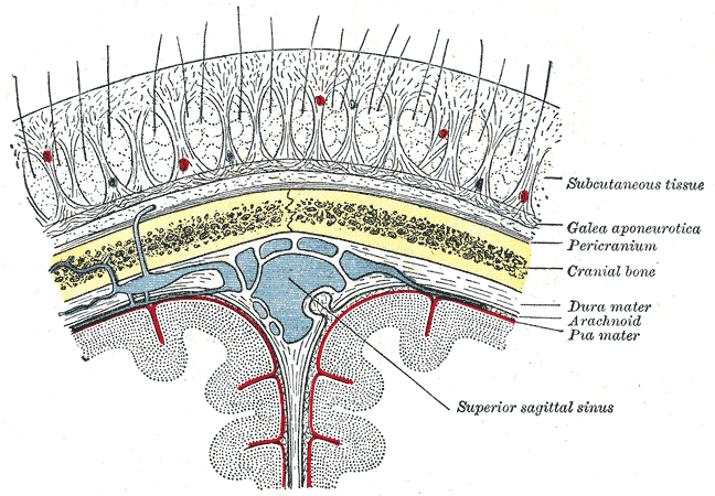
The scalp is the anatomical area bordered by the human face at the front, and by the neck at the sides and back. Structure The scalp is usually described as having five layers, which can conveniently be remembered as a mnemonic: * S: The skin on the head from which head hair grows. It contains numerous sebaceous glands and hair follicles. * C: Connective tissue. A dense subcutaneous layer of fat and fibrous tissue that lies beneath the skin, containing the nerves and vessels of the scalp. * A: The aponeurosis called epicranial aponeurosis (or galea aponeurotica) is the next layer. It is a tough layer of dense fibrous tissue which runs from the frontalis muscle anteriorly to the occipitalis posteriorly. * L: The loose areolar connective tissue layer provides an easy plane of separation between the upper three layers and the pericranium. In scalping the scalp is torn off through this layer. It also provides a plane of access in craniofacial surgery and neurosurgery. This layer i ... [...More Info...] [...Related Items...] OR: [Wikipedia] [Google] [Baidu] |
