|
Cervical Spinal Nerve 2
The cervical spinal nerve 2 (C2) is a spinal nerve of the cervical segment. Nervous System -- Groups of Nerves It is a part of the along with the C1 and C3 nerves sometimes forming part of Superior root of the ansa cervicalis. it also connects into the with the C3. It originates from the s ... [...More Info...] [...Related Items...] OR: [Wikipedia] [Google] [Baidu] |
Spinal Nerve
A spinal nerve is a mixed nerve, which carries motor, sensory, and autonomic signals between the spinal cord and the body. In the human body there are 31 pairs of spinal nerves, one on each side of the vertebral column. These are grouped into the corresponding cervical, thoracic, lumbar, sacral and coccygeal regions of the spine. There are eight pairs of cervical nerves, twelve pairs of thoracic nerves, five pairs of lumbar nerves, five pairs of sacral nerves, and one pair of coccygeal nerves. The spinal nerves are part of the peripheral nervous system. Structure Each spinal nerve is a mixed nerve, formed from the combination of nerve fibers from its dorsal and ventral roots. The dorsal root is the afferent sensory root and carries sensory information to the brain. The ventral root is the efferent motor root and carries motor information from the brain. The spinal nerve emerges from the spinal column through an opening (intervertebral foramen) between adjacent vertebrae. ... [...More Info...] [...Related Items...] OR: [Wikipedia] [Google] [Baidu] |
Cervical Segment
The spinal cord is a long, thin, tubular structure made up of nervous tissue, which extends from the medulla oblongata in the brainstem to the lumbar region of the vertebral column (backbone). The backbone encloses the central canal of the spinal cord, which contains cerebrospinal fluid. The brain and spinal cord together make up the central nervous system (CNS). In humans, the spinal cord begins at the occipital bone, passing through the foramen magnum and then enters the spinal canal at the beginning of the cervical vertebrae. The spinal cord extends down to between the first and second lumbar vertebrae, where it ends. The enclosing bony vertebral column protects the relatively shorter spinal cord. It is around long in adult men and around long in adult women. The diameter of the spinal cord ranges from in the cervical vertebrae, cervical and lumbar regions to in the thoracic vertebrae, thoracic area. The spinal cord functions primarily in the transmission of Action potentia ... [...More Info...] [...Related Items...] OR: [Wikipedia] [Google] [Baidu] |
Ansa Cervicalis
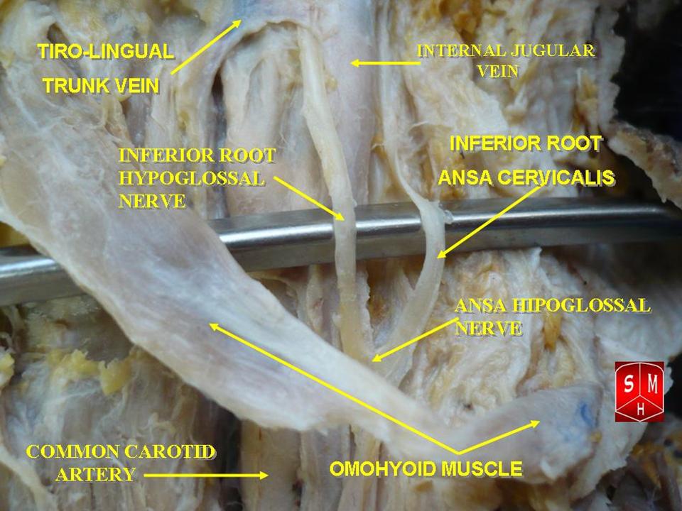
The ansa cervicalis (or ansa hypoglossi in older literature) is a loop of nerves that are part of the cervical plexus. It lies superficial to the internal jugular vein in the carotid triangle. Its name means "handle of the neck" in Latin. Branches from the ansa cervicalis innervate most of the infrahyoid muscles, including the sternothyroid muscle, sternohyoid muscle and the omohyoid muscle. Note that the thyrohyoid muscle, which is also an infrahyoid muscle and the geniohyoid muscle which is a suprahyoid muscle are innervated by cervical spinal nerve 1 via the hypoglossal nerve. Roots Two roots make up the ansa cervicalis, a superior root, and an inferior root. The superior root of the ansa cervicalis is formed from cervical spinal nerve 1 of the cervical plexus. These nerve fibers travel in the hypoglossal nerve before separating in the carotid triangle to form the superior root. The superior root goes around the occipital artery and then descends on the carotid sheath. I ... [...More Info...] [...Related Items...] OR: [Wikipedia] [Google] [Baidu] |
Superior Root Of The Ansa Cervicalis
The ansa cervicalis (or ansa hypoglossi in older literature) is a loop of nerves that are part of the cervical plexus. It lies superficial to the internal jugular vein in the carotid triangle. Its name means "handle of the neck" in Latin. Branches from the ansa cervicalis innervate most of the infrahyoid muscles, including the sternothyroid muscle, sternohyoid muscle and the omohyoid muscle. Note that the thyrohyoid muscle, which is also an infrahyoid muscle and the geniohyoid muscle which is a suprahyoid muscle are innervated by cervical spinal nerve 1 via the hypoglossal nerve. Roots Two roots make up the ansa cervicalis, a superior root, and an inferior root. The superior root of the ansa cervicalis is formed from cervical spinal nerve 1 of the cervical plexus. These nerve fibers travel in the hypoglossal nerve before separating in the carotid triangle to form the superior root. The superior root goes around the occipital artery and then descends on the carotid sheath. It ... [...More Info...] [...Related Items...] OR: [Wikipedia] [Google] [Baidu] |
Inferior Root Of The Ansa Cervicalis
The ansa cervicalis (or ansa hypoglossi in older literature) is a loop of nerves that are part of the cervical plexus. It lies superficial to the internal jugular vein in the carotid triangle. Its name means "handle of the neck" in Latin. Branches from the ansa cervicalis innervate most of the infrahyoid muscles, including the sternothyroid muscle, sternohyoid muscle and the omohyoid muscle. Note that the thyrohyoid muscle, which is also an infrahyoid muscle and the geniohyoid muscle which is a suprahyoid muscle are innervated by cervical spinal nerve 1 via the hypoglossal nerve. Roots Two roots make up the ansa cervicalis, a superior root, and an inferior root. The superior root of the ansa cervicalis is formed from cervical spinal nerve 1 of the cervical plexus. These nerve fibers travel in the hypoglossal nerve before separating in the carotid triangle to form the superior root. The superior root goes around the occipital artery and then descends on the carotid sheath. ... [...More Info...] [...Related Items...] OR: [Wikipedia] [Google] [Baidu] |
Cervical Vertebra 2
In anatomy, the axis (from Latin ''axis'', "axle") or epistropheus is the second cervical vertebra (C2) of the spine, immediately inferior to the atlas, upon which the head rests. The axis' defining feature is its strong odontoid process (bony protrusion) known as the dens, which rises dorsally from the rest of the bone. Structure The body is deeper in front or in the back and is prolonged downward anteriorly to overlap the upper and front part of the third vertebra. It presents a median longitudinal ridge in front, separating two lateral depressions for the attachment of the longus colli muscles. Odontoid Process of Axis (Dens) The dens, also called the odontoid process or the peg, is the most pronounced projecting feature of the axis. The dens exhibits a slight constriction where it joins the main body of the vertebra. The condition where the dens is separated from the body of the axis is called ''os odontoideum'' and may cause nerve and circulation compression syndrome. ... [...More Info...] [...Related Items...] OR: [Wikipedia] [Google] [Baidu] |
Rectus Capitis Anterior Muscle
The rectus capitis anterior (rectus capitis anticus minor) is a short, flat muscle, situated immediately behind the upper part of the Longus capitis. It arises from the anterior surface of the lateral mass of the atlas, and from the root of its transverse process, and passing obliquely upward and medialward, is inserted into the inferior surface of the basilar part of the occipital bone immediately in front of the foramen magnum. action: aids in flexion of the head and the neck; nerve supply: C1, C2. Additional images File:Rectus capitis anterior muscle - animation01.gif, Animation. Position of rectus capitis anterior muscle. Some bones around the muscle are shown in semi-transparent. File:Rectus capitis anterior muscle - animation02.gif, Skull has been removed (except for occipital bone The occipital bone () is a neurocranium, cranial dermal bone and the main bone of the occiput (back and lower part of the skull). It is trapezoidal in shape and curved on itself like a sh ... [...More Info...] [...Related Items...] OR: [Wikipedia] [Google] [Baidu] |
Rectus Capitis Lateralis Muscle
The rectus capitis lateralis, a short, flat muscle, arises from the upper surface of the transverse process of the atlas, and is inserted into the under surface of the jugular process of the occipital bone. Additional images File:Rectus capitis lateralis muscle - animation01.gif, Position of rectus capitis lateralis muscle (shown in red). Animation. File:Rectus capitis lateralis muscle - animation05.gif, Close up. Skull has been removed (except occipital bone). File:Rectus capitis lateralis muscle03.png, Lateral view. Still image. File:Gray129.png, Occipital bone. Outer surface. File:Gray187.png, Base of skull. Inferior surface. See also * Atlanto-occipital joint * Rectus capitis posterior major muscle * Rectus capitis posterior minor muscle * Rectus capitis anterior muscle The rectus capitis anterior (rectus capitis anticus minor) is a short, flat muscle, situated immediately behind the upper part of the Longus capitis. It arises from the anterior surface of the lateral ... [...More Info...] [...Related Items...] OR: [Wikipedia] [Google] [Baidu] |
Trapezius
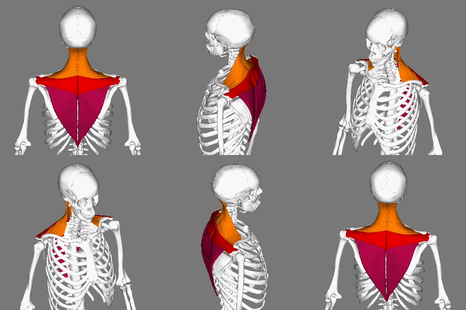
The trapezius is a large paired trapezoid-shaped surface muscle that extends longitudinally from the occipital bone to the lower thoracic vertebrae of the spine and laterally to the spine of the scapula. It moves the scapula and supports the arm. The trapezius has three functional parts: an upper (descending) part which supports the weight of the arm; a middle region (transverse), which retracts the scapula; and a lower (ascending) part which medially rotates and depresses the scapula. Name and history The trapezius muscle resembles a trapezium, also known as a trapezoid, or diamond-shaped quadrilateral. The word "spinotrapezius" refers to the human trapezius, although it is not commonly used in modern texts. In other mammals, it refers to a portion of the analogous muscle. Similarly, the term "tri-axle back plate" was historically used to describe the trapezius muscle. Structure The ''superior'' or ''upper'' (or descending) fibers of the trapezius originate from the sp ... [...More Info...] [...Related Items...] OR: [Wikipedia] [Google] [Baidu] |
Lesser Occipital Nerve
The lesser occipital nerve or small occipital nerve is a cutaneous spinal nerve. It arises from cervical spinal nerve 2, second cervical (spinal) nerve (along with the greater occipital nerve). It innervates the scalp in the lateral area of the head posterior to the ear. Structure The lesser occipital nerve is one of the four cutaneous branches of the cervical plexus. Origin It arises from the (lateral branch of the ventral ramus) of cervical spinal nerve 2, cervical spinal nerve C2; it may also receive fibres from cervical spinal nerve 3, cervical spinal nerve C3. It originates between the Atlas (anatomy), atlas, and Axis (anatomy), axis. Course and relations It curves around the Accessory nerve, accessory nerve (CN XI) to come to course anterior to it. It then curves around and ascends along the posterior border of the sternocleidomastoid muscle; rarely, it may pierce the muscle. Near the cranium, it perforates the deep fascia. It is continues upwards along the scalp po ... [...More Info...] [...Related Items...] OR: [Wikipedia] [Google] [Baidu] |
Great Auricular Nerve
The great auricular nerve is a cutaneous nerve of the head. It originates from the cervical plexus, with branches of spinal nerves C2 and C3. It provides sensory nerve supply to the skin over the parotid gland and the mastoid process of the temporal bone, and surfaces of the outer ear. Pain resulting from parotitis is caused by an impingement on the great auricular nerve. Structure The great auricular nerve is the largest of the ascending branches of the cervical plexus. It arises from the second and third cervical nerves. It winds around the posterior border of the sternocleidomastoid muscle, and, after perforating the deep fascia, ascends upon that muscle beneath the platysma muscle to the parotid gland. Here, it divides into an anterior and a posterior branch. Branches * The anterior branch (ramus anterior; facial branch) is distributed to the skin of the face over the parotid gland. It communicates with the facial nerve inside the parotid gland. * The posterior branch ( ... [...More Info...] [...Related Items...] OR: [Wikipedia] [Google] [Baidu] |
Transverse Cervical Nerve
The transverse cervical nerve (superficial cervical or cutaneous cervical) arises from the second and third spinal nerves, turns around the posterior border of the sternocleidomastoideus about its middle, and, passing obliquely forward beneath the external jugular vein to the anterior border of the muscle, it perforates the deep cervical fascia, and divides beneath the Platysma into ascending and descending branches, which are distributed to the antero-lateral parts of the neck The neck is the part of the body on many vertebrates that connects the head with the torso. The neck supports the weight of the head and protects the nerves that carry sensory and motor information from the brain down to the rest of the body. In .... It provides cutaneous innervation to this area. During dissection, the sternocleidomastoid (SCM) is the landmark. The transverse cervical nerves will pass horizontally directly over the SCM from Erb's point. Additional images File:Gray784.png, Dermatome ... [...More Info...] [...Related Items...] OR: [Wikipedia] [Google] [Baidu] |


