|
Granuloma Inguinale
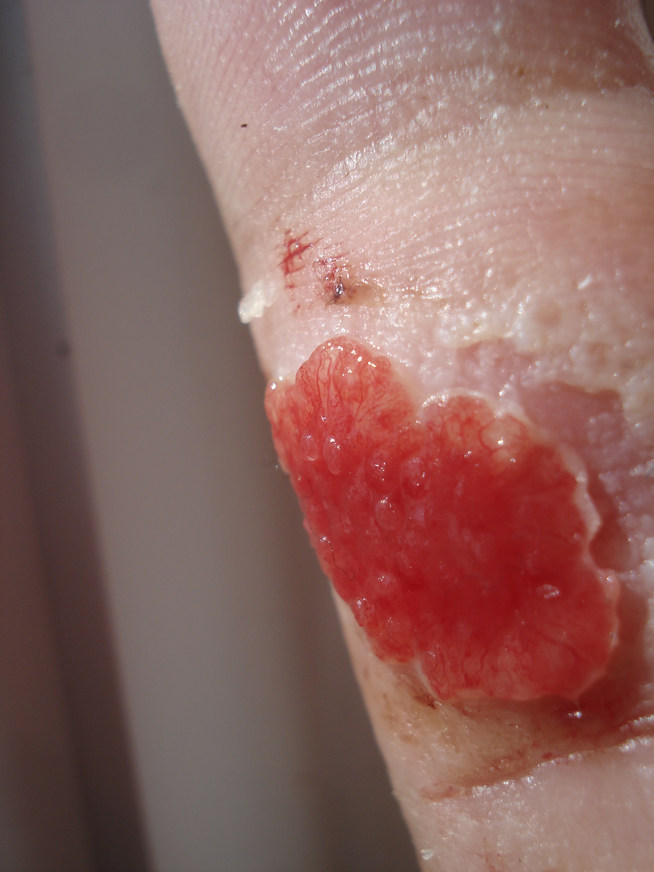
Granuloma inguinale is a bacterial disease caused by ''Klebsiella granulomatis'' (formerly known as ''Calymmatobacterium granulomatis'') characterized by genital ulcers. It is endemic in many less-developed regions. It is also known as donovanosis, granuloma genitoinguinale, granuloma inguinale tropicum, granuloma venereum, granuloma venereum genitoinguinale, lupoid form of groin ulceration, serpiginous ulceration of the groin, ulcerating granuloma of the pudendum, and ulcerating sclerosing granuloma. Oral manifestations are also notably seen. The lesions of oral cavity are usually secondary to active genital lesions. The disease often goes untreated because of the scarcity of medical treatment in the countries in which it is found. In addition, the painless genital ulcers can be mistaken for syphilis. The ulcers ultimately progress to destruction of internal and external tissue, with extensive leakage of mucus and blood from the highly vascular lesions. The destructive nature o ... [...More Info...] [...Related Items...] OR: [Wikipedia] [Google] [Baidu] |
Granuloma Annulare
Granuloma annulare (GA) is a common, sometimes chronic skin condition which presents as reddish bumps on the skin arranged in a circle or ring. It can initially occur at any age, though two-thirds of patients are under 30 years old, and it is seen most often in children and young adults. Females are two times as likely to have it than males. Signs and symptoms Aside from the visible rash, granuloma annulare is usually asymptomatic. Sometimes the rash may burn or itch. People with GA usually notice a ring of small, firm bumps (papules) over the backs of the forearms, hands or feet, often centered on joints or knuckles. The bumps are caused by the clustering of T cells below the skin. These papules start as very small, pimple looking bumps, which spread over time from that size to dime, quarter, half-dollar size and beyond. Occasionally, multiple rings may join into one. Rarely, GA may appear as a firm nodule under the skin of the arms or legs. It also occurs on the sides and c ... [...More Info...] [...Related Items...] OR: [Wikipedia] [Google] [Baidu] |
Vagina
In mammals, the vagina is the elastic, muscular part of the female genital tract. In humans, it extends from the vestibule to the cervix. The outer vaginal opening is normally partly covered by a thin layer of mucosal tissue called the hymen. At the deep end, the cervix (neck of the uterus) bulges into the vagina. The vagina allows for sexual intercourse and birth. It also channels menstrual flow, which occurs in humans and closely related primates as part of the menstrual cycle. Although research on the vagina is especially lacking for different animals, its location, structure and size are documented as varying among species. Female mammals usually have two external openings in the vulva; these are the urethral opening for the urinary tract and the vaginal opening for the genital tract. This is different from male mammals, who usually have a single urethral opening for both urination and reproduction. The vaginal opening is much larger than the nearby urethral opening, an ... [...More Info...] [...Related Items...] OR: [Wikipedia] [Google] [Baidu] |
Wright's Stain
Wright's stain is a hematologic stain that facilitates the differentiation of blood cell types. It is classically a mixture of eosin (red) and methylene blue dyes. It is used primarily to stain peripheral blood smears, urine samples, and bone marrow aspirates, which are examined under a light microscope. In cytogenetics, it is used to stain chromosomes to facilitate diagnosis of syndromes and diseases. It is named for James Homer Wright, who devised the stain, a modification of the Romanowsky stain, in 1902. Because it distinguishes easily between blood cells, it became widely used for performing differential white blood cell counts, which are routinely ordered when conditions such as infection or leukemia are suspected. The related stains are known as the buffered Wright stain, the Wright-Giemsa stain (a combination of Wright and Giemsa stains), and the buffered Wright-Giemsa stain, and specific instructions depend on the solutions being used, which may include eosin Y, azure B, ... [...More Info...] [...Related Items...] OR: [Wikipedia] [Google] [Baidu] |
Histiocyte
A histiocyte is a vertebrate cell that is part of the mononuclear phagocyte system (also known as the reticuloendothelial system or lymphoreticular system). The mononuclear phagocytic system is part of the organism's immune system. The histiocyte is a tissue macrophage or a dendritic cell (histio, diminutive of histo, meaning ''tissue'', and cyte, meaning ''cell''). Part of their job is to clear out neutrophils once they've reached the end of their lifespan. Development Histiocytes are derived from the bone marrow by multiplication from a stem cell. The derived cells migrate from the bone marrow to the blood as monocytes. They circulate through the body and enter various organs, where they undergo differentiation into histiocytes, which are part of the mononuclear phagocytic system (MPS). However, the term ''histiocyte'' has been used for multiple purposes in the past, and some cells called "histocytes" do not appear to derive from monocytic-macrophage lines. The term Histioc ... [...More Info...] [...Related Items...] OR: [Wikipedia] [Google] [Baidu] |
Mononuclear Phagocyte
Monocytes are a type of leukocyte or white blood cell. They are the largest type of leukocyte in blood and can cellular differentiation, differentiate into macrophages and conventional dendritic cells. As a part of the vertebrate innate immune system monocytes also influence Adaptive immune system, adaptive immune responses and exert tissue repair functions. There are at least three subclasses of monocytes in human blood based on their phenotypic receptors. Structure Monocytes are amoeba, amoeboid in appearance, and have nongranulated cytoplasm. Thus they are classified as Agranulocyte, agranulocytes, although they might occasionally display some azurophil granules and/or Vacuole, vacuoles. With a diameter of 15–22 Micrometre, μm, monocytes are the largest cell type in peripheral blood. Monocytes are mononuclear cells and the ellipsoidal nucleus is often lobulated/indented, causing a bean-shaped or kidney-shaped appearance. Monocytes compose 2% to 10% of all leukocytes in th ... [...More Info...] [...Related Items...] OR: [Wikipedia] [Google] [Baidu] |
Cytoplasm
In cell biology, the cytoplasm is all of the material within a eukaryotic cell, enclosed by the cell membrane, except for the cell nucleus. The material inside the nucleus and contained within the nuclear membrane is termed the nucleoplasm. The main components of the cytoplasm are cytosol (a gel-like substance), the organelles (the cell's internal sub-structures), and various cytoplasmic inclusions. The cytoplasm is about 80% water and is usually colorless. The submicroscopic ground cell substance or cytoplasmic matrix which remains after exclusion of the cell organelles and particles is groundplasm. It is the hyaloplasm of light microscopy, a highly complex, polyphasic system in which all resolvable cytoplasmic elements are suspended, including the larger organelles such as the ribosomes, mitochondria, the plant plastids, lipid droplets, and vacuoles. Most cellular activities take place within the cytoplasm, such as many metabolic pathways including glycolysis, and proces ... [...More Info...] [...Related Items...] OR: [Wikipedia] [Google] [Baidu] |
Wright-Giemsa Stain
Wright's stain is a hematologic stain that facilitates the differentiation of blood cell types. It is classically a mixture of eosin (red) and methylene blue dyes. It is used primarily to stain peripheral blood smears, urine samples, and bone marrow needle aspiration biopsy, aspirates, which are examined under a light microscope. In cytogenetics, it is used to stain chromosomes to facilitate diagnosis of syndromes and diseases. It is named for James Homer Wright, who devised the stain, a modification of the Romanowsky stain, in 1902. Because it distinguishes easily between blood cells, it became widely used for performing differential white blood cell counts, which are routinely ordered when conditions such as infection or leukemia are suspected. The related stains are known as the buffered Wright stain, the Wright-Giemsa stain (a combination of Wright and Giemsa stain, Giemsa stains), and the buffered Wright-Giemsa stain, and specific instructions depend on the solutions being us ... [...More Info...] [...Related Items...] OR: [Wikipedia] [Google] [Baidu] |
Biopsy
A biopsy is a medical test commonly performed by a surgeon, interventional radiologist, or an interventional cardiologist. The process involves extraction of sample cells or tissues for examination to determine the presence or extent of a disease. The tissue is then fixed, dehydrated, embedded, sectioned, stained and mounted before it is generally examined under a microscope by a pathologist; it may also be analyzed chemically. When an entire lump or suspicious area is removed, the procedure is called an excisional biopsy. An incisional biopsy or core biopsy samples a portion of the abnormal tissue without attempting to remove the entire lesion or tumor. When a sample of tissue or fluid is removed with a needle in such a way that cells are removed without preserving the histological architecture of the tissue cells, the procedure is called a needle aspiration biopsy. Biopsies are most commonly performed for insight into possible cancerous or inflammatory conditions. History T ... [...More Info...] [...Related Items...] OR: [Wikipedia] [Google] [Baidu] |
Lymphadenopathy
Lymphadenopathy or adenopathy is a disease of the lymph nodes, in which they are abnormal in size or consistency. Lymphadenopathy of an inflammatory type (the most common type) is lymphadenitis, producing swollen or enlarged lymph nodes. In clinical practice, the distinction between lymphadenopathy and lymphadenitis is rarely made and the words are usually treated as synonymous. Inflammation of the lymphatic vessels is known as lymphangitis. Infectious lymphadenitis affecting lymph nodes in the neck is often called scrofula. Lymphadenopathy is a common and nonspecific sign. Common causes include infections (from minor causes such as the common cold and post-vaccination swelling to serious ones such as HIV/AIDS), autoimmune diseases, and cancer. Lymphadenopathy is frequently idiopathic and self-limiting. Causes Lymph node enlargement is recognized as a common sign of infectious, autoimmune, or malignant disease. Examples may include: * Reactive: acute infection (''e.g.,'' ba ... [...More Info...] [...Related Items...] OR: [Wikipedia] [Google] [Baidu] |
Syphilitic
Syphilis () is a sexually transmitted infection caused by the bacterium ''Treponema pallidum'' subspecies ''pallidum''. The signs and symptoms of syphilis vary depending in which of the four stages it presents (primary, secondary, latent, and tertiary). The primary stage classically presents with a single chancre (a firm, painless, non-itchy skin ulceration usually between 1 cm and 2 cm in diameter) though there may be multiple sores. In secondary syphilis, a diffuse rash occurs, which frequently involves the palms of the hands and soles of the feet. There may also be sores in the mouth or vagina. In latent syphilis, which can last for years, there are few or no symptoms. In tertiary syphilis, there are gummas (soft, non-cancerous growths), neurological problems, or heart symptoms. Syphilis has been known as "the great imitator" as it may cause symptoms similar to many other diseases. Syphilis is most commonly spread through sexual activity. It may also be transmitte ... [...More Info...] [...Related Items...] OR: [Wikipedia] [Google] [Baidu] |
Granulation Tissue
Granulation tissue is new connective tissue and microscopic blood vessels that form on the surfaces of a wound during the healing process. Granulation tissue typically grows from the base of a wound and is able to fill wounds of almost any size. Examples of granulation tissue can be seen in pyogenic granulomas and pulp polyps. Its histological appearance is characterized by proliferation of fibroblasts and new thin-walled, delicate capillaries (angiogenesis), infiltrated inflammatory cells in a loose extracellular matrix. Appearance During the migratory phase of wound healing, granulation tissue is: * light red or dark pink, being perfused with new capillary loops or "buds"; * soft to the touch; * moist; * bumpy (granular) in appearance, due to punctate hemorrhages; * pulsatile on palpation; * painless when healthy; Structure Granulation tissue is composed of tissue matrix supporting a variety of cell types, most of which can be associated with one of the following functions: ... [...More Info...] [...Related Items...] OR: [Wikipedia] [Google] [Baidu] |
Diagnosis
Diagnosis is the identification of the nature and cause of a certain phenomenon. Diagnosis is used in many different disciplines, with variations in the use of logic, analytics, and experience, to determine " cause and effect". In systems engineering and computer science, it is typically used to determine the causes of symptoms, mitigations, and solutions. Computer science and networking * Bayesian networks * Complex event processing * Diagnosis (artificial intelligence) * Event correlation * Fault management * Fault tree analysis * Grey problem * RPR Problem Diagnosis * Remote diagnostics * Root cause analysis * Troubleshooting * Unified Diagnostic Services Mathematics and logic * Bayesian probability * Block Hackam's dictum * Occam's razor * Regression diagnostics * Sutton's law copy right remover block Medicine * Medical diagnosis * Molecular diagnostics Methods * CDR Computerized Assessment System * Computer-assisted diagnosis * Differential diagnosis * Medical di ... [...More Info...] [...Related Items...] OR: [Wikipedia] [Google] [Baidu] |


.png)


